Introduction
The obturator artery usually arises as one of the branches of the internal iliac artery. It courses anteriorly on the lat- eral wall of pelvis to the obturator foramen. Within the pelvis, the obturator artery gives off several branches, which supply the surrounding structures including the iliacus muscle and urinary bladder. After the obturator artery leaves the pelvic cavity through the obturator fora- men and obturator canal, it supplies the adductor muscles of the thigh and head of the femur [1-3].
It has been widely known that there is variation in the origin of obturator artery, which has been reported in cad- averic studies from several countries [4-10]. According to
these reports, the obturator artery arises from the external iliac arterial system instead of the internal iliac artery in approximately 8 to 40% of cases. These anatomic investi- gations demonstrated that the most common form of varia- tion was its origin from the inferior epigastric artery, while direct origin from the external iliac artery was not very common. Although variations in the origin of obturator artery are commonly observed, such cadaveric study has not been performed in Korea. During the past two years of dissection during the anatomy course for first-year med- ical students, we have observed the presence of variant obturator arteries. Hence, we have studied the variation in the origin of the obturator artery along with characteristics of the artery, such as bilaterality, sidedness, gender relation, and frequency.
Materials and Methods
We examined 36 pelvic halves from 18 Korean cadav-
Variant Origin of Obturator Artery: A Branch of Inferior Epigastric Artery from External Iliac Artery
Eun Young Lee, Ji Young Kim, Hoo Nam Kim, Hyun-Joon Sohn, Je Hoon Seo
Department of Anatomy, Chungbuk National University School of Medicine
(Received 19 July 2013, revised 16 September 2013, accepted 17 September 2013, Published Online 30 September 2013)
Abstract : The obturator artery normally originates from the internal iliac artery. However, variation in the origin of obturator artery has been reported in many countries. Since no such case has been reported in Korea, we examined variations in the origin of obturator artery in cadavers donated to the medical school at the Chungbuk National University. Thirty-six pelvic halves from 18 cadaveric subjects (13 males and 5 females) were studied in this study.
Normal origin of the obturator artery from the internal iliac artery was observed in 88.9% (16/18) of cadavers or in 91.7% (33/36) of pelvic halves. A variation in the origin of obturator artery was observed in 11.1% (2/18) of cadavers or in 8.3% (3/36) of pelvic halves. All of the variant obturator arteries originated from external iliac arteries as branches of inferior epigastric arteries. Bilateral presence of variant obturator arteries was observed in 5.6%(1/18) of cadavers. The obturator artery arose from inferior epigastric artery at a distance of 1 to 2.4 cm from origin point of inferior epigastric artery, and then the obturator artery ran inferiorly and medially with the inferior epigastric artery running superiorly and laterally. Presence of variant obturator artery would be important to clinical fields with interest to pelvic anatomy, such as radiology and surgery.
Keywords:Obturator artery, Inferior epigastric artery, External iliac artery
The author (s) agree to abide by the good publication practice guideline for medical journals.
The author (s) declare that there are no conflicts of interest.
Correspondence to : Je Hoon Seo (Department of Anatomy Chungbuk National University School of Medicine, Cheongju 361-763, Korea)
E-mail : [email protected]
Korean J Phys Anthropol Vol. 26, No. 3 (2013) pp. 125~130
http://dx.doi.org/10.11637/kjpa.2013.26.3.125 Original Article
ers, which had been donated to the Chungbuk National University for medical education and research. The male adult cadavers (n==13) were aged between 53 and 81 years, and female adult cadavers (n==5) were aged between 65 and 85 years. All cadavers were fixed with embalming fluid (10% formalin, 50% ethanol, 5% glycerin, and 5%
phenol), and routine dissections were carried out by first- year medical students at Chungbuk National University during the anatomy course between 2012 and 2013. The origins of obturator artery were examined macroscopically, and detailed dissections were performed. Photographs were taken using a Nikon D90 digital camera.
Results
In most of cadavers, a common anatomic pattern of ob- turator arteries was observed. The obturator arteries origi-
nated from the internal iliac arteries in 88.9% (16/18) of cases or in 91.7% (33/36) of pelvic halves (Fig. 1A). Among all cadaveric subjects, one female subject was found to have bilateral variations in obturator artery anatomy. In that cadaver, the obturator arteries originated from the external iliac arteries as a branch of the inferior epigastric arteries (Fig. 1B). One male subject had the same anatomic variation. However, this variation existed only on the left side of pelvis. The obturator artery arose from inferior epi- gastric artery at a distance of 1 to 2.4 cm from origin point of inferior epigastric artery, and the obturator artery ran inferiorly and medially, with the inferior epigastric artery running superiorly and laterally (Fig. 1B). However, addi- tional obturator arteries such as those from the internal iliac artery were not observed. The normal obturator arteries laid posterior to, and ran parallel along, the obturator nerve, towards the obturator foramen (Fig. 1A). In contrast to the normal topographic relationship, the variant obturator artery
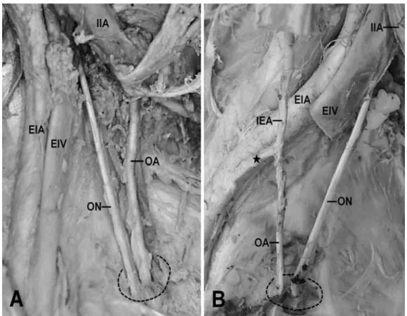
Fig. 1. The origin of the obturator artery. A: Normal obturator artery (OA) originating from the internal iliac artery (IIA) on the right side of pelvis in a male (73 years of age). The obturator artery enters the obturator foramen (dotted circle) with the obturator nerve (ON). B: A variant obturator artery originating from the proximal portion (asterisk) of inferior epigastric artery (IEA), a branch of the external iliac artery (EIA), on the right side of pelvis in a female (72 years of age). The length of proximal portion is 2.4 cm, and the obturator artery lies anterior to the obturator nerve in this case. EIV, external iliac vein.
laid anterior to the obturator nerve (Fig. 1B).
In summary, the variant obturator artery was observed in 11.1% (2/18) of cadavers or in 8.3% (3/36) of pelvic halves.
It was observed in 7.7% (1/13) of male cadavers and in 20% (1/5) of female cadavers. Bilateral presence of the variant obturator artery was observed in 5.6% (1/18) of cadavers, and was observed in 20% (1/5) of the female cadavers, whereas this was not observed in male cadavers.
Variant obturator artery was observed more frequently on the left side (2/18) than on the right side (1/18).
Discussion
In the present study, the variant obturator arteries were found in 11.1% (2/18) of cadavers or in 8.3% (3/36) of pelvic halves. The only variant origin of the obturator art- ery observed in the present study was that from the exter- nal iliac artery as a branch of the inferior epigastric artery.
It has been reported that the obturator artery could originate from the external iliac artery and from the femoral artery [4]. Of these, the latter was very rare [4]. It is widely known that in cases when the obturator artery originates from the external iliac artery, it usually arises from the inferior epi- gastric artery [4-10]. The frequency of this variant obturator artery has been reported in several studies (Table 1). Alth- ough investigators have found the incidence of variant obturator artery to be different across races and countries [4,6], the incidence (8.3%) of pelvic halves of Korean cadavers in the present study was similar to the incidence reported from a Japanese study [8]. No variant obturator artery was found to originate directly from the external iliac artery in the present study. The incidence of direct origin was reported to be considerably lower than that of origin from the inferior epigastric artery, which had been reported in 1.3% to 5.1% of pelvic halves [4,5,9,10] and in 25% of whole pelvis [6].
By the relative diameters of these arteries, Jakunowicz and Czarniawska-Grzesinska [5] clearly categorized the obturator artery variants into two types: one arising from a common trunk with the inferior epigastric artery and the other arising from the inferior epigastric artery. In the for- mer variant, the two arteries have same diameters, and the diameter of inferior epigastric artery is larger than that of the obturator artery in the latter variant. According to this classification, all cases from our study belonged to the type
arising from a common trunk. However, the use of the term
‘common trunk’ is likely to be improper from an embryo- logical point of view. The obturator artery and the inferior epigastric artery have individual pubic branches that are connected to each other at early developmental stages (Fig.
2A). During normal development, this connection between the pubic branches should disappear (Fig. 2B). However, if the proximal part of obturator artery is resorbed instead of the pubic branches, variant obturator arteries could be formed by the pubic branches of inferior epigastric artery and the obturator artery, or by the distal part of the obtu- rator artery (Fig. 2C). Consequently, the variant obturator artery might originate from the external iliac artery as a branch of the inferior epigastric artery (Fig. 2D). Although very rare, if the pubic branches remain and the proximal part of inferior epigastric artery disappears, the inferior epigastric artery could arise from the obturator artery that originated from the internal iliac artery [8,11].
With regard to the incidence of bilaterality of variant obturator artery, bilateral presence of variant obturator arteries was less frequent than unilateral presence [4,12].
According to a large study from the US, 16.9% of cadavers showed bilateral presence of variant obturator artery, while 30.9% of cadavers had unilateral presence of variant obtu- rator artery [4]. In that study, bilateral presence of variant obturator artery was more frequently observed in female subjects (31.8%) than in male subjects (15.8%). Further, variant obturator arteries were more frequently observed on the left side than on the right side of pelvis [4]. Although we studied only 36 pelvic halves, the trend observed in Korean cadavers was consistent with that of American cadavers in terms of incidence of bilaterality and sidedness, and differential frequency in both sexes.
It was speculated that variation in the origin of obturator artery does not cause clinical problems in individuals and Table 1. Frequencies of the variant obturator artery originating from the inferior epigastric artery in several countries
Number of Frequency of
Country pelvic halves variant obturator Reference
examined artery
Poland 75 6.6% [5]
Japan 560 10.5% [8]
India 98 14.3% [9]
US 640 26.7% [4]
UK 238 29.0% [10]
South Korea 36 8.3%
is occasionally detected during cadaveric dissections or autopsies [13]. Many authors have mentioned that variation in the origin of obturator artery might have to be considered by surgeons treating femoral or obturator hernia [7,9] and controlling hemorrhage in the pelvic region [14], and by radiologists interpreting angiograms of the pelvis [13]. The clinical importance of variant obturator arteries is high- lighted by a recent angiographic study in which variant obturator artery was observed at a higher frequency than previously found in cadaveric studies [12]. Using angio- graphy in pelvic trauma patients, the incidence of variant obturator arteries, bilateral or unilateral, was 55.1% in all American patients [12]. Considering this clinical impor- tance, further studies with larger number of Korean cada- vers are required to further elucidate anatomic characteris- tics, such as bilaterality, sidedness, gender relation, and frequency of the variation.
In conclusion, variant obturator arteries were observed in 8.3% of the pelvic halves from Korean cadavers, and
all of the variant obturator arteries arose from the external iliac arteries as branches of the inferior epigastric arteries.
Acknowledgements
We are deeply grateful to the deceased who had donated their bodies for medical education and research. This work was supported by the research grant of the Chungbuk National University in 2011.
References
1. Clemente CD. Gray’s Anatomy. 30th American ed. Philadel- phia: Lea & Febiger; 1985.
2. Williams PL, Warwick R, Dyson M, Bannister LH. Gray’s Anatomy. 37th ed. London and NY: Churchill Livingstone;
1989.
3. Moore KL, Agur AMR. Essential Clinical Anatomy. 3rd Fig. 2. Illustrations depicting developmental processes of variant obturator artery formation. The obturator artery (OA) and inferior epigas- tric artery (IEA) have individual pubic branches, which communicate with each other at early developmental stages (A). During normal development, the communication between pubic branches disappears (B). The proximal portion of obturator artery disappears instead of the pubic branches (C). Consequently, a variant obturator artery originates from the external iliac artery (EIA) as a branch of the inferior epigas- tric artery (D). CIA, common iliac artery; IIA, internal iliac artery.
ed. Lippincott Williams & Wilkins; 2007.
4. Pick JW, Anson BJ, Ashley FL. The origin of the obturator artery. A study of 640 body-halves. Am J Anat. 1942; 70:
317-43.
5. Jakubowicz M, Czarniawska-Grzesinska M. Variability in origin and topotraphy of the inferior epigastric and obtura- tor arteries. Folia Morphol. 1996; 2:121-6.
6. Missankov AA, Asvat R, Maoba KI. Variations of the pubic vascular anastomoses in black South Africans. Acta Anato- mica. 1996; 155:212-4.
7. Gilroy AM, Hermey DC, DiBenedetto LM, Marks SC, Page DW, Lei QF. Variability of the obturator vessels. Clin Anat.
1997; 10:328-32.
8. Kawai K, Honma S, Koizumi M, Kodama K. Inferior epiga- stric artery arising from the obturator artery as a terminal branch of the internal iliac artery and consideration of its rare occurrence. Ann Anat. 2008; 190:541-8.
9. Pai MM, Krishnamurthy A, Prabhu LV, Pai MV, Kumar SA, Hadimani GA. Variability in the origin of the obturator
artery. Clinics. 2009; 64:897-901.
10. Sañudo JR, Mirapeix R, Rodriguez-Niedenführ M, Maranil- lo E, Parkin IG, Vázquez T. Obturator artery revisited. Int Urogynecol J. 2011; 22:1313-8.
11. Won HS, Won HJ, Oh CS, Han SH, Chung IH, Kim DH.
The inferior epigastric artery arising from the internal iliac artery via a common trunk with the obturator artery. Anat Cell Biol. 2012; 45:285-7.
12. Requarth JA, Miller PR. Aberrant obturator artery is a com- mon arterial variant that may be a source of unidentified hemorrhage in pelvic fracture patients. J Trauma. 2011;
70:366-72.
13. Jusoh AR, Rahman NA, Latiff AA, Othman F, Das S, Ghafar NA, et al. The anomalous origin and branches of the obtu- rator artery with its clinical implications. Rom J Morphol Embryol. 2010; 51:163-6.
14. Daeubler B, Anderson SE, Leunig M, Triller J. Hemorrhage secondary to pelvic fracture: Coil embolization of an aber- rant obturator artery. J Endovasc Ther. 2003; 10:676-80.
폐쇄동맥의 이는곳 변이: 바깥엉덩동맥에서 일어난 아래배벽동맥의 가지
이은영, 김지영, 김후남, 손현준, 서제훈
충북대학교 의학대학 해부학교실
간추림 : 폐쇄동맥은 흔히 속엉덩동맥으로부터 일어난다. 여러 나라의 많은 연구 사례를 통해 폐쇄동맥의 이는 곳에 다양한 변이가 있음이 알려졌다. 우리나라에서는 이 변이에 대해 아직 보고된 바 없기에, 본 연구에서는 충북대학교 의과대학에 기증된 시신을 대상으로 폐쇄동맥의 이는곳에 대한 변이를 조사하였다. 총 18구 (남성 13, 여성 5)의 시신으로부터 36쪽의 골반을 연구 대상으로 하였다. 연구 대상인 18구의 시신 중 16구 (88.9%), 36쪽의 골반 중 33쪽(91.7%)에서는 속엉덩동맥에서 일어나는 정상적인 폐쇄동맥이 관찰되었다. 폐쇄동맥의 이 는곳에 대한 변이는 총 18구의 시신 중 2구(11.1%), 36쪽의 골반 중 3쪽(8.3%)에서 관찰되었다. 이러한 변이가 좌우 양쪽에 모두 존재하는 경우는 총 18구의 시신 중 1구(5.6%)였다. 변이를 보이는 경우, 폐쇄동맥의 이는곳 은 모두 바깥엉덩동맥에서 분지한 아래배벽동맥에서 일어났으며 폐쇄동맥은 아래안쪽으로, 아래배벽동맥은 위 가쪽으로 주행하였다.
찾아보기 낱말 : 폐쇄동맥, 아래배벽동맥, 바깥엉덩동맥
교신저자 : 서제훈(충북대학교 의과대학 해부학교실) 전자우편 : [email protected]