Received August 12, 2014, Revised November 4, 2014, Accepted for publication December 15, 2014
Corresponding author: Do Won Kim, Department of Dermatology, Kyungpook National University Hospital, 130 Dongdeok-ro, Jung-gu, Daegu 700-721, Korea. Tel: 82-53-420-5838, Fax: 82-53-426-0770, E-mail: [email protected]
*These authors contributed equally to this work.
This is an Open Access article distributed under the terms of the Creative Commons Attribution Non-Commercial License (http://
creativecommons.org/licenses/by-nc/4.0) which permits unrestricted non-commercial use, distribution, and reproduction in any medium, provided the original work is properly cited.
ORIGINAL ARTICLE
Alopecia Areata in the Elderly: A 10-Year Retrospective Study
Yong Hyun Jang*, Kyung Hea Park*, Sang Lim Kim, Hyun Jung Lim1, Weon Ju Lee, Seok-Jong Lee, Do Won Kim
Department of Dermatology, Kyungpook National University School of Medicine, 1BL Plastic Surgery and Dermatology Clinic, Daegu, Korea
Background: Alopecia areata (AA) is an organ-specific auto- immune disease that typically occurs in young adults. AA in the elderly is relatively rare, thus little data have been reported. Objective: This study aimed to understand the clin- ical characteristics of AA in the elderly. Methods: We per- formed a 10-year retrospective study of AA in the elderly who visited our dermatologic clinic from January 2002 to December 2011. A clinical review of medical records and telephone interviews were performed by two dermatologists.
Results: Among 1,761 patients with newly diagnosed AA, 61 (3.5%) were older than 60 years at the first visit. Among those who completed a telephone interview, 74.3% (26/35) had less than 50% of scalp-localized hair loss. There was no asso- ciation between the extent of AA and hair graying (p=0.679).
Favorable therapeutic response was observed in 62.9%
(22/35) of cases. Conclusion: AA in the elderly shows mild disease severity and favorable treatment response. There is no association between graying and the extent of AA.
However, the influence of aging on the pathogenesis of AA in the elderly deserves further investigation. (Ann Dermatol 27(4) 411∼416, 2015)
-Keywords-
Aged, Alopecia areata, Autoimmune diseases
INTRODUCTION
Alopecia areata (AA) is a common immune-mediated dis- ease typified by multiple round areas of hair loss on the scalp. It presents as asymptomatic well-defined patches of non-scarring alopecia1. The lifetime prevalence of AA is 1.7%∼2% in the United States2,3. The disease usually oc- curs in young adults, and its incidence peaks between 20 and 25 years of age2. The first disease episode typically presents before 20 years of age2. One study reports that 85.5% of Asian patients with AA present before 40 years of age4. AA in the elderly is relatively rare, thus there are few reports. A previous study indicated that late-onset AA is characterized by a marked female predominance and milder disease activity with increasing age5. The aim of our study is to better understand the demographics and clinical characteristics of AA in the elderly. In addition, we investigated the relationship between graying and the ex- tent of AA, because the AA process preferentially targets pigmented hair, and increasing evidence indicates that the melanogenic follicular melanocytes are a principal target in AA6.
MATERIALS AND METHODS
Patient population
From January 2002 to December 2011, 1,761 patients newly diagnosed with AA were retrospectively identified who had visited the Department of Dermatology, Kyungpook National University Hospital, Daegu, Korea. Among them, 61 patients 60 years of age or older were included for analysis. However, 9 patients were lost to follow-up, 2
Table 1. Age and sex distribution of alopecia areata patients at first visit
Age (yr) Male Female Total
60∼64 11 (18.0) 18 (29.5) 29 (47.5) 65∼69 9 (14.8) 4 (6.6) 13 (21.3) 70∼74 3 (4.9) 9 (14.8) 12 (19.7)
75∼79 4 (6.6) 1 (1.6) 5 (8.2)
80∼84 - 1 (1.6) 1 (1.6)
≥85 - 1 (1.6) 1 (1.6)
Total 27 (44.3) 34 (55.7) 61 (100)
Values are presented as number (%).
passed away, 10 had difficulty communicating because of impaired hearing or severe illness, and 5 refused to be interviewed. Therefore, 35 patients were finally analyzed.
Study design
We performed a retrospective study using clinical medical records and telephone interview performed by two der- matologists. The following characteristics were evaluated:
sex, age of onset, duration of disease, AA severity (extent) and type, family history, past history, coexisting systemic and/or dermatologic disease, gray hair distribution at first visit, therapeutic response, and clinical course. Most data were obtained from medical records, but some were sub- jectively assessed by telephone interview, particularly gray hair distribution at first visit and clinical course.
For analysis of the chronological changes, the patients were divided into six age groups: 60∼64, 65∼69, 70∼
74, 75∼79, 80∼84, and >84 years. The types of AA were classified as basic AA (less than five patches), AA multiplex (five patches or more), alopecia totalis (entire scalp involved), and alopecia universalis (total body in- volved). The severity of scalp hair loss at first visit was categorized as <50%, 50%∼99%, or 100% of the scalp.
Hair loss on other parts of the body and nail involvement at the first consultation were evaluated in accordance with the investigative guidelines for AA by Olsen et al.7: no body hair loss (B0), some body hair loss (B1), and 100%
body hair loss (B2); nail involvement was rated as none or some (N0 and N1, respectively). Treatment response with respect to baseline (i.e., first visit) was classified as better, unchanged, worse, or wax and wane. These 4 grades were also used during telephone interview to rate patients’
mental and physical health status. The grade “better” was further separated into 3 subgroups according to the per- centage of improved hair loss area: complete recovery (i.e., “cured”), improved area >50%, and improved area
≤50% of the alopecic area.
Statistical analysis
The data were analyzed by SPSS Statistics ver. 17.0 for Windows (SPSS Inc., Chicago, IL, USA). The association between gray hair and the extent of AA was analyzed by the χ2 test. A p-value of less than 0.05 was considered as statistically significant.
RESULTS
Sex ratio
The mean age of the 61 patients was 71 years. There were 27 males (44.3%) and 34 females (55.7%) with the male-to-female ratio approximating 1:1.26 (Table 1).
Disease onset of AA and duration from onset to first visit
The age of onset of AA varied greatly. Among 61 patients, 29 (47.5%) had an age of onset from 60∼64 years, which was the most common onset age group (Table 1). The du- ration from the recognition of initial hair loss to first hospi- tal visit was <3 months in 16 patients (26.2%) and 3∼5 months in 12 patients (19.7%).
Severity of AA at first visit
Of the 35 patients who finished the full survey, 26 (74.3%) had <50% hair loss with scalp involvement at first visit, followed by seven (20.0%) and two (5.7%) with 50%∼99% and 100% hair loss with scalp involvement, respectively. Thirty-one (88.6%) patients had no hair loss on other parts of the body, and 29 (82.9%) patients also had no nail involvement (Table 2).
Clinical type of AA at first visit and ophiasis
Among the 35 AA patients, 25 (71.4%) had the basic form of AA with less than five hair loss patches; this group in- cluded two patients with ophiasis. Eight (22.9%) patients had AA multiplex, including one with ophiasis. None of the patients had alopecia totalis, and only two (5.7%) had alopecia universalis.
Past and family history of AA, and stressful events before AA onset
Three (8.6%) patients had a past history of AA. Thirty-three (94.3%) patients had no family history of AA. Fourteen (40.0%) patients reported that a stressful event occurred before AA onset.
Coexisting systemic and dermatologic diseases
Twenty-three (65.7%) patients had coexisting systemic dis- eases, with hypertension being the most common. Thyroid disease (hypothyroidism) was present in only one patient.
Table 3. Coexisting systemic and dermatologic diseases (n=35) Coexisting diseases Male Female Total Systemic diseases
No 5 7 12 (34.3)
Yes 12 11 23 (65.7)
Hypertension 5 5 10 (35.7)
Diabetes mellitus 4 - 4 (14.3)
Hepatic disease 1 2 3 (10.7)
Thyroid disease 1 - 1 (3.6)
Others 6 4 10 (35.7)
Dermatologic diseases
No 13 16 29 (82.9)
Yes 4 1 5 (14.3)
Pruritus 2 1 3 (8.6)
Urticaria 1 - 1 (2.9)
Seborrheic capitis 1 - 1 (2.9)
Values are presented as number only or number (%).
Others: angina, myocardial infarction, osteoarthritis, osteopo- rosis, disc herniation, gastric cancer, and benign prostatic hyperplasia.
Table 2. Alopecia areata (AA) severity by age at first visit (n=35)
Extent of AA Age (yr)
60∼64 65∼69 70∼74 75∼79 ≥80 Total
Hair loss of scalp involvement (%)
<50 10 6 6 4 - 26 (74.3)
50∼99 4 2 1 - - 7 (20.0)
100 1 1 - - - 2 (5.7)
Hair loss on other parts of the body
B0 (none) 14 8 6 3 - 31 (88.6)
B1 (some) - 1 1 1 - 3 (8.6)
B2 (all) 1 - - - - 1 (2.9)
Area of hair loss
Whole body 1 - - - - 1 (2.9)
Eyebrow only - 1 1 - - 2 (5.7)
Beard - - - - - -
Pubic hair - - - 1 - 1 (2.9)
Other parts - - - - - -
None 14 8 6 3 - 31 (88.6)
Nail involvement
N0 (none) 11 9 6 3 - 29 (82.9)
N1 (some) 4 - 1 1 - 6 (17.1)
Values are presented as number only or number (%).
Table 4. Association between gray hairs and the extent of alopecia areata (AA) at first visit (n=35)
Extent of AA (%)
Gray hairs (%)
0 1∼49 50∼99 100 Total
<50 7 (20.0) 13 (37.1) 2 (5.7) 4 (11.4) 26 (74.3) 50∼99 3 (8.6) 2 (5.7) 1 (2.9) 1 (2.9) 7 (20.0)
100 1 (2.9) - - 1 (2.9) 2 (5.7)
Total 11 (31.4) 15 (42.9) 3 (8.6) 6 (17.1) 35 (100) Values are presented as number (%). p=0.679, χ2 test.
Five (14.3%) patients presented with coexisting dermato- logic diseases: pruritus cutanea, urticaria, and seborrheic capitis were observed in three, one, and one, respectively (Table 3).
Gray hair in AA patients
At first visit, 11 (31.4%) patients had dark hair, while 21 (68.6%) had gray hair (from 1%∼100%). There was no
significant association between graying and the extent of AA (p=0.679; Table 4).
Treatment modalities
All patients except one received a combination of two or more different treatment modalities; 30 of the 34 treated patients were treated with topical steroids. Other treat- ment modalities included intralesional corticosteroids, minoxidil, diphenylcyclopropenone (DPCP) immunotherapy, oral medication such as a systemic steroid (low-dose con- tinuous or minipulse), immunosuppressants (cyclosporine), antihistamines, and vitamins. Only one patient received no treatment (Table 5).
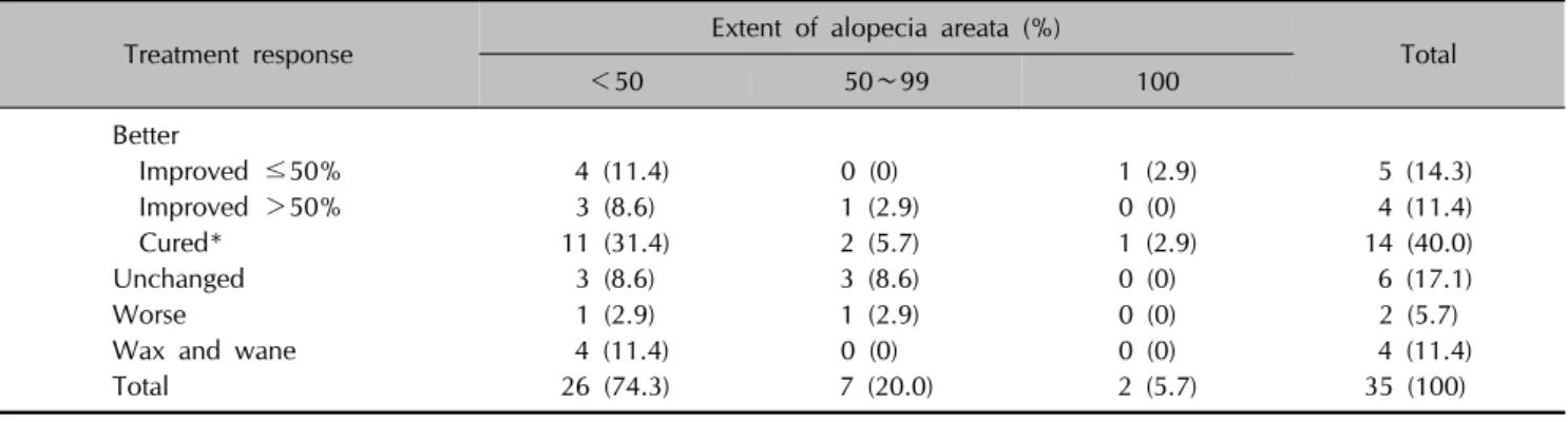
Therapeutic response and clinical course
Of the 35 patients, 22 (62.9%) showed a positive response in clinical course. Fourteen (40.0%) patients were cured,
Table 5. Treatment modalities (n=35)
Treatment modalities* Value
Topical treatments Corticosteroid Minoxidil
Intralesional corticosteroid injection Diphenylcyclopropenone (DPCP)
immunotherapy Excimer laser
Topical calcineurin inhibitor
30 (85.7) 9 (25.7) 25 (71.4) 2 (5.7) 2 (5.7) 2 (5.7) Systemic treatments
Corticosteroid (PO) Corticosteroid pulse (IV) Cyclosporine
Other systemic agents (antihistamines, vitamins, and hair nutrients)
3 (8.6) 2 (5.7) 1 (2.9) 20 (57.1)
No treatment 1 (2.9)
Values are presented as number (%). *Thirty-four of 35 patients received combination therapy with 2 or more different treatment modalities.
Table 6. Therapeutic response and clinical course according to the severity of hair loss (n=35)
Treatment response Extent of alopecia areata (%)
Total
<50 50∼99 100
Better
Improved ≤50% 4 (11.4) 0 (0) 1 (2.9) 5 (14.3)
Improved >50% 3 (8.6) 1 (2.9) 0 (0) 4 (11.4)
Cured* 11 (31.4) 2 (5.7) 1 (2.9) 14 (40.0)
Unchanged 3 (8.6) 3 (8.6) 0 (0) 6 (17.1)
Worse 1 (2.9) 1 (2.9) 0 (0) 2 (5.7)
Wax and wane 4 (11.4) 0 (0) 0 (0) 4 (11.4)
Total 26 (74.3) 7 (20.0) 2 (5.7) 35 (100)
Values are presented as number (%). *Includes 1 patient with spontaneous recovery.
and five (14.3%) showed ≥50% improvement of the alo- pecic area. Four (11.4%) patients showed <50% improve- ment of the alopecic area. Six (17.1%) patients demon- strated unchanged clinical course, two (5.7%) worsened, and four (11.4%) showed a waxing and waning response (Table 6).
DISCUSSION
The frequency of AA ranges from 0.7%∼3.8% among pa- tients attending dermatology clinics4,8. AA is thought to be mediated by an autoimmune process and manifests as pat- chy non-scarring hair loss. AA is most commonly diag- nosed in people between 20 and 25 years old2; thus, re- ports of AA in the elderly are rare. Previous reports of the overall population with AA show that it affects males and females equally9. However, female predominance was ap-
parent in the present study, with a male/female ratio of 1:1.26. Wu et al.5 also reported a female predominance (male/female ratio=1:2) in late-onset AA, with first onset at age 50 years and above. However, Statistics Korea an- nounced that overall the male/female ratio in the elderly population older than 60 years (n=7,606,903) was 1:1.34 in 2010; this rate is similar to the male/female ratio in the present study, suggesting that the sex distribution in AA in the elderly is similar to the sex distribution of the elderly in general population.
It is well documented that the prognosis of AA is propor- tional to the severity of the disease at onset10. The present results show that 74.3% (26/35) had an extent of hair loss
<50%. This mild severity is concordant with a previous report of late-onset AA in patients aged age 50 years and above5. Yang et al.11 also report that the early-onset AA group (age of onset ≤30 years) showed a greater severity and longer duration than the late-onset group (age of onset
>30 years). In the present study, 62.9% of elderly AA cas- es responded well to treatment. Therefore, AA in the eld- erly is characterized by mild clinical severity and better treatment response.
A hypothesis from a previous report on AA suggests that hair follicle melanocytes may be a primary target of im- munologic attack6. In line with this hypothesis, the phe- nomenon of AA preferentially affecting pigmented hair but sparing graying/white hair is commonly observed5. This hypothesis can also explain the “turn white overnight”
phenomenon such as ‘Marie Antoinette syndrome’ for the condition afflicting women and ‘Thomas More syndrome’
for men by selectively affecting dark hairs and leaving gray hairs12. In the present study, 31.4% (11/35) of AA pa- tients had dark hair without graying. This rate is quite high compared to our previous report in which graying scalp hair was present in 94.2% (49/52) of AA patients with Korean ancestry at age 60∼69 years13. In relation with this result, it is presumed that gray hair could be one of
the reasons for the rarity of AA in the elderly. However, no significant association between gray hair and extent of AA was found in the present study.
AA is well known to be associated with atopic disease and various autoimmune diseases, particularly thyroid disease and vitiligo1,4,14,15. The prevalences of atopy and thyroid diseases in AA patients are estimated to be as high as 46%
and 19%, respectively5,8. In the present study, there were no patients with atopic dermatitis (AD) or vitiligo, and on- ly one patient (3%) had thyroid abnormalities among 35 elderly AA patients. One-year prevalence rates of AD are much lower in the elderly than young people16,17. Becerril Angeles et al.16 reported that the prevalence of AD during the senile phase (≥60 years) is 0.6%; in comparison, Rodríguez Orozco and Núñez Tapia17 reported that it is 10.1% in people aged 6∼10 years. The low prevalence of older AD patients with AA observed in the present study suggests a positive relationship between the rareness of AD and old age. In contrast to the general AA population, in our study, there was only one case of thyroid disease and no cases of vitiligo among the elderly. Although such low prevalences of these diseases may be one of the fea- tures of elderly patients and the reasons for the few cases of coexisting AD and thyroid disease in the present study are unclear, the tendency of the low prevalence in the present study may be related to aging. However, a large-scale study is required to clarify this. Immunosene- scence is defined as all the changes occurring in the aged immune system. Two contrasting phenomena coexist in immunosenescence: a decreasing immune response, and increased autoantibody production. The former may ex- plain why the elderly population has decreased suscepti- bility to AA, like the results of our study, and the latter may demonstrate the pathogenesis of immunobullous dis- orders such as bullous pemphigoid, which is charac- terized by the production of either antibodies that react with host tissue or autoreactive immune effector T cells18,19. AA and AD are immune-mediated skin diseases associated with cutaneous immune system malfunction. Therefore, the decreased ability of immune system, immunosenescence may be associated with lower incidences of both diseases.
In conclusion, this study demonstrates that AA in the eld- erly is characterized by mild disease severity and favor- able treatment response. Dark scalp hair is commonly ob- served in elderly AA patients. The limitations of this study include a limited review which was performed using only medical records and telephone interview and the small numbers of patients involved. The influence of aging on the pathogenesis of AA in the elderly deserves to be stud- ied further, and larger-scale studies are required for further evaluation.
ACKNOWLEDGEMENT
This work was supported by Biomedical Research Institute grant, Kyungpook National University Hospital (2014).
REFERENCES
1. Alkhalifah A, Alsantali A, Wang E, McElwee KJ, Shapiro J.
Alopecia areata update: part I. Clinical picture, histo- pathology, and pathogenesis. J Am Acad Dermatol 2010;62:
177-188.
2. Alzolibani AA. Epidemiologic and genetic characteristics of alopecia areata (part 1). Acta Dermatovenerol Alp Pan- nonica Adriat 2011;20:191-198.
3. Chu SY, Chen YJ, Tseng WC, Lin MW, Chen TJ, Hwang CY, et al. Comorbidity profiles among patients with alopecia areata: the importance of onset age, a nationwide popul- ation-based study. J Am Acad Dermatol 2011;65:949-956.
4. Tan E, Tay YK, Goh CL, Chin Giam Y. The pattern and profile of alopecia areata in Singapore--a study of 219 Asians. Int J Dermatol 2002;41:748-753.
5. Wu MC, Yang CC, Tsai RY, Chen WC. Late-onset alopecia areata: a retrospective study of 73 patients from Taiwan. J Eur Acad Dermatol Venereol 2013;27:468-472.
6. Trautman S, Thompson M, Roberts J, Thompson CT. Me- lanocytes: a possible autoimmune target in alopecia areata.
J Am Acad Dermatol 2009;61:529-530.
7. Olsen E, Hordinsky M, McDonald-Hull S, Price V, Roberts J, Shapiro J, et al. Alopecia areata investigational assessment guidelines. National Alopecia Areata Foundation. J Am Acad Dermatol 1999;40:242-246.
8. Sharma VK, Dawn G, Kumar B. Profile of alopecia areata in Northern India. Int J Dermatol 1996;35:22-27.
9. Wasserman D, Guzman-Sanchez DA, Scott K, McMichael A. Alopecia areata. Int J Dermatol 2007;46:121-131.
10. McDonagh AJ, Tazi-Ahnini R. Epidemiology and genetics of alopecia areata. Clin Exp Dermatol 2002;27:405-409.
11. Yang S, Yang J, Liu JB, Wang HY, Yang Q, Gao M, et al.
The genetic epidemiology of alopecia areata in China. Br J Dermatol 2004;151:16-23.
12. Trüeb RM, Navarini AA. Thomas More syndrome. Der- matology 2010;220:55-56.
13. Kim DW, Shin DJ, Lee SJ, Chung SL, Kim JC. Statistical and clinical study of gray hair. Korean J Dermatol 1999;37:
1567-1575.
14. Barahmani N, Schabath MB, Duvic M; National Alopecia Areata Registry. History of atopy or autoimmunity increases risk of alopecia areata. J Am Acad Dermatol 2009;61:
581-591.
15. Hordinsky M, Ericson M. Autoimmunity: alopecia areata. J Investig Dermatol Symp Proc 2004;9:73-78.
16. Becerril Angeles M, Vázquez Merino CL, Angeles Garay U, Alvarado Moctezuma LE, Vilchis Guízar E. Prevalence of allergic diseases in the elderly. Rev Alerg Mex 2008;55:
85-91.
17. Rodríguez Orozco AR, Núñez Tapia RM. Prevalence of atopic dermatitis in 6-14 year old children in Morelia,
Michoacan, Mexico. Rev Alerg Mex 2007;54:20-23.
18. Loo WJ, Burrows NP. Management of autoimmune skin disorders in the elderly. Drugs Aging 2004;21:767-777.
19. Lew BL, Cho HR, Haw S, Kim HJ, Chung JH, Sim WY.
Association between IL17A/IL17RA gene polymorphisms and susceptibility to alopecia areata in the Korean po- pulation. Ann Dermatol 2012;24:61-65.