간혈관종은 간의 가장 흔한 양성 종양으로서, 대개는 선별 초음파검사나 다른 목적으로 C T나 MR 검사를 시행했을 때 우연히 발견되는 경우가 많다.그럴 경우 초음파에서 간혈관종 의 전형적 소견을 보이면 감별진단에 큰 문제가 없으나, 비전 형적으로 보일 경우, 원발간암이나 전이암, 임파종 등의 악성 종양과 구별이 힘들며, 어떤 경우는 불필요한 생검을 시행하 기도 한다. 다양하게 보일 수 있는 간혈관종의 초음파소견을 이해함으로써 이런 오류를 줄일 수 있다.
간혈관종의 전형적 초음파 소견과 다른 영상소견
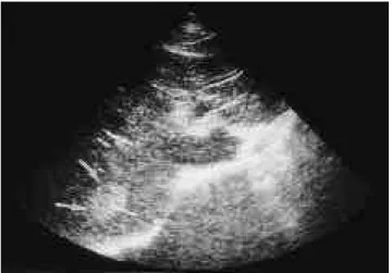
조직학적으로 작은 크기의 간혈관종은 다수의 섬유격막에 의해 나누어 지는 큰 혈액점유공간으로, 한층의 편평한 내피 세포로 싸여있다. 내부의 이런 다중계면에 의해서‘전형적’인 간혈관종의 초음파소견이 나타나게 된다. 간혈관종의 가장 흔 한 초음파소견은 크기가 보통 4 c m이하이며 균일한 고에코의 경계가 좋은 병변이고 간피막하에 위치하는 것이다. 약간의 후방 투과증강을 가지며 추적검사에서 크기의 변화가 없는 것이 보통이다 (Fig. 1). 그러나 크기가 큰 간혈관종은 내부에 다양한 변화가 나타나서 다양한 초음파소견을 보일 수 있으 며 약 2 0 - 40% 정도의 간혈관종이 이에 해당되는 것으로 알려 져 있다 ( 1 ) .
이런 비전형초음파소견을 보이는 혈관종의 감별진단을 위 한 다른 영상 진단 방법으로는 T c - 99m RBC SPECT, 역동적 간 CT, MR 등이 있다. Tc-99m RBC SPECT에서는, 초기관
류단계에서는 방사능이 감소되어 있으나 시간이 지남에 따라 점차적으로 방사능이 증가되어, 5분내지 2시간 지연혈액풀영 상(delayed blood pool image)에서 결국 열소로 나타난다.
Jacob- son등 ( 2 )은 2cm 이상의 크기를 갖는 혈관종의 예민도 는 1 00%, 2cm 이하의 혈관종에서의 예민도는 초음파상 전형 적 간혈관종은 38%, 비전형적 간혈관종은 75%로 보고한 바 있다. 이는 초음파소견이 T c - 99m RBC SPECT의 예민도와도 연관성이 있음을 시사하며, 크기가 2cm 이하일 경우는 확진이 쉽지 않다는 것을 시사한다. 역동간C T에서는 조영제주입전에 정상 간보다 저음영으로 보이고 조영제주입후에는 주변부에 초기조영증강을 보이며, 점차로 병변의 내부로 조영증강이 진 행되고 3분에서 6 0분 사이에는 완전히 채워져서 간실질과 같
비전형간혈관종의 초음파분류
1배상진・윤권하2・김표년・하현권・이문규・오용호
간혈관종은 간의 가장 흔한 양성 종양으로 알려져 있다. 간혈관종의 가장 흔한 초음파 소 견은 크기가 보통 4 c m이하이며 균일한 고에코의 경계가 좋은 병변이고 간피막하에 위치하 는 것이다. 약간의 후방 에코증강을 가지며 추적검사에서 크기의 변화가 없는 것이 보통이 다. 그러나 그 크기가 큰 경우나 다수의 혈관종이 있을 때 초음파에서 다양한 소견을 보일 수 있다. 따라서 간암이나 전이성 종양, 임파종 등의 다른 종양과의 감별이 어려울 경우가 있다. 간혈관종의 다양한 초음파 소견을 이해하는 것이 조기 진단과 불필요한 생검의 방지 에 도움을 줄 수 있으며, 이 임상화보에서는 간혈관종의 초음파 소견을 중심으로 CT, MR 과 Tc-99m RBC SPECT 소견을 기술하고자 한다.
1울산의대 서울중앙병원 진단방사선과학교실
2원광대학교 의과대학병원 방사선과
이 논문은 1 9 9 9년 9월 3 0일 접수하여 1 9 9 9년 1 2월 3일에 채택되었음.
Fig. 1. Typical hemangioma on US.
There is a 3.3 cm sized, uniformly hyperechoic lesion(arrows) in the right lobe.
은 음영을 보이게 되지만 작은 혈관종인 경우에는 이러한 소 견에 맞지 않는 경우도 있다. MRI는 혈관종을 진단하는데 예 민도와 특이도가 매우 높은 방법이다. 특히 T2 강조영상에서 신호강도가 매우 높게 나타나며 역동적 조영증강에서는 C T와 비슷한 조영증강을 보인다 ( 3 , 4 ) .
비전형 간혈관종의 초음파 소견
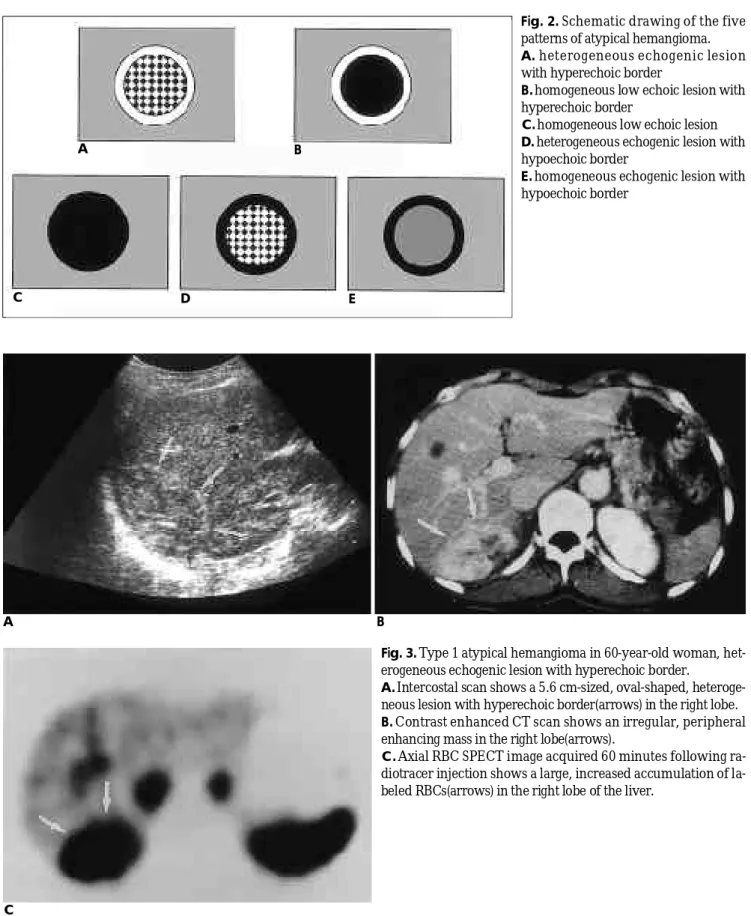
초음파에서 비전형 간혈관종의 소견은 다음과 같은 5가지 로 나타날 수 있다: (1) 고에코경계의 비균일한 병변( h e t e r o-
Fig. 2. Schematic drawing of the five patterns of atypical hemangioma.
A. heterogeneous echogenic lesion with hyperechoic border
B. homogeneous low echoic lesion with hyperechoic border
C. homogeneous low echoic lesion D. heterogeneous echogenic lesion with hypoechoic border
E. homogeneous echogenic lesion with hypoechoic border
A B
C
Fig. 3. Type 1 atypical hemangioma in 60-year-old woman, het- erogeneous echogenic lesion with hyperechoic border.
A. Intercostal scan shows a 5.6 cm-sized, oval-shaped, heteroge- neous lesion with hyperechoic border(arrows) in the right lobe.
B. Contrast enhanced CT scan shows an irregular, peripheral enhancing mass in the right lobe(arrows).
C. Axial RBC SPECT image acquired 60 minutes following ra- diotracer injection shows a large, increased accumulation of la- beled RBCs(arrows) in the right lobe of the liver.
A
C D E
B
A B
Fig. 6. Type 3 atypical hemangioma in 67-year-old man, homogeneous low echoic lesion.
A . US with right intercostal scan shows a 4 cm-sized, homogeneous low echoic lesion(arrows) in the right lobe.
B . T2W MRI(FSE, TR/TE 2800/100) shows a lesion with ‘bright bulb’appearance of high signal intensity(arrows) in the corresponding area.
Fig. 4. Type 1 atypical hemangioma in 30-year-old man.
Transverse scan shows a heterogeneous echoic lesion with hy- perechoic border(arrows) and contour bulging out beyond the liver surface.
Fig. 5. Type 2 Atypical hemangioma, homogeneous low echoic lesion with hyperechoic border.
US with transverse scan shows a 3 cm-sized, homogeneous low echoic lesion with hyperechoic border(arrows) in the right lobe of the liver.
A B
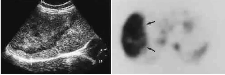
Fig. 7. Type 4 atypical hemangioma in 40-year-old woman, heterogeneous echogenic lesion with hypoechoic border.
A. US with subcostal scan shows a huge, heterogeneous echoic lesion with a thick hypoechoic border(arrows).
B. RBC SPECT image shows a large accumulation of labeled RBCs in the corresponding area(arrows) .
geneous echogenic lesion with hyperechoic border) (2) 고에코경 계의 균일한 저에코병변(homogeneous low echoic lesion with hyperechoic border) (3) 균일한 저에코병변(homogeneous low echoic lesion) (4) 저에코경계의 비균일한 병변( h e t e r o g e n e o u s echogenic lesion with hypoechoic border) (5) 저에코경계의 균 일한 병변(homogeneous echogenic lesion with hypoechoic bor- der) (Fig. 2).
제 1형, 고에코 경계의 비균일한 병변(Fig. 3, 4)
비균일한 내부에코를 가지는 병변으로 나타나는 혈관종으 로, 내부의 고에코부위가 병변 크기의 2 0 - 80%정도를 차지하 는 형이다 (Fig. 3.A-C). 이런 내부의 비균일성은 혈관종의 다 양한 변성, 즉 괴사를 동반한 내부출혈, 혈전증, 점액변성, 섬
유화와 석회화 등에 기인한다 (1,5,6). 초음파에서 혈관종은 병 변가장자리에 고에코의 테두리가 있는 경우가 많은데 일정한 두께로 나타나거나 편측으로 더 두껍게 보일 수도 있다. 이 고 에코의 테두리는 아마도 혈관종 자체 내부정맥동 벽들의 계면 ( i n t e r f a c e )에서 보이는 전형적인 혈관종의 고에코가 이차적내 부변성 이후에도 종괴가장자리를 따라 남아있기 때문으로 추 정된다 (5). 이런 병변의 에코도와 무관하게 때로는 간의 경계 바깥으로 돌출하여 악성종양으로 오인할 수도 있다 (Fig. 4).
제 2 형, 고에코 경계의 균일한 저에코 병변(Fig. 5)
고에코 경계를 가지면서 균일한 저에코로 보이는 혈관종이 며, 내부의 고에코 부위가 병변 크기의 20%미만을 차지하는 형이다. 초음파에서 간혈관종의 에코는 주위 간에코와 비교하
A B
Fig. 8. Type 4 atypical hemangioma with a cystic portion within it.
A . 7 cm-sized heterogeneous echoic lesion with hypoechoic border(arrows) and an echo-free area(arrow heads) B. Contrast enhanced CT scan shows a large mass and a low density portion within it.
A B
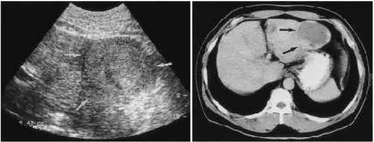
Fig. 9. Type 5 atypical hemangioma, homogeneous echogenic lesion with hypoechoic border.
A. US with transverse scan shows 6 cm-sized, homogeneous echogenic lesion with hypoechoic border(arrows) in left lateral seg- ment of liver.
B. CT scan shows a low density mass with peripheral, nodular enhancement in the same area.
여 기술하게 되는데, 종양 내부의 변화뿐 아니고 지방간 등이 있어 주위 간에코가 증가하였을 때에도 저에코의 종양으로 나타나게 된다 ( 7 ) .
제 3형, 균일한 저에코 병변(Fig. 6)
고에코의 경계가 없으면서 내부가 균일한 저에코로 보이는 혈관종으로, 역시 내부의 고에코부위가 병변 크기의 20%미만 을 차지하는 형이다.
제 4형, 저에코 경계의 비균일한 병변(Fig. 7, 8)
저에코의 경계를 가지면서 내부는 비균일한 에코로 보이는 혈관종이며, 내부의 고에코부위가 병변 크기의 2 0 - 80%를 차 지하는 형이다. 혈관종주위의 경계가 저에코로도 보일 수 있 는데 이는 조직학적으로, 섬유조직으로 구성된 얇은 위막 ( p s e u d o c a p s u l e )때문에 생긴다고 보고된 바 있으며 극히 드물 게는 액체를 함유한 중앙공동을 가질 수도 있다 ( 6 ) .
제 5형, 저에코 경계의 균일한 병변(Fig. 9)
저에코의 경계를 가지면서 내부는 그보다 약간 높은 균일 한 에코로 보이는 혈관종이다.
이러한 혈관종의 비전형소견은 드물게 추적 초음파에서 내 부 에코가 변할 수 있으며 (1,8) 이는 주변 간에코의 변화도 관계 있을 것으로 생각된다.
간혈관종의 이런 다양한 초음파 소견을 이해하는 것이 조
기 진단과 불필요한 생검의 방지, 적절한 다음 검사의 선택에 도움이 된다.
참 고 문 헌
1 . Moody AR, Wilson SR. Atypical hepatic hemangioma: a suggestive sonographic morphology. R a d i o l o g y 1993; 188: 413-417
2. Jacobson AF, Teefey SA. Cavernous hemangiomas of the liver: as- sociation of sonographic appearance and results of Tc-99m labeled red blood cell SPECT. Clin Nucl Med 1993; 19: 96-99
3 . Nelson RC, Chezmar JL. Diagnostic approach to hepatic heman- giomas. Radiology 1990; 176: 11-13
4 . Bree RL, Schwab RE, Glazer GM, Fink-Bennett D. The varied ap- pearance of hepatic cavernous hemangiomas with sonography, computed tomography, magnetic resonance imaging and scintigra- phy. R a d i o g r a p h i c s 1987; 7: 1153-1175
5 . Lee CH, Ko YT, Lee DH, Lim JW, Yoon Y. The significance of e- chogenic rim of atypical hepatic hemangioma on ultrasonogram. J Korean Radiol Soc 1996; 35: 751-755
6 . Takayasu K, Moriyama N, Shima Y, et al. Atypical radiographic findings in hepatic cavernous hemangioma: correlation with histo- logic features. AJR 1986; 146: 1149-1153
7 . Marsh JI, Gibney RG, Li DKB. Hepatic hemangioma in the pres- ence of fatty infiltration: an atypical sonographic appearance.
Gastrointest Radiol 1989; 14: 262-264
8 . Gibney RG, Hendin AP, Cooperberg PL. Sonographically detected hepatic hemangiomas: absence of change over time. A J R 1 9 8 7 ; 149: 953-957
J Korean Radiol Soc 2000;42:3 17- 3 2 1
Address reprint requests to : Pyo Nyun Kim, M.D., Department of Diagnostic Radiology, Asan Medical Center, University of Ulsan, College of Medicine, #388-1, Pungnap-dong, Songpa-gu, Seoul 138-736, Korea.
Tel. 82-2-224-4376 Fax. 82-2-476-4719
U l t ra s o n o g raphic Classification of Atypical Hepatic Hemangiomas
1Sang-Jin Bae, M.D., Kwon-Ha Yoon, M.D.2, Pyo Nyun Kim, M.D., Hyun Kwon Ha, M.D., M o o n - Gyu Lee, M.D., Yong Ho Auh, M.D.
1Department of Diagnostic Radiology, Asan Medical Center, University of Ulsan, College of Medicine
2Department of Radiology, Wonkwang University Hospital
Cavernous hemangioma is the most common benign hepatic tumor. Typically, the most common features revealed by ultrasound(US) include its small size(4cm or less in diameter), uniform hyperechogenicity, well- defined margins, position in the subcapsular region of the right lobe of the liver, and some posterior echo en- hancement. In addition, follow-up scanning may reveal changes in size, though this is rare. The US findings of hepatic hemangiomas may vary, however, especially when lesions are large and/or multiple. For that reason, differential diagnosis between this condition and hepatocellular carcinomas, metastatic lesions, lymphomas and other tumors is difficult. An understanding of the various sonographic findings of hepatic hemangioma can facilitate the early detection of the condition.
Index words :Liver, neoplasms Liver, US
경희의대 진단방사선과에서는 3월 1일(수요일) 방사선해부학(흉부 및 복부) 연수교육을 시행하려 합니다.
수강하시는 전공의와 전문의에게 많은 도움이 되리라 생각합니다.
체부방사선해부학 연수교육
09 : 0 0 - 0 9 : 3 0 Embryology of abdominal organ 이 선 화(이 화 의 대) 09 : 3 0 - 1 0 : 0 0 Hepatic segmental anatomy 이 문 규(울 산 의 대)
1 0 : 0 0 - 1 0 : 3 0 Perihepatic space 박 철 민(고 려 의 대)
1 0 : 3 0 - 1 0 : 5 0 휴 식
1 0 : 5 0 - 1 1 : 2 0 Lesser sac 고 영 태(경 희 의 대)
1 1 : 2 0 - 1 1 : 5 0 Omentum and mesentery 이 동 호(경 희 의 대)
1 1 : 5 0 - 1 3 : 1 0 점 심
1 3 : 1 0 - 1 3 : 4 0 Imaging of airways 임 정 기(서 울 의 대)
1 3 : 4 0 - 1 4 : 1 0 Imaging of pericardium 성 동 욱(경 희 의 대) 1 4 : 1 0 - 1 4 : 4 0 Diaphragm: anatomic, radiologic 이 경 수(성균관의대)
and pathologic consideration 1 4 : 4 0 - 1 5 : 0 0 휴 식
1 5 : 0 0 - 1 5 : 3 0 Radiology of thoracic catheter, 이 기 남(동 아 의 대) wire and tube
1 5 : 3 0 - 1 6 : 0 0 Retroperitoneal anatomy 임 주 원(경 희 의 대) 1 6 : 0 0 - 1 6 : 3 0 Pelvic extraperitoneal space 조 경 식(울 산 의 대)
일 시 : 2000년 3월 1일(수요일)
장 소 : 경희대학교 중앙도서관 시청각교육실
연수책임교수 : 윤 엽 연수담당교수 : 이동호
연수평점 : 6점
신청방법 : 사전등록 후 송금 전 화 : 02)958-8625, 8621 FAX : 02)968-0787
사전등록마감 : 2000 2월 2 9일 (연수당일 환불 불가) 수 강 료 : 전문의 및 일반의 - 5만원
전공의 - 3만원
송금구좌 : 서울은행 3 5 1 0 4 - 1 0 3 1 3 2 1 (예금주 : 오 주 형)
☞ 중식제공, 대학교 무료주차(병원주차 불가)
☞ 비디오 촬영불가