Goblet cell carcinoid tumor is a rare neoplasm that shows histological features of both adenocarcinoma and carcinoid tumors. Goblet cell carcinoid tumor of the ap- pendix (GCTA) primarily arises from the appendix, but can also occur in other parts of the gastrointestinal tract (1-5). The common clinical presentations in order of fre- quency are acute appendicitis (22.5%), asymptomatic (5.4%), non-localized abdominal pain (5.15%) and the presence of an appendiceal mass (3.09%) (5). Small bow- el obstruction is one of the rare clinical presentations of GCTA (1-5). To the best of our knowledge, the CT find- ing of GCTA with small bowel obstruction has not yet been reported.
Here we present a case report of GCTA with small bowel obstruction in an 80-year-old man.
Case Report
An 80-year-old man was admitted to our hospital for an abdominal pain. He had been admitted to our hospi- tal on two prior occasions and had been diagnosed with obstructive small bowel ileus two months earlier. The patient did not undergo abdominoplevic CT during the previous admissions. His medical history included hy- pertension, but he had no history of previous abdominal operations. Physical examination showed diffuse mild abdominal tenderness with hypoactive bowel sound.
Laboratory tests were unremarkable except for the pres- ence of an increased white blood cell count (14.3×
103/mm3). Plain abdominal radiographs revealed dilata- tion of the small bowel loops with air-fluid levels com- patible with a small bowel obstruction.
Abdominoplevic Multi Detector Row Computed Tomography (MDCT) was performed prior to the ad- ministration of intravenous contrast material, and a por- tal venous phase CT scan was taken after IV contrast ad- ministration. No oral or rectal endoluminal opacification
Goblet Cell Carcinoid Tumor of the Appendix with Small Bowel Obstruction: A Case Report1
Su Yeon Hwang, M.D., Kyung Mi Jang, M.D., Min Jeong Kim, M.D., Sung Hye Koh, M.D., Eui Yong Jeon, M.D., Kwang Seon Min, M.D.2, Jin Won Seo, M.D.2, Hyoung Chul Park, M.D.3
1Department of Radiology, Hallym University, College of Medicine, Korea
2Department of Pathology, Hallym University, College of Medicine, Korea
3Department of Surgery, Hallym University, College of Medicine, Korea Received May 4, 2009 ; Accepted May 25, 2009
Address reprint requests to : Kyung Mi Jang, M.D., Department of Radiology, Hallym University, College of Medicine 896 Pyungchon-dong, Dongan-gu, Anyang-city, Kyungki-do 431-070, Korea.
Tel. 82-31-380-3885 Fax. 82-31-380-3878 E-mail: [email protected]
Goblet cell carcinoid tumor of the appendix (GCTA) is a tumor with histological fea- tures of both adenocarcinoma and carcinoid tumors. The most common clinical pre- sentation of GCTA is acute appendicitis, although small bowel obstruction has been re- ported as a rare clinical symptom of GCTA. However, to the best of our knowledge, the CT feature of small bowel obstructions in patients with GCTA has not been report- ed to date. Here, we present a case of small bowel obstruction in a patient with GCTA caused by extensive tumor infiltration at the terminal ileum and distal ileum.
Index words :Carcinoid tumor Appendix
Tomography, X-Ray computed Intestine, small
was administered. The CT scan showed dilated small bowel loops with a transition point at the medial aspect of the cecum and a narrowed terminal ileum (Fig. 1). At the transition point, the distal ileum was collapsed and adhered to the terminal ileum (Fig. 1B). The proximal portion of the appendix was also adhered to the terminal ileum at the inferior aspect of the transition point (Fig.
1C). The appendix was not thoroughly traced on the CT scan. The cecum near the orifice of the appendix and the portion of the appendix that was visible showed mild diffuse wall thickening with enhancement (Figs.
1B, C). The CT also showed mild diffuse wall thickening with enhancement at the distal ileum around the transi-
tion point (Figs. 1B, C). Mutual adhesions among the ter- minal ileum, distal ileum, and appendix with mild wall thickening were visualized around the transition point without demonstrable pathologic process, and a smooth transition zone was noted. We could not predict the cause of the small bowel obstruction precisely and be- lieved that the adhesion itself provoked narrowing of the terminal ileum and the small bowel obstruction. We considered the mild wall thickening at the cecum, ap- pendix, and distal ileum as secondary findings of the small bowel obstruction.
The patient underwent conservative medical treat- ment for five days. However, the small bowel obstruc- tion was not improved upon serial follow-up plain ab- dominal radiographs, and he complained of aggravating abdominal pain; therefore, he underwent laparotomy.
During surgery, severe inflammation at the appendix that was adhered to the terminal ileum was observed.
Multiple subcentimeter peritoneal seeding nodules were also observed. Because these findings suggested appen- diceal carcinoma, the intraoperative frozen biopsy of the peritoneal seeding nodule was performed. Pathological examination of the frozen section revealed poorly differ- entiated metastatic adenocarcinoma. Based on the pathological results, an extended right hemicolectomy was performed.
Upon pathological examination, the mucosal surface of the resected specimen showed a small nodular lesion at the appendiceal orifice and mild to moderate narrow- ing of the terminal ileum (Figs. 2A, B). The serosal sur- C
A B
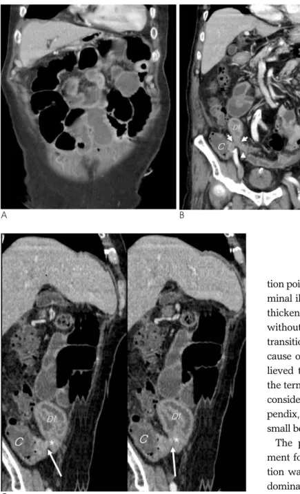
Fig. 1. Goblet cell carcinoid tumor of the appendix in an 80-year-old man.
A, B. Reformatted coronal contrast en- hanced CT images show dilated small bowel loops with a transition point (short arrows) at the medial aspect of the cecum (C) and narrowed terminal ileum (arrowheads). At the transition point, distal ileum (DI) is adhered to the terminal ileum (arrowheads).
C. Two serial curved planar reforma- tion images of a transition point show the partially visible appendix (long ar- row) placed at the inferior aspect of the transition point and adhered to the terminal ileum (asterisk). There is mild diffuse wall thickening with enhance- ment at the appendix and cecum (C) near the orifice of the appendix.
face of the small intestine and cecum was diffusely fi- brotic. Except for the appendiceal orifice, the appendix was not identified. The pericecal soft tissue showed dif- fuse fibrotic changes without discrete mass formation.
Upon microscopic examination, there were diffusely in- filtrated islands of tumor cells distended by mucus that resembled signet-ring cells in the appendix (Fig. 2C).
The tumor involved the serosa, subserosal soft tissue, proper muscle, and submucosa of the cecum, and there were scattered islands of tumor cells in the subserosal soft tissue of the small intestine. Immunohistochemical studies revealed that the tumor cells were reactive for chromogranin, synaptophysin, and CD56, and the final pathologic diagnosis was goblet cell carcinoid of the ap- pendiceal orifice.
Discussion
GCTA accounts for less than 5% of primary tumors of the appendix, whereas the classic carcinoid tumor of the appendix accounts for 32% to 57% of all appendiceal tu- mors (5, 6). The classic carcinoid tumor of the appendix demonstrates a uniquely indolent clinical course and has a 5-year survival rate of greater than 90%. In addi- tion, carcinoid tumors metastasize in 2 to 5% of all cases (5, 6). In contrast, GCTA is a low-grade malignancy with a 5-year survival rate of between 60 and 84%.
Moreover, GCTA metastasizes in 15 to 30% of all cases (5, 6). Therefore, right hemicolectomy is usually per- formed to reduce the risk of metastatic disease (2).
However, the vast majority of goblet cell carcinoid tu- mors are rarely diagnosed preoperatively and are usual- ly diagnosed during operation or the pathologic exami- nation of the appendiceal specimen (2). GCTA is an infil- trative tumor that typically involves the entire appendix.
These characteristics are manifested as a diffuse mural thickening of the appendix with or without peritoneal seeding lesions or ovarian metastasis upon CT (7). The infiltrative nature of GCTA was demonstrated as a par- tially visible appendix with mild diffuse enhancing wall thickening in this case (Figs. 1B, C). Moreover, the ex- traappendiceal tumor infiltration into the terminal ileum caused a small bowel obstruction in this case.
In the evaluation of small bowel obstructions on CT, it is important to define the cause of the obstruction. If the cause is not identified, a diagnosis of adhesions is usual- ly inferred. Adhesive ileus is common in patients who C
A B
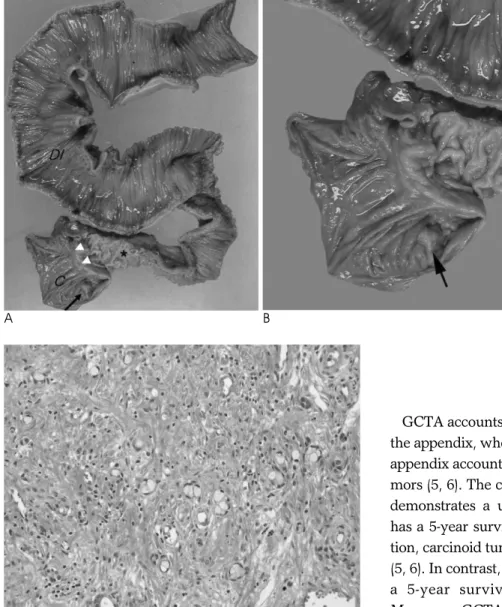
Fig. 2. Pathologic findings of the re- sected specimen following right hemi- colectomy.
A, B. The resected gross specimen of the right hemicolectomy shows mild to moderate narrowing of the terminal ileum (asterisk) and dilated distal ileum (DI). The appendiceal orifice of the resected cecum (C) shows a small elevated submucosal lesion (long ar- row). Arrowheads = Ileocecal valve C. On the photomicrograph (original magnification, ×200; H-E stain) there are scattered islands of tumor cells showing abundant intracytoplasmic mucin in the appendix.
have undergone previous abdominal operations (8). In this case, the patient had no history of previous abdomi- nal operations. However, we suggested adhesive ileus preoperatively because we thought that the small bowel obstruction had no cause and we overlooked the possi- bility of an infiltrative appendiceal lesion. In addition to GCTA, the classic carcinoid tumor, lymphoma, and nonmucinous adenocarcinoma of the appendix can show an infiltrative growth pattern (7, 9, 10). Classic car- cinoid tumors of the appendix are not typically detected by direct imaging studies due to their small size and lo- cation in the distal appendix. Lymphoma manifests as marked enlargement of the appendix with diffuse wall thickening but relative maintenance of its vermiform shape. Thus, in this case, the possibilities of lymphoma and classic carcinoid tumor are low. However, nonmu- cinous adenocarcinoma of the appendix can manifest as an infiltrative appendiceal lesion with extraappendiceal infiltration and direct invasion of the adjacent organs (7).
Nonmucinous adenocarcinomas rarely occur in the ap- pendix and tend not to form mucoceles. Its reported CT finding is a subtle infiltrative appendiceal lesion with surrounding periappendiceal infiltration with or without direct invasion of the adjacent organs (5).
In summary, GCTA in this case presented as a small bowel obstruction. On pathological examination, GCTA was found to be associated with diffuse extraappen- diceal infiltration of the terminal ileum and distal ileum, which caused a small bowel obstruction. However, we did not notice the malignant appendiceal cause of the small bowel obstruction preoperatively. When small bowel obstructions with a transition point at the right lower quadrant abdomen are encountered, it is impor-
tant to evaluate the appendix thoroughly. If the appen- dix is partially visible at this transition point, the possi- bility of an infiltrative appendiceal lesion such as a gob- let cell carcinoid tumor should be considered, especially in patients with no previous abdominoplevic operation.
References
1. Maes M, Segers K, Cheyns P. Goblet cell carcinoid of the appen- dix: laparoscopic appendectomy or right hemicolectomy? Acta Chir Belg 2008;108:447-450
2. Toumpanakis C, Standish RA, Baishnab E, Winslet MC, Caplin ME. Goblet cell carcinoid tumors (adenocarcinoid) of the appen- dix. Dis Colon Rectum 2007;50:315-322
3. Tang LH, Shia J, Soslow RA, Dhall D, Wong WD, O’Reilly E, et al.
Pathologic classification and clinical behavior of the spectrum of goblet cell carcinoid tumors of the appendix. Am J Surg Pathol 2008;32:1429-1443
4. van Eeden S, Offerhaus GJ, Hart AA, Boerrigter L, Nederlof PM, Porter E, et al. Goblet cell carcinoid of the appendix: a specific type of carcinoma. Histopathology 2007;51:763-773
5. Pahlavan PS, Kanthan R. Goblet cell carcinoid of the appendix.
World J Surg Oncol 2005;20:36
6. Tchana-Sato V, Detry O, Polus M, Thiry A, Detroz B, Maweja S, et al. Carcinoid tumor of the appendix: a consecutive series from 1237 appendectomies. World J Gastroenterol 2006;12:6699-6701 7. Pickhardt PJ, Levy AD, Rohrmann CA Jr, Kende AI. Primary neo-
plasms of the appendix: radiologic spectrum of disease with patho- logic correlation. Radiographics 2003;23:645-662
8. Hayanga AJ, Bass-Wilkins K, Bulkley GB. Current management of small-bowel obstruction. Adv Surg 2005;39:1-33
9. Pickhardt PJ, Levy AD, Rohrmann CA Jr, Abbondanzo SL, Kende AI. Non-Hodgkin’s lymphoma of the appendix: clinical and CT findings with pathologic correlation. AJR Am J Roentgenol 2002;
178:1123-1237
10. Bittle MM, Chew FS. Radiological reasoning: recurrent right lower quadrant inflammatory mass. AJR Am J Roentgenol 2005;185:S188- S194
대한영상의학회지 2009;61:173-177
소장폐쇄를 일으킨 충수돌기의 술잔세포 유암종:
증례 보고1
1한림대학교 성심병원 영상의학과
2한림대학교 성심병원 병리과
3한림대학교 성심병원 일반외과
황수연∙장경미∙김민정∙고성혜∙전의용∙민광선2∙서진원2∙박형철3
충수의 술잔세포 유암종은 유암종과 선암종 두 종양의 조직학적 특징을 모두 가지는 종양이며, 충수염이 가장 흔 한 합병증이다. 드물게 충수의 술잔세포 유암종에 의해 장폐색이 유발될 수 있으며 보고된 바 있다. 그러나, 현재까 지 충수의 술잔세포 유암종에 의한 장폐색의 전산화단층촬영 소견에 대한 보고 및 고찰은 없었다. 이에 저자는 충수 의 술잔세포 유암종이 말단회장 및 원위부 회장 침윤으로 인해 소장의 폐색을 초래한 사례를 보고하고자 한다.