C ASE R EPORT J Kor Neurotraumatol Soc 2(2):124-127, 2006
124 J Kor Neurotraumatol Soc
Monoplegia after Anterior Cervical Discectomy and Fusion
- A Case Report -
Ki Suk Choi, M.D., Il Tae Jang, M.D., Hyang Kwon Park, M.D., Yong C Sheen, M.D., and Jae Hyeon Lim, M.D.
Department of Neurosurgery, Nanoori Hospital, Seoul, Korea
Spinal cord injury is one of the complications of the anterior cervical surgery. Motor weakness is occurred as a result of spinal cord injury and most of the motor weaknesses are accompanied by sensory deficit. We experienced a case of monoplegia of the lower extremity without sensory deficit after anterior cervical discectomy and fusion (ACDF). A 36-year-old male patient had multiple cervical disc protrusion at C4-5, 5-6 and 6-7. He had no neurologic deficit. Monoplegia was developed at the early postoperative period of ACDF. Reoperation was performed, but a definite spinal cord compression was not showed on operative field. We thought that spinal cord ischemia was the cause of monoplegia and was related to the operative posture of the hyperextension of the neck and poor blood supply to the spinal cord. A case of monoplegia without sensory deficit has not been reported. Therefore, we discuss the pathophysiology of monoplegia without sensory deficit.
Key Words: Anterior cervical discectomy and fusion․Monoplegia․Spinal cord ischemia
Corresponding Author: Ki Suk Choi, M.D.
Department of Neurosurgery, Nanoori Hospital, 63-8, Nonhyun-dong Kangnam-gu, Seoul, 135-010, Korea Tel: 82-2-3446-9797, Fax: 82-2-3448-0210 E-mail: [email protected]
*본 논문의 요지는 대한신경외과학회 2004년도 추계학술대회 에서 발표 되었슴.
INTRODUCTION
Complications of anterior surgery for cervical radiculopathy or myelopathy include carotid or vertebral artery laceration, esophageal tears, recurrent laryngeal nerve injury, spinal cord injury, upper air way difficulties from edema or hematoma formation, and dis- placement of grafted bone or implant system2,3).
Spinal cord injury of anterior surgery for cervical spine was reported as 1.04% at cervical spine research society(6)reference. The weakness of upper and/or lower extremities is one of the com- plications after cervical operation. Most of motor weaknesses are accom- panied by sensory deficit. We operated a 36-year-old man who had cervical disc protrusions at C4-5, 5-6 and 6-7.
After anterior cervical discectomy and fusion, he complained of right leg monoplegia without sensory deficit of any extremities.
Reoperaion was performed. But on operative field, spinal cord compression was not noted.
Motor weakness was recovered finally. Monoplegia without sensory deficit after anterior cervical surgery is a rare complication.
Therefore, we discuss the pathophysiology.
CASE REPORT
A 36-year-old man, who complained of neck pain, numbing sensation on four extremities and the subjective weakness of both legs while walking, visited our hospital.
The physical examination showed no motor and sensory deficits.
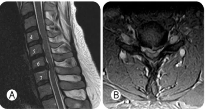
Magnetic resonance (MR) imaging showed multiple cervical disc protrusions at C4-5, 5-6 and 6-7 (Fig. 1). A standard microsurgical anterior approach to C4-5, 5-6 and 6-7 and a discectomy and interbody fusion with Smith-Robinson technique were performed.
The patient’s position was supine and neck was hyperextended.
A vertical skin incision along the anterior border of SCM muscle was done. Dissection was performed medial to SCM muscle, carotid sheath and Longus coli muscle. The discectomy was done from caudal to cranial level of disc. After decom- pression of neural structure, the interbody fusion was performed with an allograft bone and fixed with Zephir plate (Sofamor
KS Choi, et al.
Volume 2, No 2 December, 2006 125 Fig. 1. A: Sagittal T2-weighted MR imaging showing disc
protrusion at C4-5, 5-6 and 6-7. B: Axial T2-weighted MR imaging showing severe central protrusion of disc material at C6-7.
Fig. 2. A: Postoperative sagittal T2-weighted MR imaging showing high signal intensity of spinal cord at C6-7. B:
Postoperative axial T2-weighted MR imaging showing high signal intensity of spinal cord at right C6-7.
Danek, Memphis. TN). The operation time was about 160 minutes.
Before closure of operation wound, an intraoperative lateral radio- graph control was done, and the correct position of the implant and allograft bone was checked.
The patient complained of right leg weakness after recovery from general anesthesia.
The physical examination showed monoplegia of right leg, with intact sensory function. The other extremities had no neurologic deficits. The follow up MR imaging showed spinal cord compre- ssion and high signal intensity of spinal cord on T2-weighted image at C6-7 (Fig. 2).
Megadose steroid therapy was started and reoperation was done. However on operative field, a definite cord compression was not showed. Right leg monoplegia was recovered finally.
DISCUSSION
Causes of spinal cord injury are known as mechanical compre- ssion by operative instruments, posterior displacement of grafted bone, and ischemia of spinal cord.
The mcidence of spinal cord injury with resultant quadriplegia, tetraplegia or paraplegia after ACDF is known as 0.1%11). Two- thirds of these results in tetraplegia and one-third in Brown- Sequard syndrome.5,11) Monoplegia of lower extremity was not reported.
We experienced monoplegia of lower extremity without sensory deficits as an early postoperative complication. In this case, the patient had multilevel disc protrusions at C4-5, 5-6 and C6-7, but motor and sensory functions were intact except subjective weakness of both leg while walking.
Intraoperative mechanical trauma was absent and duration of operative time was relatively short as 160 minutes. Therefore the cause of monoplegia of the leg was thought to be the spinal cord ischemia. The operative posture of the neck and circulation of blood supply can be considered as the factor affecting spinal cord ischemia.
The operative posture of the patient was supine and hyperex- tension of neck with a pillow under shoulder. Athletes possessing a narrow cervical canal have a high incidence of transient quadri- plegia secondary to hyperflexion, hyperextension and compressive forces12). Between C3 and C7, the diameter of the canal is 17 to 18 mm10). If the canal 13 mm or less, it is stenotic by defini- tion2). A sagittal diameter of below 12 mm is critical to the pro- duction of cervical myelopathy11).
Chen CJ et al.3) studied a functional cord impingement at flexion-extension cervical MR imaging. MR imaging in 31%
showed functional cord impingement at extension MR imaging.
If the patient had both stabilization degeneration (disc protrusion or osteophytic formation with hypertrophy of the ligamentum flavum) and C7 canal space of 10 mm or less, probability of functional cord impingement at extension MR imaging could increase to 79%. Spinal canal space was defined as the diameter of C7 at the midvertebral level of the most midsagittal section.
C7 vertebra was chosen because it was less involved with spondylosis and might represent the true canal diameter of the
Monoplegia after Anterior Cervical Discectomy and Fusion
126 J Kor Neurotraumatol Soc
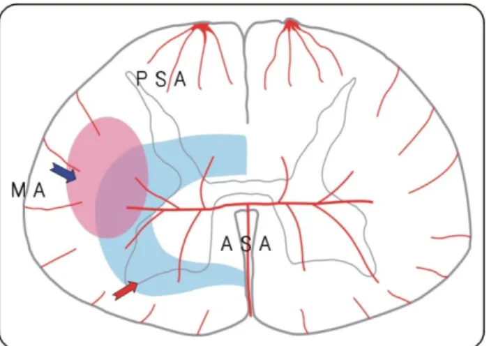
Fig. 3. Blood supply of cervical spinal cord, PSA: pos- terior spinal artery, ASA: anterior spinal artery, MA: margi- nal artery, lateral corticospinal tract (blue arrow), water- shed area of vascular territory (red arrow).
middle and lower cervical spine. In this case, an actual sagittal diameter at C6-7 was narrow due to disc protrusion.
The hyperextension of neck can aggravate decreasing of spinal canal space. This can develop the spinal cord ischemia due to compression of spinal arteries.
Blood supply to the cervical spinal cord is from anterior spinal artery, posterior spinal artery and marginal artery (Fig. 3). Anterior spinal artery gives rise to a number of sulcal branches, and supplies the anterior two-thirds of the spinal cord. This includes the lateral and anterior funiculi, the anterior third of the pos- terior funiculi, and all of the gray matter anterior to the subs- tantia gelatinosa. The posterior spinal arteries are paired and supply, via the posterior pial plexus, the posterior third of the cord. This includes the posterior two-thirds of the posterior funiculi, the dorsolateral fasciculus of Lissauer, and the marginal and sub- stantia gelatinosa parts of the dorsal horn of the gray matter. The pial arterial plexus supples the peripheral parts of the cord, whereas the sulcal arteries supply the cental gray matter7,9,11).
Overlap between the two systems occurs in the inner third to quarter of the white matter, and outer edge of the gray matter except the posterior half of the posterior horn7,8,9,11). This is a
“water-shed” zone of vascular territories in the spinal cord (Fig. 3)11). Within the cervical spinal cord, there is a differential vulner- ability to direct compression of descending white fiber tracts.
The corticospinal tract is most vulnerable to direct compression.
There is followed by the anterior horn cells, anterior funiculus and posterior funiculus11).
We think that the lateral corticospinal tract is belong to watershed area of these arteries, and therefore was most vulnerable to the ischemia than the other tracts of the cervical spinal cord.
CONCLUSION
Monoplegia of the lower extremity after anterior surgery of cervical disc protrusion was not reported. We tried to explain this phenomenon as spinal cord ischemia due to mechanical com- pression of spinal cord. The surgical position of neck and cervi- cal blood supply are considered as related factors.
The hyperextension of neck is responsible for spinal cord ischemia when spinal canal stenosis is severe. The lateral cortico- spinal tract is most vulnerable to the ischemia because it is belong to the water-shed area of the cervical spinal cord partly.
Therefore monoplegia of the lower extremity without sensory deficit was possible.
REFERENCES
1. Arthur HW: Spine care, ed 1. Louis: Mosby pp1368-1378, 1995
2. Bohlman H: Cervical spondylosis and myelopathy. Instr Course Lect 44:81-97, 1995
3. Chen CJ, Hsu JL, Niu CC, Chen TY, Chen MC, Tseng YC, et al: Cervical degenerative disease at flexion-extension MR imaging: prediction criteria. Radiolgy 227(1):136-142, 2003 4. Emery SE, Smith MD, Bohlman HH: Upper-airway obstruction
after multilevel cervical corpectomy. J Bone Joint Surg 73(4):
544-551. 1991
5. Flynn TB: The neurologic complications of anterior cervical interbody fusion. Spine 7:536-539, 1982
6. Graham JJ: Complications of cervical spine surgery. Sping 14:1046-1050, 1989
7. Hassler O: Blood supply to the human spinal cord. A micro- angiographic study. Arch Neurol 15:302-307, 1996
8. Jurnball IM, Brieg A, Hassler O: Blood supply of cervical spinal cord in man. A microangiographic cadaver study. J Neurosci 24:951-965, 1996
KS Choi, et al.
Volume 2, No 2 December, 2006 127 9. Lazorthes B, Gouaze A, Zadek JO, Santini JJ, Lazorthes Y,
Burdin P: Arterial vascularization of the spinal cord: Recent studies of the anastomotic substitution pathways. J Neurosurg 35:253-262, 1971
10. Matsunaga S, Sakou T, Taketomi E, Yanaguchi M, Okano T: The natural course of myelopathy caused by ossification of the posterior longitudinal ligament in the cervical spine.
Clin Orthop Relat Res 308:168-177, 1994
11. Paul HY: Microsurgery of cervical spine, New York: Raven pp185-190, 1991
12. Torg JS, Pavlov H, Genuario SE, Sennett B, Wisneski RJ, Robie BH, et al: Neuropraxia of the cervical spinal cord with transient quadriplegia. J Bone Joint Surg 68A:1354- 1370, 1986