대한소화기학회지 2009;53:206-210
접수: 2008년 4월 10일, 승인: 2008년 10월 6일 연락처: 박 혁, 540-719, 전남 순천시 조례동 1742번지
성가롤로병원 소화기내과
Tel: (061) 720-2111, Fax: (061) 720-6160 E-mail: [email protected]
Correspondence to: Hyuk Park, M.D.
Department of Gastrointestinal Medicine, Saint Callolo Hos- pital, 1742, Jorye-dong, Suncheon 540-719, Korea
Tel: +82-61-720-2111, Fax: +82-61-720-6160 E-mail: [email protected]
육안으로는 보이지 않고 현미경 검사로만 진단된 점액과분비 담관유두종증
순천 성가롤로병원 소화기내과, 외과*
정종혁ㆍ박 혁ㆍ문승원ㆍ문장식ㆍ명보현ㆍ김도현ㆍ김호동ㆍ한 철*
A Case of Macroscopically Unvisualized Mucin-hypersecreting Biliary Papillomatosis Diagnosed by Microscopy
Jong Hyeok Jeong, M.D., Hyeuk Park, M.D., Seung Won Moon, M.D., Jang Sik Mun, M.D., Bo Hyun Myoung, M.D., Do Hyun Kim, M.D., Ho Dong Kim, M.D., and Chul Han, M.D.*
Departments of Gastrointestinal Medicine and General Surgery*, Saint Carollo Hospital, Suncheon, Korea
The mucin-hypersecreting biliary papillomatosis is a premalignant neoplasm characterized by intraductal papillary proliferation involving extensive areas of the intrahepatic and/or extrahepatic bile duct. We report a case of mu- cin-hypersecreting biliary papillomatosis manifested as obstructive jaundice and diagnosed only by microscopy, with a review of literatures. A 74-year-old female, who had a past history of cholecystectomy about 13 years ago, was admitted to our hospital with jaundice. A CT scan showed marked dilatation of intrahepatic and extra- hepatic bile duct without intraductal filling defect or extrabiliary mass. During endoscopic retrograde chol- angiopancreatography, mucin extrusion from the duodenal major papilla and dilated common bile duct with amor- phous filling defects was noted. Percutaneous transhepatic biliary drainage for cholangioscopy was failed. In the operation field, there was a lot of mucin but was no visible mass at the common bile duct with bare eyes and cholangioscopy. However, papilloma was detected at the random biopsy specimen by microscopy. The patient un- derwent partial resection of common bile duct and choledocho-jejunal anastomosis. (Korean J Gastroenterol 2009;53:206-210)
Key Words: Mucin-hypersecreting biliary papillomatosis; Obstructive jaundice
서 론
점액과분비 담관유두종증은 담관 내에 유두모양 종양들이 다양한 분포와 범위로 발생하는 것을 특징으로 하는 양성 종양으로, 육안으로 확인할 수 있는 다량의 점액이 담관 내 로 분비되어 담관 폐쇄를 유발함으로써 독특한 임상 및 영 상의학 소견을 나타낸다.1-3 이 종양들은 선종에서부터 선암 종으로 진행하는 전암 병변으로 최근 그 보고가 증가하고
있다.1-9
점액과분비 담관유두종증은 담관 폐쇄로 인한 황달, 발 열, 복통 등의 증상을 보이고, 초음파나 복부 CT 등으로 담 관의 종양이나 담관 내에 존재하는 점액의 발견이 어려워 보고된 대부분의 증례들은 경피경간담관경으로 담관 점막 의 변화를 육안으로 직접 관찰함으로써 확진이 가능하였 다.1-3
저자들은 폐쇄 황달이 발생하여 임상 및 영상의학 검사로
정종혁 외 7인. 현미경 검사로만 진단된 점액과분비 담관유두종증 207
Fig. 1. Abdominal CT (A) and MRCP (B) findings. Markedly dilated intrahepatic and extra- hepatic bile ducts are noted, al- though there is no intraductal filling defect.
Fig. 2. ERCP findings. On en- doscopic view, mucin flows out from duodenal major papilla (A), and there are amorphous mutiple filling defects at common bile duct on cholangiogram (B).
담관유두종증이 의심되었던 환자에서 경피경간담관경과 수 술 시 육안으로는 유두종이 관찰되지 않았으나 조직검사에 서만 확진된 점액과분비 담관유두종증 1예를 경험하였기에 문헌 고찰과 함께 보고한다.
증 례
74세 여자 환자가 내원 약 1개월 전부터 시작된 복통을 동반하지 않은 황달과 소양증으로 내원하였다. 환자는 13년 전 담석으로 담낭절제술을, 10년 전에 급성 충수염으로 충 수절제술을 시행 받은 것 외에 건강하였다. 외래에서 시행 한 진찰에서 혈압 140/100 mmHg, 맥박 80회/분, 호흡수 18 회/분, 체온 36.0oC였고, 급성 병색을 보이며 의식은 명료하 였다. 공막과 피부에 황달 소견이 관찰되었고, 복부진찰에 서 통증이나 반발압통은 없었고, 간이나 비장 종대 및 종괴 도 촉지되지 않았다. 입원 당시 시행한 말초 혈액 검사에서 백혈구 6,900/mm3, 혈색소 11.1 g/dL, 혈소판 253,000/mm3이 었고, 혈청 생화학 검사에서는 총 단백 8.2 g/dL, 알부민 4.1
g/dL, AST 98 IU/L, ALT 118 IU/L, alkaline phosphatase 605 IU/L, r-GTP 746 IU/L, 총 빌리루빈 10.3 mg/dL, 직접 빌리루 빈 8.0 mg/dL이었다.
복부 CT 및 자기공명 담췌관조영술(magnetic resonance cholangiopancreatography, MRCP)에서 간내외 담관의 분명한 확장과 좌측 간내 담관에 결석으로 의심되는 병변이 발견되 었으나 간내외 담관 전체를 확장시킬 만한 총담관 말단부의 음영 결손은 보이지 않았다(Fig. 1). 내시경역행담췌관조영 술(endoscopic retorgrade cholangiopancreatography, ERCP)에서 십이지장의 주유두 개구부를 통해 다량의 점액이 배출되었 고, 늘어난 총담관 내에 부정형의 음영 결손이 다수 확인되 었다(Fig. 2).
ERCP 후, 내시경 비담관배액술(enodoscopic nasobiliary drai- nage, ENBD)을 시행하였으나 담즙은 전혀 배출되지 않았다.
점액과분비 담관유두종증이 의심되어 병변을 육안으로 확 인하고 병변의 범위를 파악하기 위해 경피경간담관경 검사 를 염두에 두고 경피경간담관배액술(percutaneous transhepa- tic biliary drainage, PTBD)을 시도하였으나 실패하였다.
208 The Korean Journal of Gastroenterology: Vol. 53, No. 3, 2009
Fig. 4. Operation finding. There is large amount of mucin, but is no tumor in the biliary tract by naked eye.
Fig. 5. Microscopic findings. (A) Papillary proliferation and adenoma are seen at the mucosal surface of common bile duct (H&E stain,
×80), (B) lined by tall columnar epithelial cells having minimal pleomorphism and regular oval nulei (H&E stain, ×200).
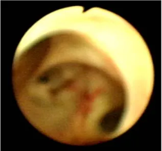
Fig. 3. Cholangioscopic finging. There is no tumor at the biliary tract.
이후 병변의 확진을 위해 수술 중 담관경 검사를 시행하 여 그 결과에 따라 바로 수술을 포함한 치료 계획을 세우기 로 하였다. 수술실에서 시행한 담관경 검사에서 총담관과 우측 간내 담관에 다량의 점액이 가득 찬 것 이외에 종괴는 관찰되지 않았으며, 좌측 간내 담관에 있는 결석을 전기수 압쇄석술 후에 제거하고 좌측 간내 담관을 관찰하였으나 역 시 종괴는 보이지 않았다(Fig. 3). 수술 시야에서 총담관에 다량의 점액이 존재하는 것이 관찰되었으나 종괴는 보이지 않았고(Fig. 4), 잔류 담낭관 등 병변으로 의심되는 담관 부 위를 절제하고 담관-공장 문합술을 시행하였다. 절제된 담 관의 병리조직검사에서 점막에 국한된 유두모양의 증식 및 유두선종이 다발성으로 관찰되었다(Fig. 5). 수술 후 환자의
증상은 호전되고, 총 빌리루빈도 2.2 mg/dL로 감소하였으며 퇴원 후 현재 외래 추적관찰 중이다.
고 찰
담관의 양성 종양은 선종, 유두종 등의 상피 종양과 섬유 종, 지방종, 신경종 및 염증과 관련된 과형성 용종 등이 있 으며 드물게 진단된다.5 이 중 담관유두종은 담관 내강 내로 돌출하는 많은 유두모양의 돌기를 특징으로 하며, 점액분비 유무에 따라 담관 내로 점액을 다량 분비하는 군과 그렇지 않은 군으로 나눌 수 있고,1-3 조직학적으로 점막의 과증식, 경도나 고도의 형성이상, 선종, 상피내 선암종 또는 침윤 선
Jeong JH, et al. A Case of Macroscopically Unvisualized Mucin-hypersecreting Biliary Papillomatosis Diagnosed by Microscopy 209
암종과 같은 다양한 조직 소견들이 한 환자의 점막 상피에 서 동시에 관찰되어 선종-선암의 이행하는 전암 병변으로 알려져 있다.1-9
담관유두종은 췌관내 유두상 점액종양(intraductal papillary mucinous neoplasm, IPMN)과 육안, 영상의학, 조직 특성이 유사하며, 담관과 췌관의 생태학적인 기원이 같아서 담관에 서 발생한 IPMN로 간주하기도 한다.6,9
점액과분비 담관유두종증은 50-60대에 호발하고 남녀의 비율은 비슷하거나 남자에서 약간 더 호발하며,1,2,8 담관 어 디에서나 발생할 수 있으나 주로 간내 담관, 특히 좌엽에 많 이 발생하고 총담관을 포함한 간외 담관을 침범한 경우는 상대적으로 드물다.1,2,10
점액과분비 담관유두종증의 원인은 아직 명확하게 알려 져 있지 않지만 담관 결석, 간흡충 감염, 담관 상피의 반응 성 증식, 지속적인 췌장 분비액의 역류, 궤양 결장염, 장폴 립증, 총담관낭종 등과 관련이 있다.3,4 이러한 환자에서 담 즙의 정체, 담즙과 점액 성분의 변화, 세균 감염 등의 원인 으로 만성 담관염이 발생하고 이러한 장기간의 염증으로 인 해 상피세포의 과형성 및 담관 점막 점액선의 증식이 일어 나며 이 과정에서 점막상피의 탈락이나 치유가 반복되면서 유두모양 병변이 형성된다.4,8,10
점액과분비 담관유두종증은 오랜 기간 동안 무증상으로 지속될 수 있으나 대부분 소화장애, 비특이적인 통증 등이 재발된다. 주요 증상은 다량의 점액 분비로 인한 담관 폐쇄 로 황달, 발열·오한과 같은 급성 담관염, 담관 통증 등이 발 생하고, 증상 발현기간은 수주일에서부터 수년까지 다양하 며, 서서히 나타나는 경우가 대부분이나 급성으로 발생할 수도 있다.1-9 이번 증례에서도 서서히 발생한 폐쇄 황달을 주소로 내원하였다.
점액과분비 담관유두종증은 초음파나 CT에서 간내 혹은 간외 담관이 확장된 소견을 보이는데, 종양의 근위부는 물 론 원위부도 확장되는 특징이 있으며, 뚜렷한 종괴를 관찰 할 수 없는 경우도 많다.6,8,11 ERCP에서는 총담관의 현저한 확장과 다양한 모양의 다발 충만 결손이 보이고, 내시경으 로 확장된 담도에서 십이지장 내로 분비되는 점액을 관찰할 수도 있다.2,11 이와 같이 담관 내에 존재하는 다량의 점액으 로 인해 담관 병변의 정확한 진단이 어렵다. 경피경간담관 경 검사는 미세한 담관 점막의 변화를 관찰하고 종양의 범 위를 확정할 수 있으며 조직 생검을 시행할 수 있다는 점에 서 담관유두종증을 진단하는 데 가장 유용하다.1,2,7 담관경 검사로 담관 점막에서 돌출된 다양한 높이의 다발, 유두모 양 성장을 하는 병변이 미만성으로 퍼져있는 것을 관찰할 수 있다.8 조직학적으로 보면 원주형 세포들로 이뤄진 유두 모양 종양들이 늘어난 담관을 부분적으로 또는 전체적으로 가득 채우는 것이 특징이며, 종양세포는 키가 크고 핵이 세
포의 바닥 쪽에 위치한다.10 핵/세포질 비율, 핵의 과염색성, 핵의 다형성 및 이형성, 유사분열의 정도, 핵들이 중첩된 정 도 등에 따라 저등급 이형성(low-grade dysplasia), 고등급 형 성이상(high-grade dysplasia), 상피내 선암종(in situ adeno- carcinoma), 침윤성 선암종(invasive adenocarcinoma) 등으로 구분할 수 있다.8,10
치료는 조기에 간엽 절제 및 총담관 절제를 포함한 근치 절제술을 시행해야 하며 술 전 경피경간담관경을 통하여 병 변의 범위를 파악하는 것이 중요하다.1-8 광범위한 병변일 경우 간이식이 유일한 치료방법이다.8 그 외에 보존 치료로 소파술, 담관-장관 문합술, 담관내 스텐트 삽입 등을 고려할 수 있으나 대개 담관의 재폐쇄로 인해 수술 치료가 필요하 다.4
점액과분비 담관유두종증의 예후는 통상적인 담관암에 비해 양호한 것으로 알려져 있다.2,4-6 이는 점액 생산 담관 종양이 호발하는 위치가 간좌엽이고 성장속도가 느리며 조 직학적으로 고분화된 선암종이 많고 담관벽을 뚫고 침윤하 기보다는 점막을 따라서 확장하는 경우가 많기 때문이며, 점액으로 인한 담관 폐쇄 증상이 비교적 조기에 발현되는 것도 주된 이유로 추정된다.2
이번 증례에서와 같이 육안으로는 보이지 않고 현미경 검 사로만 진단된 담관유두종증은 진단이나 수술 범위를 확정 하기 어려울 것으로 생각하며, 아직까지 학회에 보고된 예 가 없어 진단과 치료를 위한 연구가 더 필요할 것이다.
참고문헌
1. Jung JH, Kim MH, Lee SS, et al. A case of mucin-hyper- secreting gallbladder papillary carcinoma manifested as ob- structive jaundice. Korean J Gastrointest Endosc 2006;32:
235-238.
2. Kim DK, Kim MH, Park KM, et al. A case of mucin-hyper- secreting biliary papillomatosis: including review of Korean literatures. Korean J Gastrointest Endosc 2003;26:167-171.
3. Kim HJ. Biliary papillomatosis. Korean J Gastroenterol 2005;46:149-151.
4. Kim HK, Shin ES, Choe JW, et al. A case of biliary papil- lomatosis which underwent curative resection. Korean J Gastrointest Endosc 1998;18:111-114.
5. Yoo HM, Chung JB, Song SY, et al. A case of multiple bili- ary papillomatosis with focal adenocarcinoma. Korean J Gastrointest Endosc 1998;18:625-629.
6. Chang HS, Kim HJ, Na JO, et al. Eight cases of mucin-hy- persecreting cholangiocarcinoma similar to intraductal papil- lary mucinous tumor of the pancreas. Korean J Gastrointest Endosc 2000;20:33-40.
210 대한소화기학회지: 제53권 제3호, 2009
7. Kim YS, Myung SJ, Kim HJ, et al. An analysis of nine cases of multiple biliary papillomatosis. Korean J Gastrointest Endosc 1998;18:681-687.
8. Gu MJ, Choi JH. Biliary papillomatosis: a report of two cases. Korean J Pathol 2003;37:446-450.
9. Jeen YM, Jin SY. Papillary cholangiocarcinoma arising from
biliary papillomatosis. Korean J Hepatol 2007;13:239-242.
10. Koo HL, Yu ES. Biliary papillomatosis. Korean J Hepatol 2002;8:336-339.
11. Lim JH, Yi CA, Lim HK, Lee WJ, Lee SJ, Kim SH.
Radiological spectrum of intraductal papillary tumors of the bile ducts. Korean J Radiol 2002;3:57-63.