Ⅰ. Introduction
The selection of a proper treatment method in the treatment of cleft lip and palate (CLP) patients with severe maxillary hypoplasia is much complicated. This may be explained by palatal scar contracture, upper lip tension, and the fact that postoperative stability decreases due to large discrepancy of horizontal distance in these patients1. Ross2has reported approximately 25% of CLP patients with class III malocclu- sion need surgical treatment. Although Le Fort I osteotomy has been widely performed, advancement of the maxilla in cleft patients is extremely difficult due to scars3and a deficiency in bone and soft tissue may cause technical problems during surgery. Furthermore, 4-40% of patients undergoing Le Fort I osteotomy show various relapse patterns, which increases with long-term follow-up4. This may be attributed to scar contrac- ture and remarkable interference of the nasal septum in CLP patients5. In contrast, special attention has been focused on dis- traction osteogenesis as a new surgical treatment which can compensate for the drawbacks of conventional orthognathic surgery techniques by inducing new bone formation by using
tension through gradual traction force. Since distraction osteo- genesis was first described by Codivilla in 1905, it has been developed through clinical applications by Ilizalov. Polley and Figueroa6applied this technique to patients with severe defi- ciencies in the maxilla and midface.
Since distraction osteogenesis has been applied to the maxil- la and midface, it has been widely performed on CLP patients with class III malocclusion as a new treatment option.
However, there have been few studies on the long-term follow- up results of maxillary growth after distraction osteogenesis, especially transoral approach. Whereas the mandible mainly shows normal growth in CLP patients, long term follow-up of maxillary growth in CLP patients after distraction osteogenesis has been relatively neglected, which may bring problems with predictability and stability. Therefore, we report the treatment outcomes of transoral distraction and the follow-up results of the growth of the distracted maxilla in terms of cephalometric parameters.
Ⅱ. Cases report
We analyzed the clinical and cephalometric data from 2 patients with maxillary cleft deformities related to CLP who underwent distraction osteogenesis by a single clinician at Department of Oral and Maxillofacial Surgery, Ewha Womans University Mokdong Hospital. These two patients underwent cheiloplasty within one year of birth. The ages of the patients at distraction were 10 years 8 months and 12 years 10 months, 김 선 종
158-710 서울 양천구 목동911-1
이화여자대학교 의과대학 이화여자대학교 목동병원 구강악안면외과 Sun-Jong Kim
Department of Oral & Maxillofacial Surgery, Ewha Womans University Mockdong Hospital, Ewha Womans University School of Medicine
911-1, Mok-dong, Yangcheon-gu, Seoul, 158-710, Korea TEL: +82-2-2650-5631 FAX: +82-2-2650-5764 E-mail: [email protected]
Maxillary distraction osteogenesis in the management of cleft lip and palate: report of 2 cases
Jin-Woo Kim, Sung-Ho Park, Jin-Hyun Jang, Myung-Rae Kim, Sun-Jong Kim Department of Oral and Maxillofacial Surgery, Ewha Womans University Mokdong Hospital,
Ewha Womans University School of Medicine, Seoul, Korea
This study is to evaluate the growth and development of the maxilla advanced by transoral distraction osteogenesis of cleft lip and palate children.
Subjects are two patients diagnosed as maxillary hypoplasia with cleft lip and palate, and followed up over 5 years after distraction. At the age of 11.4 years (mean), the distraction had been rendered and periodically taken lateral cephalograms were analysed to trace the growth of the maxilla. This cephalometric study showed continuous growth and development of the distracted maxilla to be stable through long term follow-up.
Key words:Distraction osteogenesis, Cleft lip and palate, Maxillary growth, Cephalometrics
[paper submitted 2011. 5. 2 / revised 2011. 7. 21 / accepted 2011. 7. 26]
Abstract (J Korean Assoc Oral Maxillofac Surg 2011;37:321-8)
respectively.(Table 1) Bone defect of hard palate and alveolar bonewas reconstructed by iliac bone graft prior to distraction osteogenesis.
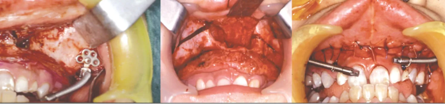
At surgery of distraction, a horizontal incision was made 5- 10 mm superior to the mucogingival junction between bilateral first molar teeth. The anterior aspect of the maxilla and zygo- matic buttress areas were exposed. The anticipated vector of the maxillary distraction was determined. The distractor was temporarily fixed at both zygomatic buttresses. To avoid dam- age to unerupted permanent tooth germ and previously repaired alveolar bone, the horizontal maxillary osteotomy was conducted superior to the level of conventional Le Fort I osteotomy. Minimal pterygomaxillary junction osteotomy was performed and the maxilla was not down-fractured. The dis- tractor was then fixed to the zygomatic buttresses in the pre- planned position, and the maxilla was checked for sufficient mobility by activation of the distractor. After suture, the turn- ing arm of distractor was fixed to the area between the incisor and canine teeth with microscrews and steel wires so that the patients or their parents might easily access and activate the
distractor. After the maxilla was checked for mobilization by rotating the screw clockwise, the distractor was returned to its original position.(Fig. 1)
After a latency period of 7 days, the maxilla was distracted 0.5 mm twice daily. The distractor was removed after a consol- idation period of 12 weeks. The patients were observed with periodic follow-up for supervision of orthodontist and oral sur- geon per three months and no additional surgeries were per- formed. Lateral cephalograms were taken immediately before surgery (T0), and immediately after distraction (T1), 6 months (T2), 1 year (T3), 2 years (T4) and 5 years (T5) after surgery.(Fig. 2)
To standardize the amount of maxillary movement and growth of the maxilla and mandible, the Natural Head Position (NHP) horizontal and vertical reference lines, as proposed by Madsen et al.7, were used. The horizontal reference line was defined as a line through the nasion rotated 7�upward from the sella-nasion, and the vertical reference line was defined as a line perpendicular to the horizontal line passing through the sella. The magnitude of the horizontal and vertical movement
Table 1.Patient information
Patient I Patient II
Age at the time of distraction 10 yr 8 mo 12 yr 10 mo
Cleft classification Unilateral complete cleft lip and palate, Lt Unilateral incomplete cleft lip and palate, Rt
Previous cheiloplasty 3 mo after birth 4 mo after birth
Previous cleft palate surgery 15 mo after birth 16 mo after birth
Previous reconstruction of alveolar cleft anterior iliac bone graft reconstruction anterior iliac bone graft reconstruction
(9 yr 8 mo) (12yr 3mo)
Follow-up period 5 yr 8 mo 5 yr 3 mo
Preoperative overjet (mm) -4.35 -5.84
Amount of advancement done (mm) 8.41 9.08
Postperative overjet / last follow-up overjet (mm) 4.06/2.20 3.24/-1.75
Actual distracted amount (mm) 10/10 (Rt/Lt) 10/6 (Rt/Lt)
Orthodontic treatment after distraction Camouflage treatment after Orthognathic surgery is planned due to distraction osteogenesis remarkable growth of mandible (yr: year[s], mo: month[s])
Fig. 1.Clinical photographs at surgery.
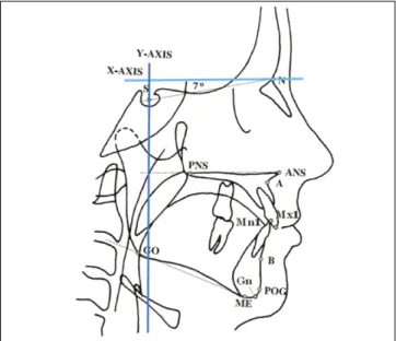
of point A and B were measured on each lateral cephalogram, and SNA, SNB, ANB, upper incisor to SN, nasolabial angle, distance from the upper and lower lips to the E line, overjet and overbite were also measured.(Fig. 3) All measurements
were made by an oral surgeon and an orthodontist using the same program (V-ceph 5.5, Osstem, Seoul, Korea).
1. Case 1
The patient first reported to our department with multiple missing teeth at 5 years old. Cheiloplasty and hard palate surgery was done previously by plastic surgeons, and they referred the patient to our clinic for dental problems. He was diagnosed as complete unilateral CLP on the left side and skeletal Class III due to maxillary deficiency. Distraction of maxilla was planned and autogenous anterior iliac bone graft was performed 13 months prior to distraction due to bony defect of left side of hard palate and alveolar bone.
The patient’s preoperative overjet was -4.35 mm and we intended for an overjet of +4 mm after distraction considering post-operative relapse. And since the direction of action of transoral distractor is not only forwards but also downwards, we decided to distract total of 10 mm in order to gain + 4 mm overjet. Distraction osteogenesis was used in same amounts (10 mm) on both left and right sides. The maxilla showed increase in SNA (4.34�) and ANB (6.74�). From the horizon- tal and vertical reference lines, Point A moved a mean of 6.72 mm forward and a mean of 2.21 mm downward. The mandible showed posterior and downward rotation movements. Overjet increased 8.41 mm (-4.35-4.06 mm). During a period of 5 years and 8 months, decrease in ANB (1.58�) and SNA (2.30�) was observed. This decreasing tendency was particularly noticeable during the T2-T3 period rather than the T1-T2 peri- od. From the horizontal and vertical reference lines, Point A
Fig. 2.Treatment protocol for maxillary distraction using an intraoral distractor.
Operation
Distraction strat
Distractor removal
7 days 12 weeks
0.5 mm×2 per day 10 days
Latency period
Distraction period
Consolidation period
TO: Preoperative T1: At the end of distraction T3: At 1 year after T1 T5: At 5 years after T1
T2: At 6 months after T1 T4: At 2 years after T1
Fig. 3. Representation of landmarks and constructed lines used to identify craniofacial and dental parameters on lat- eral cephalometric radiograph. The following points were assessed; N: nasion, S: sella, ANS: anterior nasal spine, PNS: posterior nasal spine, A: subspinale, B: supramen- tale, Mx1: incisal edge of maxillary central incisor, Pg:
pogonion, Gn: gnathion, Go: gonion, Me: menton, Mn1:
incisal edge of mandibular central incisor, X-Axis: horizon- tal reference line-line through nasion rotated 7� upward from sella-nasion line, Y-Axis: vertical reference line-line perpendicular to horizontal reference line through sella.
moved 2.80 mm forward and 7.88 mm downward. Overjet decreased by 1.86 mm (within 22% of baseline, 2.20 mm) which showed a Class I relationship. The maxillary anterior teeth to SN line angle increased by 10.34�due to camouflage
treatment. There were no complications from previous alveolar reconstruction and distraction osteogenesis. No extra follow up surgery is scheduled, and the patient will only be under ortho- dontic supervision until growth is finished.(Figs. 4-7, Table 2)
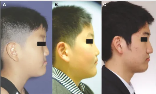
Fig. 4. Comparison of clinical photo- graph. A. Preoperative, B. After dis- traction osteogenesis, C. Post-op 5 years 8 months.
Fig. 5. Comparison of Lateral cephalo- gram. A. Preoperative, B. After distrac- tion osteogenesis, C. Post-op 5 years 8 months.
A B C
A B C
Fig. 6. Comparison of Introral photo- graph, frontal view. A. Preoperative, B.
after distraction osteogenesis.
A B
2. Case 2
The patient first reported to our department with anterior cross-bite at 6 years old. Cheiloplasty and hard palate surgery was done previously by plastic surgeons. He was diagnosed as incomplete unilateral CLP on the right side and skeletal Class
III due to maxillary deficiency. Distraction of maxilla was planned and autogenous anterior iliac bone graft was per- formed 7 months prior to distraction due to bony defect of right side of hard palate and alveolar bone.
The patient’s preoperative overjet was -5.84 mm and an overjet of +3 mm was planned according to the orthodontist’s recommendation. Because right cleft resulted in collapse of maxillary dental arch with the dental midline offset to the right, we decided to correct maxillary yawing by distraction of different amount on each side. Distraction osteogenesis was done in different amounts on left (6 mm) and right (10 mm) side. The maxilla showed increases in SNA (3.44�) and ANB (7.23�), and post-operative dental midline showed similarity with facial midline. From the horizontal and vertical reference lines, Point A moved 7.05 mm forward and 1.98 mm down- ward. Overjet increased by 9.08 mm. During a period of 5 years and 3 months, increase in SNA (0.14�) and decrease in ANB (6.49�) were observed. From the horizontal and vertical reference lines, Point A moved 0.28 mm posteriorly and 7.20 mm downward. The mandible showed a significant increase in growth, leading to crossbite in T2 period and negative ANB values in T5 period. Overjet decreased by 4.99 mm (55% of baseline). No complication was observed on the maxillary right alveolar ridge which was treated with alveolar recon- struction. The facial midline was offset 1 mm to the left, but this will be treated through orthognatic surgery once growth is finished.(Figs. 8-10, Table 3)
Table 2.Cephalometric analysis of patient I
T0 T1 T2 T3 T4 T5 T1-T0 T5-T1
SNA (�) 74.11 78.45 78.14 76.54 76.97 76.87 4.34 -1.58
SNB (�) 80.10 77.70 77.57 79.12 79.59 78.42 -2.40 0.72
ANB (�) -5.99 0.75 0.57 -2.58 -2.62 -1.55 6.74 -2.30
Point A to ver. reference line1(mm) 60.19 62.40 62.33 65.54 68.48 70.28 2.21 7.88
Point A to hor. reference line1(mm) 61.52 68.24 67.28 68.32 69.48 71.04 6.72 2.80
Point B to ver. reference line1(mm) 98.82 97.42 100.03 100.62 114.81 118.42 -1.40 21.00
Point B to hor. reference line1(mm) 77.72 77.04 77.82 80.48 80.42 79.02 -0.68 1.98
U1 to SN (�) 99.37 97.08 98.63 94.50 106.24 107.42 -2.29 10.34
Nasolabial angle (�) 83.35 95.62 93.56 95.31 91.13 89.42 12.27 -6.20
Upper lip to E-line (mm) -7.42 -1.79 -1.70 -2.36 -4.24 -3.28 5.63 -1.49
Lower lip to E-line (mm) -2.45 -1.60 -1.36 -1.23 -1.84 -1.89 0.85 -0.29
Incisal overjet (mm) -4.35 4.06 5.68 3.10 2.40 2.20 8.41 -1.86
Incisal overbite (mm) 5.96 2.85 2.12 2.04 2.18 1.98 -3.11 -0.87
(T0: before distraction osteogenesis [DO], T1: after DO, T2: 6 months after DO, T3: 1 year after DO, T4: 2 years after DO, T5: 5 years after DO, ver: vertical, hor: horizontal)
1: The natural head position (NHP) horizontal and vertical reference lines7were used for measuring amount of Point A and B to reference lines.
Refer to Fig. 3.
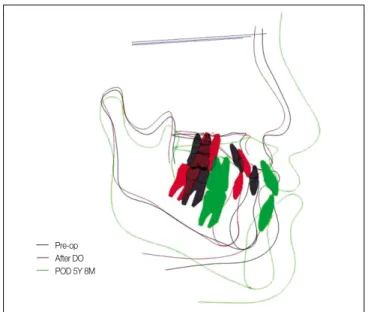
Fig. 7. Superimposition of Lateral cephlogram.(T0-Black, T1-Red, T5-Green)
Pre-op After DO POD 5Y 8M
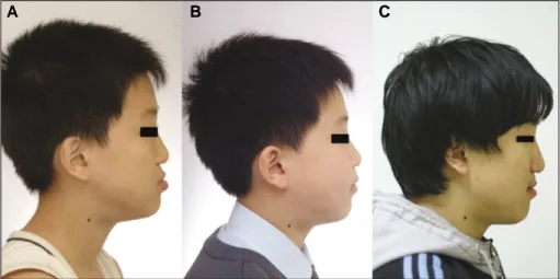
Fig. 8. Comparison of clinical photo- graph. A. Preoperative, B. After dis- traction osteogenesis, C. Post-op 5 years 3 months.
A B C
Fig. 9. Comparison of Lateral cephalo- gram. A. Preoperative, B. After dis- traction osteogenesis, C. Post-op 5 years 3 months.
A B C
Table 3.Cephalometric analysis of patient II
T0 T1 T2 T3 T4 T5 T1-T0 T5-T1
SNA (�) 73.8 77.24 76.78 76.79 77.08 77.38 3.44 0.14
SNB (�) 75.58 71.79 72.89 74.24 75.27 78.42 -3.79 6.63
ANB (�) -1.78 5.45 3.89 2.55 1.81 -1.04 7.23 -6.49
Point A to ver. reference line1(mm) 79.24 81.22 82.49 84.42 86.28 88.42 1.98 7.20
Point A to hor. reference line1(mm) 87.24 94.29 94.38 92.18 94.44 94.01 7.05 -0.28
Point B to ver. reference line1(mm) 142.48 146.42 148.89 150.24 149.42 152.37 3.94 5.95
Point B to hor. reference line1(mm) 79.42 77.71 79.89 80.42 80.84 84.24 -1.71 6.53
U1 to SN (�) 94.76 103.9 96.12 96.42 94.13 93.42 9.14 -10.48
Nasolabial angle (�) 92.48 88.51 82.1 80.93 89.61 88.42 -3.97 -0.09
Upper lip to E-line (mm) -3.22 1.73 1.24 1.11 -1.85 -1.94 4.95 -3.67
Lower lip to E-line (mm) 5.42 4.83 4.88 4.72 1.76 2.42 -0.59 -2.41
Incisal overjet (mm) -5.84 3.24 -0.01 -0.48 -1.24 -1.75 9.08 -4.99
Incisal overbite (mm) -1.42 0.6 -0.22 -0.83 -0.42 -1.24 2.02 -1.84
(T0: before distraction osteogenesis [DO], T1: after DO, T2: 6 months after DO, T3: 1 year after DO, T4: 2 years after DO, T5: 5 years after DO, ver: vertical, hor: horizontal)
1: The natural head position (NHP) horizontal and vertical reference lines7were used for measuring amount of Point A and B to reference lines.
Refer to Fig. 3.
Ⅲ. Discussion
Distraction osteogenesis has been developed through improve- ments of osteotomy techniques and fixation of bone fragments.
As Polley and Figueroa6 first performed distraction osteogenesis on patients with cleft deformities in the maxilla and midface, there have been numerous reports on successful forward move- ment of the maxilla by distraction osteogenesis4,8-10. This method not only has the advantages of simplicity, safety and pre- dictability, but also showed satisfactory outcomes in patients who have difficulty in undergoing conventional orthognatic surgery8. Distraction osteogenesis produces skeletal changes through traction force on callus, which accelerates tissue regeneration, especially in the soft tissue-deficient area sur- rounding the bone9,10.
In conventional orthognathic surgery undergoing maxillary advancement, 5-80% of CLP patients showed post-operative relapse and long-term instability14-17. Cheung et al.11reported that the relapse rate of orthognathic surgery with respect to horizontal and vertical reference lines was 22% with long-term follow-up. Posnick and Dagys4reported a vertical relapse of 19%, a horizontal relapse of 23% and a mean relapse of 6.9 mm. Thongdee and Samman12reported a horizontal relapse of 31% and a vertical relapse of 52% after maxillary surgical
movement in unilateral CLP with preceding alveolar bone grafting.
Louis et al.13have shown that the relapse rate of orthognathic surgery becomes higher as the amount of maxillary advance- ment increases. Some investigators have demonstrated that the maximum of maxillary advancement achieved by conventional orthognathic surgery techniques is about 10 mm in CLP patients, therefore distraction osteogenesis can be performed when advancement over 10 mm is required13,14. Even other investigators have stated that the maximum advancement by conventional orthognathic surgery techniques in CLP patients is 5 mm due to scar contracture14,15. Based on these results, we have performed distraction osteogenesis in cases that require maxillary advancement of ≥5 mm. In our cases, we performed distraction osteogenesis with advancement of 10 mm in con- sideration of post-operative relapse and quantity we were to gain.
Cheung et al.18have indicated that skeletal stability is better in distraction osteogenesis than in Le Fort I osteotomy, regard- less of the magnitude of maxillary movement, because skeletal relapse occurs more frequently in Le Fort I osteotomy due to insufficient soft tissue. A previous meta-analysis in cleft chil- dren has suggested that the distraction is more effective in the treatment of severe cleft patients16. Rachmiel et al.17 had demonstrated that the relapse rate of distraction was smaller because of regeneration of membranous bone between the bone segments.
Kusnoto et al.25have proposed that the consolidation period should be adequately maintained because active bone forma- tion occurs in the pterygoid region 6 weeks after maxillary dis- traction. In our cases, an adequate consolidation period of 12 weeks was given. In addition, after adequate maxillary move- ment was identified at surgery by activating distractors, the maxilla was not down-fractured with minimal dissection of soft tissue in the pterygomaxillary area and around the nasal cavity, which was based on the results reported by previous in vitro and in vivo studies18,19.
In our cases, from the horizontal and vertical reference lines, Point A moved a mean of 6.89 mm forward and a mean of 2.10 mm downward after distraction. Because vector of intraoral distraction is anterior and inferior, about 7 mm of anterior movement was acquired. After a period of 5 years and 6 months, Point A moved a mean of 1.26 mm forward and a mean of 7.54 mm downward. This implies that a lower relapse rate in distraction osteogenesis than in orthognathic surgery may be attributed to the persistent growth of the maxilla after distraction osteogenesis, which is similar to the results reported by previous studies17,20,22.
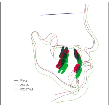
Fig. 10. Superimposition of Lateral cephlogram.(T0-Black, T1-Red, T5-Green)
Pre-op After DO POD 5Y 8M
Distraction osteogenesis has been developed through numer- ous clinical and experimental studies and has various advan- tages over orthognathic surgery in cleft patients. Although postoperative normal maxillary growth was difficult to achieve due to scar contracture and tension from the upper lip in con- ventional method, distraction osteogenesis improved bone and soft tissue, and dentofacial structure through persistent maxil- lary growth.
In summary, distraction osteogenesis is thought to be able to provide improvements in facial aesthetic, a stable intermaxil- lary relationship and occlusion status due to persistent maxil- lary growth.
References
1. Rachmiel A, Aizenbud D, Peled M. Long-term results in maxil- lary deficiency using intraoral devices. Int J Oral Maxillofac Surg 2005;34:473-9.
2. Ross RB. Treatment variables affecting facial growth in complete unilateral cleft lip and palate. Cleft Palate J 1987;24:5-77.
3. Erbe M, Stoelinga PJ, Leenen RJ. Long-term results of segmental repositioning maxilla in cleft palate patients without previously grafted alveolo-palatal cleft. J Craniomaxillofac Surg 1996;24:
109-17.
4. Posnick JC, Dagys AP. Skeletal stability and relapse patterns af- ter Le Fort I maxillary osteotomy fixed with miniplates: the uni- lateral cleft lip and palate deformity. Plast Reconstr Surg 1994;
94:924-32.
5. McCarthy JG, Schreiber J, Karp N, Thorne CH, Grayson BH.
Lengthening the human mandible by gradual distraction. Plast Reconstr Surg 1992;89:1-8.
6. Polley JW, Figueroa AA. Management of severe maxillary defi- ciency in childhood and adolescence through distraction osteoge- nesis with an external, adjustable, rigid distraction device. J Craniofac Surg 1997;8:181-5.
7. Madsen DP, Sampson WJ, Townsend GC. Craniofacial reference plane variation and natural head position. Eur J Orthod 2008; 30:
532-40.
8. Kanno T, Takahashi T, Takano H, Funaki K, Ichida T.
Simultaneous maxilla-mandibular distraction osteogenesis using a subcutaneous device for a bilateral cleft lip and palate patient.
Asian J Oral Maxillofac Surg 2006;18:303-8.
9. Figueroa AA, Polley JW, Friede H, Ko EW. Long-term skeletal stability after maxillary advancement with distraction osteogene- sis using a rigid external distraction device in cleft maxillary de- formities. Plast Reconstr Surg 2004;114:1382-92.
10. Harada K, Sato M, Omura K. Maxillary distraction in patients with cleft deformity using a rigid external distraction device: a pilot study on the distraction ratio of the maxilla to the device.
Scand J Plast Reconstr Surg Hand Surg 2004;38:277-80.
11. Yamauchi K, Mitsugi M, Takahashi T. Maxillary distraction os-
teogenesis using Le Fort I osteotomy without intraoperative down-fracture. Int J Oral Maxillofac Surg 2006;35:493-8.
12. Rachmiel A, Rozen N, Peled M, Lewinson D. Characterization of midface maxillary membranous bone formation during distrac- tion osteogenesis. Plast Reconstr Surg 2002;109:1611-20.
13. Satoh K, Mitsukawa N, Tosa Y, Kadamatsu K, Hosaka Y.
Simultaneous hybrid of maxillary Le Fort I halo distraction and mandibular set-back for patients with severe cleft jaw deformity.
J Craniofac Surg 2006;17:962-9.
14. Proffit WR, Phillips C, Prewitt JW, Turvey TA. Stability after surgical orthodontic correction of skeletal Class III malocclusion.
2. Maxillary advancement. Int J Adult Orthodon Orthognath Surg 1991;6:71-80.
15. Hoffman GR, Brennan PA. The skeletal stability of one-piece Le Fort 1 osteotomy to advance the maxilla; Part 1. Stability result- ing from non-bone grafted rigid fixation. Br J Oral Maxillofac Surg 2004;42:221-5.
16. Harada K, Sato M, Omura K. Long-term skeletal and dental changes in patients with cleft lip and palate after maxillary dis- traction: a report of three cases treated with a rigid external dis- traction device. Cranio 2005;23:152-7.
17. Cheung LK, Chua HD, Ha¨gg MB. Cleft maxillary distraction versus orthognathic surgery: clinical morbidities and surgical re- lapse. Plast Reconstr Surg 2006;118:996-1008.
18. Cheung LK, Chua HD, Bendeus M. Distraction or osteotomy for the correction of maxillary cleft deformities: which is better?
Ann R Australas Coll Dent Surg 2004;17:57-63.
19. Thongdee P, Samman N. Stability of maxillary surgical move- ment in unilateral cleft lip and palate with preceding alveolar bone grafting. Cleft Palate Craniofac J 2005;42:664-74.
20. Louis PJ, Waite PD, Austin RB. Long-term skeletal stability after rigid fixation of Le Fort I osteotomies with advancements. Int J Oral Maxillofac Surg 1993;22:82-6.
21. Wang XX, Wang X, Yi B, Li ZL, Liang C, Lin Y. Internal mid- face distraction in correction of severe maxillary hypoplasia sec- ondary to cleft lip and palate. Plast Reconstr Surg 2005;116:51- 60.
22. Cho BC, Kyung HM. Distraction osteogenesis of the hypoplastic midface using a rigid external distraction system: the results of a one to six-year follow-up. Plast Reconstr Surg 2006;118:1201- 12.
23. Cheung LK, Chua HD. A meta-analysis of cleft maxillary os- teotomy and distraction osteogenesis. Int J Oral Maxillofac Surg 2006;35:14-24.
24. Rachmiel A, Laufer D, Jackson IT, Lewinson D. Midface mem- branous bone lengthening: A one-year histological and morpho- logical follow-up of distraction osteogenesis. Calcif Tissue Int 1998;62:370-6.
25. Kusnoto B, Figueroa AA, Polley JW. Radiographic evaluation of bone formation in the pterygoid region after maxillary distraction with a rigid external distraction (RED) device. J Craniofac Surg 2001;12:109-17.
26. Kanno T, Takahashi T, Ariyoshi W, Tsujisawa T, Haga M, Nishihara T. Tensile mechanical strain up-regulates Runx2 and osteogenic factor expression inhuman periosteal cells: implica- tions for distraction osteogenesis. J Oral Maxillofac Surg 2005;
63:499-504.