ISSN: 2233-601X (Print) ISSN: 2093-6516 (Online)
− 239 −
Received: November 19, 2018, Revised: December 18, 2018, Accepted: February 22, 2019, Published online: August 5, 2019
Corresponding author: Hyun Oh Park, Department of Thoracic and Cardiovascular Surgery, Gyeongsang National University Hospital, Gyeongsang National University College of Medicine, 79 Gangnam-ro, Jinju 52727, Korea
(Tel) 82-55-750-8124 (Fax) 82-55-753-8138 (E-mail) [email protected]
Corresponding author: Chung Eun Lee, Department of Thoracic and Cardiovascular Surgery, Gyeongsang National University Hospital, Gyeongsang National University College of Medicine, 79 Gangnam-ro, Jinju 52727, Korea
(Tel) 82-55-750-8124 (Fax) 82-55-753-8138 (E-mail) [email protected]
© The Korean Society for Thoracic and Cardiovascular Surgery. 2019. All right reserved.
This is an open access article distributed under the terms of the Creative Commons Attribution Non-Commercial License (http://creativecommons.org/
licenses/by-nc/4.0) which permits unrestricted non-commercial use, distribution, and reproduction in any medium, provided the original work is properly cited.
Immunoglobulin G4-Related Aortitis of the Abdominal Aorta
Jae Won Choi, M.D.
1, Jun Young Choi, M.D.
1, Kyung Hyuk Go, M.D.
2,
Yun Hong Cheon, M.D.
3, Jong Woo Kim, M.D.
1, Chung Eun Lee, M.D.
1, Hyun Oh Park, M.D.
1Departments of 1Thoracic and Cardiovascular Surgery and 2Pathology, 3Division of Rheumatology, Department of Internal Medicine, Gyeongsang National University Hospital, Gyeongsang National University College of Medicine, Jinju, Korea
Noninfectious aortitis, inflammatory abdominal periaortitis, and idiopathic retroperitoneal fibrosis are chronic inflammatory diseases with unclear causes. Recent studies have shown that some cases of aortitis are asso- ciated with immunoglobulin G4 (IgG4)-related systemic disease. Herein, we report a case of IgG4-related aor- titis (IgG4-RA) that was diagnosed after surgery. Our patient was a 46-year-old man who had experienced abdominal pain for several weeks. Preoperative evaluations revealed an area of aortitis on the infrarenal aorta. He underwent surgery, and histological examination resulted in a diagnosis of IgG4-RA.
Key words: 1. Aortitis 2. Arteritis
3. Retroperitoneal fibrosis
Case report
A 46-year-old man visited Gyeongsang National University Hospital with abdominal pain in the peri- umbilical area that had lasted for 5 weeks. He had no other medical history. On examination, he pre- sented normal vital signs and had no localized signs of infection. Laboratory tests showed 8,710 leuko- cytes/mm3 with the normal formula, a C-reactive protein level of 15.8 mg/L, and an erythrocyte sed- imentation rate of 40 mm/hr. Coagulation, hepatic en- zymes, and summary urinalysis findings were normal.
A chest radiograph showed no pulmonary or media- stinal abnormal lesions. Blood culture and urine cul- ture were sterile. We investigated immunological causes, including anti-double stranded DNA antibody, anti-beta-2 glycoprotein 1 immunoglobulin G (IgG),
lupus anticoagulant, anti- cardiolipin IgG and im- munoglobulin M (IgM), fluorescent treponemal anti- body absorption IgG and IgM, anti-neutrophil cyto- plasmic antibody, and human leukocyte antigen-B51;
all findings were negative. Computed tomography (CT) of the abdomen was performed to confirm the cause of abdominal pain, and no other abnormal finding was found except a diffuse infrarenal aortic wall thickening (14 mm), with homogeneous en- hancement and mild aortic lumen stenosis (Fig. 1).
Aortic angiographic CT was taken to assess the con- dition of the ascending aorta and aortic arch, and there were no abnormal findings. Based on these findings, we suspected aortitis and performed sur- gery to obtain a biopsy to assess this possibility. We identified an irregularly-shaped area of solid chronic inflammatory tissue surrounding the aorta, with no
Korean J Thorac Cardiovasc Surg 2019;52:239-242 □ CASE REPORT □
https://doi.org/10.5090/kjtcs.2019.52.4.239
Jae Won Choi, et al
− 240 −
Fig. 2. Intraoperative finding. (A) An irregularly-shaped area of solid chronic inflammatory tissue sur- rounding the aorta (black arrow).
(B) Restoration of the aortic size after removal of periaortic tissue.
Fig. 1. Computed tomography showing diffuse infrarenal aortic wall thickening (14 mm), with homogeneous enhancement and mild aortic lumen stenosis.
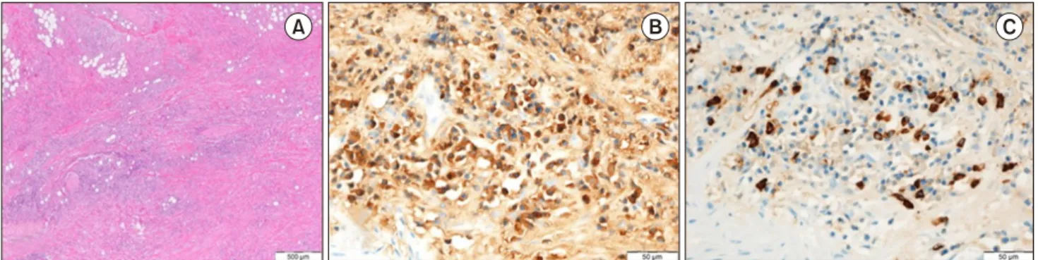
signs of acute infection (Fig. 2A). We removed the tissue surrounding the anterior wall and lateral wall of the aorta and confirmed the restoration of aortic size (Fig. 2B). On a low-power histologic image of a hematoxylin and eosin–stained section of periaortic soft tissue, diffuse inflammatory infiltration and irreg- ular fibrosis were noted (Fig. 3A). In an im- munohistochemical assessment, IgG-positive plasma cells were observed in the fibrous tissue (Fig. 3B), and IgG4-positive plasma cells were identified in the periaortic tissue (Fig. 3C). The ratio of IgG4- to IgG-positive cells was 40%, and the patient was diag- nosed with IgG4-related aortitis (RA). On hospital day 15, the patient began glucocorticoid treatment for the remaining inflammatory tissue of the aorta and discharged.
The patient provided written informed consent for publication of clinical details and images.
Discussion
Aortitis is an inflammatory disease of the aortic wall, with various etiologies that are not all well- understood. Because of the variety of causes, it may be difficult to pinpoint the cause of aortitis before starting treatment [1]. Aortitis is divided into in- fectious causes and non-infectious causes. Bacteria such as Staphylococcus and Salmonella are the main causes of infectious aortitis, although tuberculosis, syphilis, or virus that cause inflammation in humans can also be causes [1]. The major causes of non-in- fectious aortitis are connective tissue disorders, trau- ma, and neoplasms, but this category also includes IgG4-RA [2]. IgG4-RA is a newly recognized systemic inflammatory disease that must be carefully dis- tinguished from neoplasms [2,3]. Histologically, IgG4-RA has the following characteristics: thickening of the aortic wall, a lymphoplasmacytic infiltrate enriched in IgG4-positive plasma cells, and a variable degree of periaortic fibrosis referred to as a ‘storiform pattern’
[3]. Most cases of IgG4-RA are diagnosed based on histological findings after surgery, as in the case of our patient.
The diagnosis of IgG4-RA requires biopsy of the af- fected tissues, but it might be suspected based on routine laboratory tests and selected imaging tests.
Inoue et al. [4] described the CT findings associated with IgG4-RA in 17 patients. In all cases, the aortic wall was severely thickened, with an average max-
IgG4-Related Aortitis
− 241 −
Fig. 3. (A) A low-power histologic image of a hematoxylin and eosin–stained section of periaortic soft tissue. Diffuse inflammatory in- filtration (bluish area) and irregular fibrosis (pinkish area) are noted (×40). (B) Immunohistochemistry showing IgG-positive plasma cells in fibrous tissue (×400). (C) Immunohistochemistry showing numerous IgG4-positive plasma cells (×400). IgG, immunoglobulin G.
imum thickness of 5–22 mm (mean, 11 mm) [4]. All lesions were homogeneously enhanced in the late phase of contrast-enhanced CT and 19 of 22 (86%) were well-circumscribed [4]. Cystic change, calcifica- tion, or lymph node enlargement was not detected in any lesions [4]. Our patient showed similar findings;
the aortic wall thickness was 14 mm, and the lesions were homogeneously enhanced in the late phase and well-circumscribed. Cystic change, calcification, or lymph node enlargement was not detected. It is also helpful to measure serum IgG4 levels to diagnose IgG4-RA. Serum IgG4 levels were above the normal upper limit in 86% of the 114 patients in a previous study [5]. The degree of elevation of IgG4 is not per- fectly associated with the extent of disease activity, but the serum IgG4 concentration tends to increase with the number of involved organs and generally decreases after glucocorticoid therapy [6]. Unfortunately, we did not suspect IgG4-RA before surgery and did not measure the patient’s preoperative IgG levels.
Nonetheless, our patient's initial IgG4 level after sur- gery was 1,520 mg/dL (normal, 8–140 mg/dL), and we will follow up further changes in his IgG4 levels.
No optimal treatment for IgG4-RA has been established. Kasashima and Zen [7] suggested that glucocorticoid therapy ultimately will not control the disease, and long-term glucocorticoid toxicity may pose risks, such as increasing the likelihood for aort- ic aneurysm rupture. However, some points of con- sensus among experts do exist. All patients with symptomatic active IgG4-RA require treatment [8].
Glucocorticoids are the first-line treatment in all pa- tients with untreated active IgG4-RA, except when treatment is contraindicated [8]. We have started the
patient on glucocorticoid treatment, and we plan to follow the effects thereof in the future.
In conclusion, although IgG4-RA is rare, clinicians should keep in mind that aortitis could be associated with IgG4-related systemic disease. In addition, large-scale prospective studies are needed to confirm the efficacy and safety of corticosteroid therapy and to design more useful and safe treatment strategies.
Conflict of interest
No potential conflict of interest relevant to this ar- ticle was reported.
ORCID
Jae Won Choi: https://orcid.org/0000-0003-3984-0296 Jun Young Choi: https://orcid.org/0000-0001-7774-8541 Kyung Hyuk Go: https://orcid.org/0000-0002-6721-1828 Yun Hong Cheon: https://orcid.org/0000-0002-0099-6253 Jong Woo Kim: https://orcid.org/0000-0003-2578-9821 Chung Eun Lee: https://orcid.org/0000-0003-4469-7201 Hyun Oh Park: https://orcid.org/0000-0003-1302-6456
References
1. Gornik HL, Creager MA. Aortitis. Circulation 2008;117:
3039-51.
2. Stone JH, Zen Y, Deshpande V. IgG4-related disease. N Engl J Med 2012;366:539-51.
3. Okazaki K, Uchida K, Koyabu M, Miyoshi H, Takaoka M.
Recent advances in the concept and diagnosis of autoimmune pancreatitis and IgG4-related disease. J Gastroenterol 2011;
46:277-88.
4. Inoue D, Zen Y, Abo H, et al. Immunoglobulin G4-related
Jae Won Choi, et al
− 242 −
periaortitis and periarteritis: CT findings in 17 patients.
Radiology 2011;261:625-33.
5. Zen Y, Nakanuma Y. IgG4-related disease: a cross-sectional study of 114 cases. Am J Surg Pathol 2010;34:1812-9.
6. Cheuk W, Chan JK. IgG4-related sclerosing disease: a crit- ical appraisal of an evolving clinicopathologic entity. Adv Anat Pathol 2010;17:303-32.
7. Kasashima S, Zen Y. IgG4-related inflammatory abdominal aortic aneurysm. Curr Opin Rheumatol 2011;23:18-23.
8. Khosroshahi A, Wallace ZS, Crowe JL, et al. International consensus guidance statement on the management and treatment of IgG4-related disease. Arthritis Rheumatol.
2015;67:1688-99.