www.labmedonline.org 41 eISSN 2093-6338
WHO 2008년 가이드라인에 따라 10% 이상에서 형성이상이 관찰 되면 의미가 있다고 판단하지만, 정상 성인의 골수에서도 진단기 준을 넘는 형성이상이 관찰되었다는 연구결과가 발표된 바가 있다 [6]. 골수형성이상증후군에 동반된 용혈빈혈의 예가 흔하지 않지 만, 형성이상은 모양으로 판단하게 되기 때문에 용혈빈혈에서 적혈 구계의 과증식이나 형성이상이 골수형성이상증후군으로 진단될 수 있다. 이 때문에 염색체 이상이나 이상유전자의 증명은 클론성 질환임을 확인할 수 있는 증거가 될 수 있다. 저자는 용혈빈혈의 증상 및 검사소견을 보이면서 혈구세포의 형성이상 및 복합 염색 체 이상이 있는 증례를 경험하였고, 이를 문헌고찰과 함께 보고하 는 바이다.
증 례
특정병력이 없는 78세 남자가 호흡곤란으로 내원하였다. 호흡곤 란은 2-3개월 전부터 시작되었고, 신체활동 시 더 심해지는 양상이 었다. 수년 전부터 간헐적으로 붉은색의 소변이 있었던 것 이외에 는 과거력, 가족력상 특이 소견은 없었고, 이학적 검사상 비장비대 는 관찰되지 않았다.
일반혈액검사에서 혈색소 65 g/L (130-180 g/L), 백혈구 4.05×
서 론
골수형성이상증후군은 무효조혈(ineffective hematopoiesis)과 골수계 세포의 형성이상을 특징으로 하는 클론성 조혈모세포질환 이다. 드물지만 용혈빈혈이 골수형성이상증후군에서 발병되는데, 지중해 빈혈, 바이러스 감염으로 인한 만성용혈빈혈 및 자가면역 용혈빈혈이 보고된 바 있고, 자가면역용혈빈혈은 골수형성이상증 후군에서 0.4%-3% 빈도를 보인다[1-5].
골수형성이상증후군은 조혈세포의 특징적인 모양의 변화로 진 단을 하기 때문에 골수도말검사가 결정적인 역할을 하게 된다.
용혈빈혈로 나타난 골수형성이상증후군 1예
A Case of Myelodysplastic Syndrome Characterized by Hemolytic Anemia at Presentation
김유경Yu Kyung Kim, M.D.
영남대학교 의과대학 진단검사의학교실
Department of Laboratory Medicine, Yeungnam University College of Medicine, Daegu, Korea
증례보고
Lab Med Online
Vol. 6, No. 1: 41-44, January 2016
http://dx.doi.org/10.3343/lmo.2016.6.1.41 진단혈액학
Corresponding author: Yu Kyung Kim
Department of Laboratory Medicine, Yeungnam University College of Medicine, 170 Hyunchoong-ro, Nam-gu, Daegu 42415, Korea Tel: +82-53-640-6703, Fax: +82-53-653-7774, E-mail: [email protected] Received: May 20, 2015
Revision received: July 30, 2015 Accepted: December 4, 2015
This article is available from http://www.labmedonline.org 2016, Laboratory Medicine Online
This is an Open Access article distributed under the terms of the Creative Commons Attribution Non-Commercial License (http://creativecommons.org/licenses/by-nc/3.0/) which permits unrestricted non-commercial use, distribution, and reproduction in any medium, provided the original work is properly cited.
A man aged 78 yr with no history of chemotherapy or toxic exposure presented with a history of dyspnea and intermittent red urine for 3 months and several years, respectively. Hematologic data at admission were as follows: hemoglobin, 65 g/L; white blood cell count, 4.05×10⁹/L; platelet count, 96×109/L; and reticulocyte count, 10.9%. A peripheral blood smear revealed polychromasia, nucleated red blood cells, and neutrophils with a non-lobulated nucleus. The bone marrow was hypercellular and exhibited an increase in erythroid precursors with trilineage dysplasia and our findings were suggestive of refractory cytopenia with multilineage dysplasia (RCMD). Karyotype of bone marrow cells was as follows:
45,XY,der(9;17)(p10;q10),add(18)(q11.2)[ 10]/45,idem,del(3)(q21)[ 10]. Other laboratory findings showed decreased serum haptoglobin, in- creased lactate dehydrogenase, and increased indirect bilirubin levels. Moreover, results of the direct/indirect antiglobulin test (Coombs’ test) and paroxysmal nocturnal hemoglobinuria analysis with CD55, CD59, fluorescent aerolysin (FLAER), and CD24 were negative. Cold agglutinin and Don- ath-Landsteiner antibodies were not detected. This is a case of myelodysplastic syndrome (MDS) associated with hemolytic anemia and complex chromosomal abnormalities at presentation.
Key Words: Hemolytic anemia, Myelodysplastic syndrome, Refractory cytopenia with multilineage dysplasia
김유경: A Case of MDS with Hemolytic Anemia
http://dx.doi.org/10.3343/lmo.2016.6.1.41 42 www.labmedonline.org
Fig. 2. G-banded karyogram of bone marrow cells. (A) A derivative chromosome composed of the short arm of chromosome 9 and the long arm of chromosome 17. Additional material of unknown origin is attached at band 18q11.2. (B) Deletion of the long arm of chromosome 3.
A B
der(9;17)(p10;q10)
del(3)(q21)
1 1
6 6
13 13
19 19
2 2
7 7
14 14
20 20
8 8
15 15
21 21
3 3
9 9
16 16
22 22
4 4
10 10
17 17
11 11
18 18
Y Y
5 5
12 12
X X
add(18)(q11.2)
109/L (4-10×109/L), 혈소판 96×109/L (140-440×109/L), 그물적혈 구 10.9% (0.5-1.5%)였고, 응고검사는 정상이었다. 생화학 검사상 총단백, 알부민, 감마-글루타밀전이효소, 나트륨, 칼륨, 염소, 혈액 요소질소, 크레아티닌, 철, 총철결합능은 참고치 범위 내에 속하였 으나, 총 빌리루빈 31.4 mg/L (1-12 mg/L), 직접 빌리루빈 8.5 mg/L (0-5 mg/L), 젖산탈수소효소 699 IU/L (150-550 IU/L)로 증가하였 고, 합토글로빈 10 mg/L (500-3,200 mg/L)로 감소하였다. 직접 및 간접 항글로불린검사는 음성이었고, 저온응집소도 검출되지 않았 다. 류마티스인자는 참고치 범위 내에 속하였고, 항핵항체, 항혈소 판항체 및 혈소판관련항체는 모두 음성이었다.
말초혈액도말검사상 다염적혈구의 증가, 유핵적혈구가 관찰되고 화학검사를 종합하여 용혈빈혈로 생각하였으나, 호중구의 비분엽 성핵이 관찰되어 골수검사를 시행하였다. 골수검사에서 세포충실 도는 증가하였고, 골수구계와 적혈구계의 비율은 0.38:1로 적혈구 계의 증식이 관찰되었다. 적혈구, 과립구, 거대핵세포 모두에서 형 성이상이 있었고, 환상철적혈모세포는 관찰되지 않았으며 모세포 는 2.0%로 다계열형성이상불응혈구감소증에 합당하였다(Fig. 1).
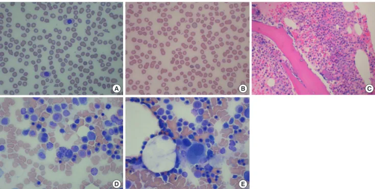
골수검체로 시행한 염색체 검사에서 45,XY,der(9;17)(p10;q10),add (18)(q11.2)[10]/45,idem,del(3)(q21)[10]의 이상소견이 발견되었다 (Fig. 2). 추가로 시행한 적혈구에 대한 CD55, CD59 항체검사에서 Fig. 1. (A and B) Peripheral blood smear showing neutrophils with a non-lobulated nucleus, poikilocytosis of red blood cells, and polychromasia. (C) Bone marrow biopsy showing hypercellularity and hypolobulated megakaryocyte. (D and E) Bone marrow smear showing dyserythropoiesis and dysplastic megakaryocytes.
A
D
B
E
C
김유경: A Case of MDS with Hemolytic Anemia
http://dx.doi.org/10.3343/lmo.2016.6.1.41 www.labmedonline.org 43
Type Ⅱ 적혈구는 0.6%, Type Ⅲ 적혈구는 0.1%로 참고범위(Type
Ⅱ+ Type Ⅲ 적혈구; 0-1%) 내에 속했고, 백혈구에 대한 CD24, fluo- rescent aerolysin (FLAER) 항체검사에서 Type Ⅱ 과립구는 0.2%, Type
Ⅲ 과립구는 0%로 정상이었다(Type Ⅱ+ Type Ⅲ 과립구; 0-1%).
Donath-Landsteiner 항체 음성이었고, 삼투압취약성 검사, 자가용혈 검사 및 혈색소 전기영동검사는 시행하지 못하였다.
환자의 혈액, 골수 및 염색체 검사소견으로 용혈빈혈을 동반한 골수형성이상증후군으로 최종 진단하였고, 이후 치료 및 추적관 찰은 환자의 전원으로 인해 종료되었다.
고 찰
골수형성이상증후군에서 B림프구, T림프구의 기능저하, 면역글 로불린 증가 또는 감소, 단클론감마병증 등 면역학적 이상이 있으 며, 이로 인한 혈관염, 결합조직장애, 무증상 혈청면역이상 등의 자 가면역질환과 면역혈소판감소증, 면역용혈빈혈이 보고되었다[5].
Novaretti 등[7]은 용혈빈혈의 임상증상이 없는 골수형성이상증후 군 환자에서도 자가항체가 34.4%의 높은 빈도로 관찰됨을 보고하 였다. 또한 Barcellini 등[8]은 초기 골수형성이상증후군, 즉 불응성 빈혈이나, 환상철적모세포를 동반한 불응성빈혈 환자군의 53.8%
에서 자가항체가 존재하고, 이 경우 caspase-3의 활성도가 높고, 종양괴사인자α (TNF-α)와 인터루킨 4의 농도가 낮음을 보고하였 다. 이처럼 골수형성이상증후군에서 나타나는 염증성 혹은 면역 조절 시토카인의 과생성과 같은 면역체계의 변화가 자가면역현상 과 관련성이 있을 것으로 추정되나, 병태생리학적인 정확한 인과 관계는 밝혀져 있지 않다[9]. 골수형성이상증후군 치료과정 중 용 혈빈혈이 발현한 환자에서 T림프구의 증식을 억제하는 mycophe- nolate mofetil을 투약하여 치료에 성공한 증례와, 골수형성이상증 후군 진단과 동시에 용혈빈혈 증상을 나타낸 환자에서 고용량 스 테로이드와 저용량의 항암치료를 시행하여 용혈증상이 개선된 증 례들은 골수형성이상증후군의 면역체계의 변화와 자가면역현상 이 관련되어 있음을 시사하고 있다[10, 11].
본 증례의 경우 적혈구에 대한 자가항체 중 면역글로불린G나 C3 외에도 면역글로불린A를 확인하지 못했고, 환자의 추적이 종료 되어 면역억제치료를 시행하지 못했으며 기타 비면역성 용혈빈혈 을 완벽하게 배제하지 못한 한계점을 가지고 있다. 이와 유사하게 골수형성이상증후군에 동반된 용혈빈혈에서 항글로불린 검사 음 성인 증례가 보고된 바 있으나, 모두 면역억제치료를 시행하여 자 가면역용혈빈혈임을 유추할 수 있었다[12, 13].
골수형성이상증후군 진단 이후 치료과정에서 보고되는 용혈빈 혈은 3% 이하의 빈도로 관찰되며 이 경우 진단에는 큰 어려움이 없다. 반면 용혈빈혈의 임상증상을 보이며 골수세포의 형성이상이
동반되는 경우 용혈빈혈에서 보일 수 있는 적혈구계의 과증식이나 형성이상이 골수형성이상증후군의 형태와 유사하여 진단 시 혼란 의 여지가 있다. Girodon 등[14]은 혈액학적 질환이 없는 54명의 노 인을 대상으로 골수세포의 형성이상을 조사하였다. 연구대상의 38.9%에서 적혈구계 10% 미만의 형성이상이 관찰되었고, 1.9- 22.2%에서 거대핵세포의 20% 이상에서 형성이상이 관찰되었다.
Bowen 등[15]은 골수형성이상증후군의 진단기준으로 최소한 200 개의 골수세포와 20개의 거대핵세포에서 각 계열마다 10% 이상의 형성이상이 관찰되는 경우로 제시하였다. 또한 pseudo-Pelger 호 중구, 환상철적모세포, 소거핵구 및 모세포의 증가는 질환의 클론 성과 밀접한 관계가 있으나, 호중구의 저과립만으로 형성이상을 판단하지 않도록 권고하였다. WHO 2008년 가이드라인은 10% 이 상에서 형성이상이 관찰되면 의미가 있다는 기준을 제시하였고, 이에 따라 골수형성이상을 판독한 연구결과가 발표되었다[6]. 4명 의 판독자가 정상인 120명의 골수검체로 형성이상을 판독하였을 때 46%에서 10% 이상의 골수형성이상이 관찰되었으며, 26%에서 는 2계열 이상, 7%에서는 3계열 이상의 형성이상이 동반되었고, 4 명의 판독자 간 일치율은 과립구계의 경우 41%, 거대핵세포는 26%, 적혈구계는 24%로 나타나 판독자에 따른 결과의 불일치가 여전히 존재하여 형태학적인 결과만으로 골수형성이상증후군을 진단하는 것은 여전히 한계가 있음을 보여주고 있다. 이러한 진단 적인 한계가 있으므로 골수형성이상증후군은 international prog- nostic scoring system (IPSS)을 이용하여 점수에 따라 고식적 치료 부터 줄기세포이식까지 광범위한 치료방침을 결정하게 된다[16].
염색체 이상을 포함한 유전학적 이상은 클론성 질환임을 확인할 수 있는 중요한 단서이며, IPSS에서도 골수의 모세포 비율 및 혈구 감소 정도와 함께 점수결정의 중요한 인자에 속한다. 본 증례와 같 이 용혈빈혈의 증상을 동반하여 골수형성이상증후군으로 보고된 증례들을 살펴보면, 염색체 이상을 동반한 경우는 매우 드물다[12, 17]. 그러므로, 용혈빈혈의 골수에서 형성이상이 관찰되는 경우, 정 상범주 내의 형성이상 소견 및 클론성과 관련된 중요한 형태학적 이상에 관한 숙지가 필요하다. 특히 빈혈만을 동반하는 경우 용혈 빈혈의 치료 이후에도 골수의 형성이상이 지속되는지 추적 관찰 하여 골수형성이상증후군의 진단을 확인하여 치료방침을 결정해 야 할 것이다.
요 약
화학요법이나 독성물질에 노출된 적 없는 78세 남자 환자가 3개 월간의 호흡곤란과 수년간의 간헐적인 붉은색 소변이 있었다. 입 원 당시 혈액검사상 혈색소 65 g/L, 백혈구 4.05×10⁹/L, 혈소판 96
×109/L, 그물적혈구 10.9%였다. 말초혈액도말검사상 다염적혈구
김유경: A Case of MDS with Hemolytic Anemia
http://dx.doi.org/10.3343/lmo.2016.6.1.41 44 www.labmedonline.org
의 증가, 유핵적혈구 및 호중구의 비분엽성핵이 관찰되었다. 골수 검사에서 세포충실도는 증가하였고, 적혈구계의 증식과 함께 적혈 구, 과립구, 거대핵세포의 형성이상이 관찰되었고 다계열형성이상 불응혈구감소증에 합당하였다. 골수검체로 시행한 염색체 검사에 서 45,XY,der(9;17)(p10;q10),add(18)(q11.2)[10]/45,idem,del(3)(q21) [10]의 이상소견이 발견되었다. 합토글로빈은 감소, 젖산탈수소효 소 및 간접빌리루빈이 증가하였으며, 직접 및 간접 항글로불린검 사는 음성이었고, 적혈구에 대한 CD55, CD59 항체검사와 백혈구 에 대한 CD24, fluorescent aerolysin (FLAER) 항체검사에서 음성 이었다. 저온응집소와 Donath-Landsteiner 항체도 검출되지 않았 다. 본 증례는 용혈빈혈증상을 나타내었으나 골수형성이상증후군 으로 진단받고 복합 염색체 이상이 동반된 증례이다.
감사의 글
이 논문은 2014년도 재단법인 천마의학연구재단 지원에 의하여 이루어 졌음.
REFERENCES
1. Gologan R. Mixed myelodysplastic syndrome associated with beta- thalassemia intermedia. Leuk Res 2010;34:e221-3.
2. Herbaux C, Badens C, Guidez S, Lacoste C, Maboudou P, Rose C. A new ATRX mutation in a patient with acquired α-thalassemia myelo- dysplastic syndrome. Hemoglobin 2012;36:581-5.
3. Yarali N, Duru F, Sipahi T, Kara A, Teziç T. Parvovirus B19 infection reminiscent of myelodysplastic syndrome in three children with chronic hemolytic anemia. Pediatr Hematol Oncol 2000;17:475-82.
4. Shimamoto T and Ohyashiki K. Immunosuppressive treatments for myelodysplastic syndromes. Leuk Lymphoma 2003;44:593-604.
5. Enright H, Jacob HS, Vercellotti G, Howe R, Belzer M, Miller W. Parane- oplastic autoimmune phenomena in patients with myelodysplastic syn- dromes: response to immunosuppressive therapy. Br J Haematol 1995;
91:403-8.
6. Parmentier S, Schetelig J, Lorenz K, Kramer M, Ireland R, Schuler U, et al. Assessment of dysplastic hematopoiesis: lessons from healthy bone
marrow donors. Haematologica 2012;97:723-30.
7. Novaretti MC, Sopelete CR, Velloso ER, Rosa MF, Dorlhiac-Llacer PE, Chamone DA. Immunohematological findings in myelodysplastic syn- drome. Acta Haematol 2001;105:1-6.
8. Barcellini W, Zaninoni A, Imperiali FG, Boschetti C, Colombi M, Iurlo A, et al. Anti-erythroblast autoimmunity in early myelodysplastic syn- dromes. Haematologica 2007;92:19-26.
9. Voulgarelis M, Giannouli S, Ritis K, Tzioufas AG. Myelodysplasia-asso- ciated autoimmunity: clinical and pathophysiologic concepts. Eur J Clin Invest 2004;34:690-700.
10. Lin JT, Wang WS, Yen CC, Chiou TJ, Liu JH, Hsiao LT, et al. Myelodys- plastic syndrome complicated by autoimmune hemolytic anemia: re- mission of refractory anemia following mycophenolate mofetil. Ann Hematol 2002;81:723-6.
11. Oren H, Uçar C, Gülen H, Duman M, Irken G. Autoimmune hemolytic anemia occurring with myelodysplastic syndrome: report of a pediat- ric case and review of the literature. Ann Hematol 2001;80:540-2.
12. Tamura S, Konya H, Miyazaki E, Inoue N, Okamoto T, Takemoto Y, et al. Coombs negative autoimmune hemolytic anemia in a patient with myelodysplastic syndrome. Rinsho Ketsueki 1991;32:132-6.
13. Shim KY, Roh MO, Kim HJ, Bae SB, Kim CK, Lee KT, et al. Autoim- mune hemolytic anemia in myelodysplastic syndrome. Korean J He- matol 2006;41:317-20.
14. Girodon F, Favre B, Carli PM, Nash N, Desbiolles N, Tatou E, et al. Mi- nor dysplastic changes are frequently observed in the bone marrow aspirate in elderly patients without haematological disease. Clin Lab Haematol 2001;23:297-300.
15. Bowen D, Culligan D, Jowitt S, Kelsey S, Mufti G, Oscier D, et al.
Guidelines for the diagnosis and therapy of adult myelodysplastic syn- dromes. Br J Haematol 2003;120:187-200.
16. Adès L, Itzykson R, Fenaux P. Myelodysplastic syndromes. Lancet 2014;383:2239-52.
17. Antic D, Impera L, Fekete MD, Djordjevic V, Storlazzi CT, Elezovic I.
Novel chromosomal translocation (17;22)(q12;q12) in a case of myelo- dysplastic syndrome characterized with signs of hemolytic anemia at presentation. Gene 2012;493:161-4.