Immediate Effects of Pulsed Magnetic Field in Subjects with Upper Trapezius Trigger Point
Purpose: This study was to determine the immediate effects of pulsed magnetic field (PMF) in subjects with upper trapezius (UT) trigger point (TrP).
Methods: Fifteen subjects with UT TrP were recruited for the study’ s PMF group (pain threshold=2.29 kg/cm2), and 15 age- , weight-, and gender-matched subjects with UT TrP were recruited for control group (pain threshold=2.25 kg/cm2). Pressure algometer was used to measure pressure pain threshold on UT TrP and, cervical range of motion (ROM) inclinometer was used to measure cervical ROM. Surface electromyography was used to record UT, lower trapezius, and serratus anterior muscle activity and relative ratio during scapular plane abduction between pre- and post-treatment.
Results: The PMF effectively improved pain threshold and concurrently increased ROM (rotation to the painful side, lateral flexion to the nonpainful side). In addition, the PMF may effectively deactivate UT activity during abduction and the muscle activity ratio between UT and serratus anterior.
Conclusion: These findings provided empirical evidence that PMF can be an effective treatment method to reduce pain threshold, to increase cervical ROM, and deactivate UT activity in individuals with TrP.
Key Words:Eletromyography, Pulsed magnetic field, Pressure pain threshold, Range of motion, Trigger point, Upper trapezius Sun-Young Kang1, Joo-Hee Park1, Ja-Eik Song1, Hye-Seon Jeon1, Hyun Sook Lee2
1Department of Physical Therapy, College of Health Science, Yonsei University, 2Department of Oriental Biomedical Engineering, College of Health Science, Sangji University
The Journal of Korean Society of Physical Therapy Original articles
I. Introduction
Myofascial pain syndrome (MPS) is one of the most frequent causes of musculoskeletal pain. It is characterized by a deep, painful sensation originating from one or more muscles and their fasciae and by the presence of one or more hypersensitive sites, referred to as myofascial trigger points (TrPs), which are situated within the same area. The TrP is surrounded by an area referred to as a “taut band”,1-2 where the muscle appears as a tight and rigid structure. The upper
Copylight ⓒ 2014 The Korea Society of Physical Therapy
This is an Open Access article distribute under the terms of the Creative Commons Attribution Non-commercial License (Http:// creativecommons.org/license/by-nc/3.0.) which permits unrestricted non-commercial use, distribution,and reproduction in any medium, provided the original work is properly cited.
Received Nov 7, 2014 Revised Dec 12, 2014 Accepted Dec 16, 2014
Corresponding author Hyun Sook Lee, [email protected]
trapezius (UT) is one of the most frequently affected muscles.2 The pathophysiology of MPS is largely unknown, although several factors including ischaemic muscle spasms, overactivity of muscle spindle-endplates, and peripheral sensitization of nociceptive and non-nociceptive fibers have been suggested as possible determinants.2-3
The UT trigger points are common soft tissue impairments that often affect neuromotor control in glenohumeral and scapulothoracic movement.4 Approximately 90% of the healthy population have latent trigger points combined with muscle shortening and decreased pressure pain threshold.4-5 These trigger point pain syndrome are caused by postural alignment impairments, muscle imbalance, and repetitive overload or cumulative traumatic disorders.6-7 This is often manifested with chronic pain and abnormal motor control patterns, leading to functional movement impairments in the shoulder
complex.8
Numerous therapeutic approaches have been used with varying success rates to treat MPS. These include injection of local anesthetics or saline, dry needling, systemic pharmacological therapy (steroidal and non-steroidal anti- inflammatory drugs) and physiotherapy rehabilitative treatments (massage therapy, ultrasounds, biofeedback, transcutaneous electrical nerve stimulation).9-10 Among physical therapies, transcutaneous electrical nerve stimulation (TENS) is one of the most frequently employed treatments.9-11 However, studies on TENS have reported beneficial effects only in the immediate post-treatment period.9,11
For many years, the use of the pulsed magnetic field (PMF) was proposed as an alternative noninvasive medical treatment for influencing human physiology, via inducing electric current in deep tissue with the rapidly changing field of the magnetic impulses.12 The PMF became a promising another technique for reducing musculoskeletal pain.13 The advantage of PMF over conventional electrical stimulation is that it can be applied at high levels of intensity, which permit the activation of deep anatomical structures without local discomfort.14 However, the PMF has not been applied to subjects with UT TrP, furthermore, the effectiveness of PMF therapy on electromyography (EMG)-related variables in subjects with UT TrP has not been studied.
Therefore, this study aims to determine if the application of PMF on subjects with UT TrP would have a significant immediate effect on pain threshold, range of motion (ROM), and EMG variables. In addition, another purpose is to compare the changed pain threshold, ROM, and EMG after real and sham PMF treatment. Our hypotheses were that (1) the pain threshold, ROM, and EMG would be improved after PMF treatment and (2) the changed pain threshold, ROM, and EMG in PMF group would greater than in placebo group.
II. Methods
1. Subjects
Thirty healthy, young male subjects (22.1±1.7 years) were recruited from Yonsei University. Inclusion requirement for participation in this study was presenting a TrP in the UT
muscle. The TrP was defined as tender point within a palpable taut band and less than 2.9 kg/cm2 pain threshold. This cutoff pressure was selected because the lowest pressure threshold in the UT of normal male adults was previously identified.4 Specific neck and shoulder pain such as radiculopathy, systematic pathology, and past or present neurologic or musculoskeletal diseases were excluded.
General characteristics including age, height, weight, body mass index, and pain threshold on UT TrP were collected (PFM group: 21.6±1.6yrs, 168.3±6.7 cm, 64.0±10.6 kg, 22.5±3.2 kg/m2, and 2.2±0.3 kg/cm2, respectively), (placebo group: 22.6±1.9 yrs, 170.8±7.2 cm, 65.9±9.8 kg, 22.5±2.8 kg/m2, and 2.3±0.5kg/cm2, respectively). All variables of general characteristics were shown no significant difference between PMF and placebo groups (p=0.12, 0.35, 0.61, 0.98, and 0.82, respectively). This study was approved by the Younsei University Wonju Institutional Review Board.
Before the testing, all participants signed an informed consent form.
2. Test instrument and procedure 1) Pressure algometer
The pressure algometer (FPK 60, Wagner Instruments Inc, Greenwich, CT, U.S.A) comprising a force gauge with a 1 cm' rubber disk, was used to measure pain threshold. The rubber disk was placed vertically to the UT TrP. Pressure was then gradually applied on the rubber disk (representing kg/cm2) and recorded when the subjects started to feel pain or discomfort.15 Measurement was repeated three times before and after treatment. Mean recorded pressure threshold values were computed and used for further analysis. The validity and reproducibility of pressure algometry to measure pressure sensitivity and pain thresholds in the evaluation of MPS has been well established by many researchers.16
2) Cervical ROM inclinometer
The cervical ROM inclinometer (CROM, Performance Attainment Associates, Roseville, USA) measures the cervical ROM for flexion, extension, lateral bending, and rotation using separate inclinometers. These inclinometers are attached to a frame resembling three eyeglasses. The inclinometers for
the sagittal (flexion and extension) and frontal planes (lateral bending) have a gravity- dependent needle, and the other for the transverse plane (rotation) has a magnetic needle. The patients wear a magnetic neck brace with the magnetic needle on the transverse plane. A previous study demonstrated the intra- and inter-rater reliability of the CROM inclinometer with an intra-class correlation coefficient range of 0.89 to 0.99.17
To measure active cervical ROM, all patients were asked to sit upright on a chair and look straight ahead. The tester explained and demonstrated neck motions to the patients. All patients performed a warm-up exercise, which involved repeating the six neck motions three times.18 To prevent compensatory trunk motions and perform accurate measurement of the active cervical ROM, the upper thoracic region was restrained on the backrest with an orthopedic belt. The CROM inclinometer was placed on the head of each participant by the investigator. The patients were instructed to place their head in the neutral position. The neutral head position was defined and confirmed by 0° on the three inclinometers, without any motions on the sagittal, frontal, and transverse plane.18 Patients were asked to perform neck flexion, extension, lateral bending, and rotation to the end range of each motion as much as possible.
3) Surface EMG
A surface EMG was used to determine shoulder muscle activity and associated ratio during scapular plane abduction.
We selected three primary scapula upward rotator muscles;
UT, lower trapezius (LT), serratus anterior (SA), and attached the electrodes parallel to the right shoulder muscles with a 20mm inter- electrode distance.19 Prior to the attachment of electrode, the skin was carefully prepared to reduce skin impedance by shaving hair and disinfecting with alcohol. The EMG data were collected at a sampling frequency of 1,024 Hz with a 60 Hz notch filter.
Then, the subjects were asked to abduct their dominant arm (right side) with 3% of their body weight (Mean=2.1 kg, SD=0.28) in sitting position.20 The target abduction was set 160°, and abduction movement was guided at 30° scapular plane using a vertical pole, which was fixed from the ground
to the ceiling. The subjects were asked to raise the shoulder with comfortable speed, and hold 5-second for maintaining abduction posture. Before the test, the subjects were given 5 times to familiarize the test, and three trials were performed to collect the data. 30 seconds of resting interval was provided between the test trials. The raw EMG data were processed into the root mean square (RMS) using window for 100 ms.
The maximal voluntary isometric contraction (MVIC) was used to normalize EMG data for each tested muscle and maximal voluntary muscle contraction test was performed according to Kendall's manual muscle testing position.21-22 The mean EMG amplitude data obtained during the middle 3-second of each trial was used for statistical analysis. In addition, the muscle activity ratio was determined: dividing normalized mean UT EMG amplitude by normalized mean EMG amplitude of LT and SA.
3. PMF device and treatment procedure
The PMF device consisted of a magnetic field generator and a flat multiple layer disk coil. Our PMF stimulator was a prototype manufactured by Nuga Medical Co., Ltd, KOREA, and was certified as MRT-II by Ministry of Food and Drug Safety in Korea. It generates time-varying PMF that has peak magnetic field intensity of 0.2 T, 5 mm away from coil.
The pulse duration was 480μs, including three micro-pulses, and pulse repetition rate was 1Hz. Since the waveforms of generated PMF show rapidly decayed sinusoidal signal, averaging magnetic fields over a period were calculated as 10.6 Gauss. Therefore, our PMF stimulator would deliver approximately 44 mW/cm2 at the skin surface.23 Since this study was designed to compare the results from the experimental group and placebo group, subjects in the experiment group received the treatment with real PMF device, and subjects in the placebo group received the treatment with sham PMF device that is exact same shape and size with real PMF stimulator, but do not generate any magnetic pulses. Real or sham PMF treatments were applied to the TrP area on the UT with duration of 15min.
4. Data analysis
Descriptive statistics were obtained for all variables. A
Shapiro-Wilk test was performed to assess whether continuous data approximated a normal distribution. The descriptive data were expressed as mean and standard deviation. A paired t-test was used to compare the pre and post values of pain threshold, ROM, EMG activity, and EMG ratio within the PMF and placebo groups. An independent t-test was used to compare on amount of changed pain threshold, ROM, EMG activity, and EMG ratio between the PMF and placebo group. Statistical Package for the Social Sciences version 21.0 (SPSS Inc, Chicago, IL, USA) was used for statistical analyses and 0.05 was considered statistically significant for all tests.
III. Results
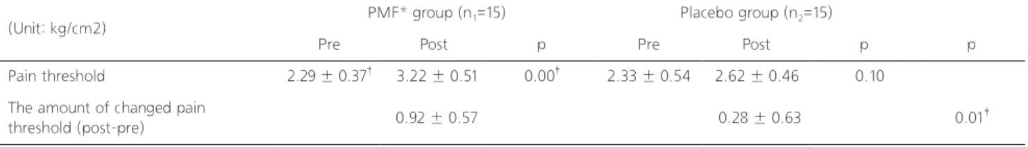
The pain threshold in post-treatment was significantly increased compared to pre-treatment in the PMF group;
however, there was no pre-post significant difference within the placebo group (p<0.05). The changed pain threshold after the treatment was significantly greater in PMF group than placebo group (Table 1). For the cervical ROM, the all ranges of motion tended to increase after the treatment in both
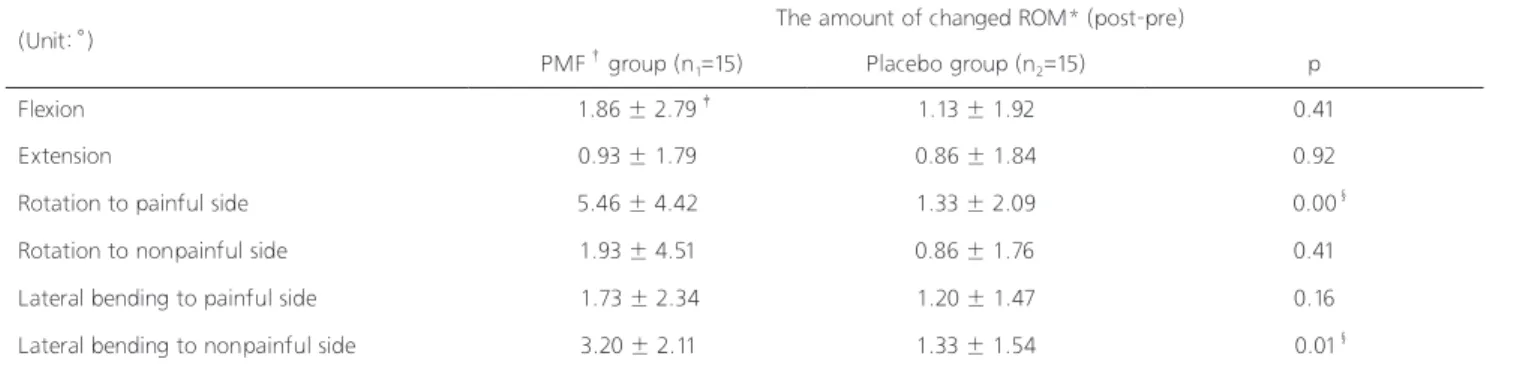
PMF and placebo group. In PMF group, the ranges of rotation toward the painful side and lateral bending toward the nonpainful side were significantly increased in post- treatment compared to pre-treatment (Table 2). In addition, in PMF group, the changed ROM for rotation toward the painful side and lateral bending toward the nonpainful side after the treatment were significantly greater than in placebo group (Table 3). However, there were no significant difference in changed ROM of flexion, extension, rotation to nonpainful side, and lateral bending to painful side between PMF and placebo group. Paired t-test showed significant decrease in UT EMG activity during scapular plane abduction between pre and post PMF treatment. Also, the muscle activity ratio between UT and SA was significantly decreased in PMF group (Table 4). The changed UT EMG activity as well as the changed UT/SA ratio after the treatment was significantly difference between PMF and placebo group (Table 5).
IV. Discussion
The results of this study show the possible immediate effects of PMF in the treatment of myofascial pain. The patients who
*pulsed magnetic field, †mean±standard deviation, ‡p<0.05.
*pulsed magnetic field, †mean±standard deviation, ‡p<0.05.
(Unit: kg/cm2) PMF* group (n1=15) Placebo group (n2=15)
Pre Post p Pre Post p p
Pain threshold 2.29 ± 0.37† 3.22 ± 0.51 0.00‡ 2.33 ± 0.54 2.62 ± 0.46 0.10
The amount of changed pain
threshold (post-pre) 0.92 ± 0.57 0.28 ± 0.63 0.01‡
(Unit: °) PMF* group (n1=15) Placebo group (n2=15)
Pre Post p Pre Post p
Flexion 34.66 ± 8.75† 36.13 ± 8.35 0.10 34.80 ± 5.23 35.93 ± 5.71 0.10
Extension 41.86 ± 14.12 42.80 ± 14.14 0.20 45.40 ± 10.98 46.26 ± 11.08 0.09
Rotation to painful side 38.33 ± 6.17 42.93 ± 7.10 0.00‡ 41.20 ± 4.94 42.06 ± 5.21 0.09 Rotation to nonpainful side 42.93 ± 6.14 44.73 ± 5.84 0.12 41.20 ± 5.22 42.06 ± 3.97 0.20 Lateral bending to painful side 29.00 ± 4.05 30.06 ± 4.19 0.21 29.40 ± 3.88 30.40 ± 3.85 0.20 Lateral bending to nonpainful side 27.33 ± 4.16 30.40 ± 4.53 0.00‡ 29.13 ± 3.71 30.26 ± 3.61 0.09 Table 1. The pain threshold in PMF and placebo group
Table 2. Comparison of ROM between pre- and post- treatment
underwent treatment with PMF showed significant benefits as measured through subjective and objective indices and through myofascial TrP characteristics. There was also evidence of improvement of the cervical ROM, particularly in rotation and contralateral bending and deactivate upper trapezius activity. On the other hand, the group receiving placebo treatment did not show any significant improvement.
The use of PMF in the treatment of musculoskeletal pain
is not new. PMF has been successfully used for the past 30 years in the treatment of pain in many osteoarticular diseases.2,14 In particular, PMF have been proven effective in the treatment of osteoarthritis of the knee and the cervical column and for tendinitis of the rotator cuff.2 As previously reported, the reduction of pain threshold of MPS resulting from magnetic stimulation could be explained both by an interference with peripheral nervous system and by triggering
*range of motion, †pulsed magnetic field, ‡mean±standard deviation, §p<0.05.
*electromyography, †pulsed magnetic field, ‡mean±standard deviation, §p<0.05.
(Unit: °) The amount of changed ROM* (post-pre)
PMF† group (n1=15) Placebo group (n2=15) p
Flexion 1.86 ± 2.79‡ 1.13 ± 1.92 0.41
Extension 0.93 ± 1.79 0.86 ± 1.84 0.92
Rotation to painful side 5.46 ± 4.42 1.33 ± 2.09 0.00§
Rotation to nonpainful side 1.93 ± 4.51 0.86 ± 1.76 0.41
Lateral bending to painful side 1.73 ± 2.34 1.20 ± 1.47 0.16
Lateral bending to nonpainful side 3.20 ± 2.11 1.33 ± 1.54 0.01§
(Unit: %MVIC) The amount of changed EMG*activity (post-pre)
PMF† group (n1=15) Placebo group (n2=15) p
Upper trapezius -5.29 ± 5.58‡ -0.88 ± 3.02 0.01§
Lower trapezius 4.43 ± 7.22 0.60 ± 2.35 0.07
Serratus anterior 1.90 ± 6.61 -1.36 ± 1.70 0.08
Upper trapezius/Lower trapezius -3.90 ± 9.05‡ -0.38 ± 0.77 0.16
Upper trapezius/Serratus anterior -0.27 ± 0.34 0.02 ± 0.06 0.01§
Lateral bending to nonpainful side 3.20 ± 2.11 1.33 ± 1.54 0.01§
Table 3. Comparison of changed ROM after treatment between the PMF and placebo group
Table 5. Comparison of changed EMG activity and EMG ratio after treatment between the PMF and placebo group
*pulsed magnetic field, †mean±standard deviation, ‡p<0.05.
(Unit: %MVIC) PMF* group (n1=15) Placebo group (n2=15)
Pre Post p Pre Post p
Upper trapezius 34.12 ± 9.45† 28.83 ± 7.31 0.00‡ 38.92 ± 9.45 38.04 ± 8.59 0.28
Lower trapezius 12.68 ± 11.21 17.12 ± 13.90 0.03‡ 10.71 ± 4.73 11.32 ± 4.77 0.33
Serratus anterior 45.66 ± 33.08 47.57 ± 32.05 0.28 42.32 ± 11.86 40.95 ± 12.26 0.11
Upper trapezius/Lower trapezius 8.27 ± 11.05 4.37 ± 5.00 0.12 4.51 ± 2.64 4.12 ± 2.23 0.18 Upper trapezius/Serratus anterior 1.12 ± 0.83 0.84 ± 0.53 0.01‡ 0.97 ± 0.28 0.99 ± 0.29 0.30 Lateral bending to nonpainful side 27.33 ± 4.16 30.40 ± 4.53 0.00‡ 29.13 ± 3.71 30.26 ± 3.61 0.09 Table 4. Comparison of the EMG activity and EMG ratio between pre- and post- treatment
of central mechanisms of pain modulation. Considering the fact that interactions of magnetic stimulation with biological tissues are largely unknown, we could hypothesize that the reduction of pain itself could possibly lead to a break of a pathological vicious circle accounting for the chronization of myofascial pain syndrome.24 In this vicious circle, pain could sustain tissue anomalies typical of MPS and tissue anomalies themselves lead to a reinforcement of pain.1 These results should encourage further research aimed at establishing the long-term clinical usefulness of this new procedure in the treatment of MPS. In particular, it would be interesting in the future to compare the effect of PMF with that of other conventional physical therapies in this pathology.
Pujol et al. (1998)25 proposed the use of peripheral PMF to reduce musculoskeletal pain. The authors evaluated patients suffering from pain with different etiologies located at different sites (e.g. epicondylitis, carpal tunnel syndrome, ulnar nerve compression syndrome, and posterior tibial tendinitis). The patients underwent one session of PMF applied to the painful site for 20 min. At the end of the treatment session, the patients treated with PMF showed a statistically significant reduction in pain as compared to a control group who received a placebo treatment.25 The results of our study extend these findings by demonstrating that PMF is effective in a group of patients with UT TrP. Another notable difference between our study and Pujol et al.’s concerns the protocol of clinical evaluation. In Pujol et al.’s study only the numerical rating scale was used to provide a subjective clinical evaluation of pain. In our study, we used multiple parameters of evaluation, including pain threshold, cervical ROM, and muscle activity of UT. All these parameters showed a significant improvement after treatment, suggesting that many aspects of myofascial pain may be modified by PMF therapy.
With reference to cervical ROM, it is important to note that the parameter which indicated a significant improvement in the PMF group was exclusively the rotation and lateral bending contralateral to the UT TrP, while other ROMs were not modified. This is not surprising since it is known that the UT muscle is much more implicated in controlateral bending and rotation than in other movements. This result is in accordance with other studies on the treatment of UT
TrP, in which it was shown that the most evident functional limitations concerned this movements.9
It is important to underline that the technique of PMF used in the present study presents noticeable differences in comparison with static magnetic therapy. First of all, classic magnetic therapy is generally applied to a large body area (the torso or the limbs), but the PMF may be used for the treatment of more focal pain, as myofascial TrPs. In addition, the advantage of magnetic flux changes is that it affects deep anatomical structures and thus may interfere with the overactivity of muscle spindle-endplate, occurring in taut band of TrP. It would explain the result of deactivation of UT muscle during scapular abduction. Finally, repetitive impulse (1Hz) with the rapidly changing field of the magnetic flux triggered the muscle contraction during treatment, resulting in repetitive UT contraction and inhibition. By muscle hold and relax mechanism, it would explain the immediate decrease of UT muscle activity; and decrease of cervical ROM related with UT.
This study has several limitations. First, our patients were male so the results cannot be generalized to women. Second, the subjects in this study were small number threatening external validity. Third, this study immediately confirmed the effects of PMF. Therefore further studies would be needed to demonstrate the long-term effects of PMF in subjects with UT TrP.
Acknowledgements
This research was financially supported by Basic Science Research Program though the National Research Foundation of Korea (NRF) funded by the Ministry of Education, Science and Technology (2011-0025231) and Sangji University Research Fund, 2014.
References
1. Travell JG, Simons DG. Myofascial pain and dysfunction. The trigger points manual. The upper parts of the body, Baltimore, MD: Williams & Wilkins, 1983:164-6.
2. Trock DH, Bollet AJ, Markoll R. The effect of pulsed electromagnetic fields in the treatment of osteoarthritis of the
knee and cervical spine. Report of randomized, double blind, placebo controlled trials. J Rheumatol 1994;21(10):1903-11.
3. Roth BJ, Saypol JM, Hallett M et al. A theoretical calculation of the electric field induced in the cortex during magnetic stimulation. Electroenceph clin Neurophysiol 1991;81(1):47-56.
4. Fischer AA. Documentation of myofascial trigger points. Arch Phys Med Rehabil 1987;69(4):266-91.
5. Simons DG. Understanding effective treatments of myofascial trigger points. J Bodyw Mov Ther. 2002;6(2):81-8.
6. Huguenin LK. Myofascial trigger points: The current evidence.
Phys Ther Sport. 2004;5(1):2-12.
7. Kwon JW, Nam SK, Choi YM et al. The Effect of Different Head Positions in Sitting on Head/Shoulder Posture and Muscle Activity. J Korean Soc Phys Ther 2013:25(4):217-23.
8. Nagrale AV, Glynn P, Joshi A et al. The efficacy of an integrated neuromuscular inhibition technique on upper trapezius trigg er points in subjects with non-specific neck pain: A randomized controlled trial. J Man Manip Ther. 2010;18(1):37-43.
9. Esenyel M, Caglar N, Aldemir T. Treatment of myofascial pain.
Am J Phys Med Rehabil 2000;79(1):48-52.
10. Smania N, Corato E, Fiaschi A et al. Therapeutic effects of peripheral repetitive magnetic stimulation on myofascial pain syndrome. Clin Neurophysiol 2003;114(2):350-8.
11. Bryant TN, Machin D. Statistical methods. In: Wilson BA, McLellan DL, editors. Rehabilitation studies handbook, Cambridge, MA: Cambridge University Press, 1997:189-204.
12. Shupak NM, Prato FS, Thomas AW. Human exposure to a specific pulsed magnetic field: Effects on thermal sensory and pain thesholds. Neurosci Lett 2004;363(2):157-62.
13. Struppler A, Jacob C, Müller-Barna P et al. A new method for the rehabilitation of central paresis by peripheral magnetic stimulation. Neurol Rehabil 1997;3(1):145-58.
14. Vavken P, Arrich F, Schuhfried O et al. Effectiveness of pulsed electromagnetic field therapy in the management of osteoarthitis of the knee: A meta-analysis of randomized controlled trials. J Rehabil Med 2009;41(6):406-11.
15. Kim MH, Kim SH, Kim HJ. Comparison of Ultrasonography Images on Normal Muscle and Myofascial Trigger Points Activated Muscle. J Korean Soc Phys Ther 2013:25(2):76-80.
16. Levoska, S. Manual palpation and pain threshold in female office employees with and without neck-shoulder symptoms.
Clin J Pain 1993;9(4):236-41.
17. Tousignant M, Smeesters C, Breton AM et al. Criterion validity study of the cervical range of motion (CROM) device for rotational range of motion on healthy adults. J Orthop Sports Phys Ther. 2006;36(4):242-8.
18. Dall'Alba PT, Sterling MM, Treleaven JM et al. Cervical range of motion discriminates between asymptomatic persons and those with whiplash. Spine (Phila Pa 1976). 2001;26(19):2090-4.
19. Nam KS, Kwon JW. The Effects of Head Position in Different Sitting Postures on Muscle Activity with/without Forward Head and Rounded Shoulder. J Korean Soc Phys Ther 2014;26(3):140- 6.
20. Thigpen CA, Padua DA, Michener LA et al. Head and shoulder posture affect scapular mechanics and muscle activity in overhead tasks. J Electromyogr Kines, 2010;20(4):701-9.
21. Weon JH, Jung DY. Comparison of the Muscle Activities of Upper Trapezius and Middle Deltoid between Subjects with and without Elevation of Shoulder Girdle during Arm Elevation. J Korean Soc Phys Ther 2012:24(6):388-92.
22. Kendall FP. Muscles: Testing and function with posture and pain. 5th ed. Baltimore, MD, Lippincott Williams & Wilkins, 2005:165-86.
23. Jeon HS, Kang SY, Park JH et al. Effects of pulsed electromagnetic field therapy on delayed-onset muscle soreness in biceps brachii. Phys Ther Sport. 2014:1-6.
24. Porta M, Perretti A, Gamba M et al. The rationale and results of treating muscle spasm and myofascial syndromes with botulinum toxin type A. Perfusion Dig. 1998;8(1):346-52.
25. Pujol J, Pascual-Leone A, Doltz C et al. The effect of repetitive magnetic stimulation on localized musculoskeletal pain.
NeuroReport. 1998;9(8):1745-8.