CONTENTS
Ⅰ. INTRODUCTION
Ⅱ. METERIALS AND METHODS
Ⅲ. RESULTS
Ⅳ. DISCUSSION
Ⅴ. CONCLUSION REFERENCES KOREAN ABSTRACT
Ⅰ. INTRODUCTION
Internal derangement of the temporomandibular joint (TMJ) has been defined as an abnormal positional relationship of the disc relative to the mandibular condyle, fossa and/or articular eminence, and is a major cause of jaw pain, clicking and/or crepitus as well as limitation of opening.1)
In the TMJ, the disc functions as a true articular surface during the mandibular movement. The normal shape of the disc is biconcave. The central part of the disc is considerably thinner than its peripheral part. Its posterior band is considerably thicker.2) The form of the undersurface of the disc and the top of the condyle fit so well together. Such a disc morphology provides a self-positioning feature
that, in conjunction with the interarticular pressure, centers the disc on the condyle. The normal position of the disc has traditionally been described as a 12 o'clock relationship between the superior aspect of the condyle and posterior band of the disc.3-6) The disc maintains its morphology unless destructive forces or structural changes occur in the joint. Factors that lead to the elongation or tearing of the discal ligaments can cause internal derangement disorders. Although causes of internal derangement of the TMJ remain unknown, the greatest contributing factor of internal derangement is trauma, which may be either macrotrauma (e.g., blow) or microtrauma (e.g., muscle hyperactivity).
Disc deformation most commonly begins with thickening and enlargement of the posterior band.
Then, the posterior disc attachment appears stretched and gets longer and thinner.5)
If the morphology of the disc is altered and the discal ligaments become elongated, the disc is permitted to slide forward across the articular surface of the condyle. As the posterior border of the disc becomes thinned, it is pulled further into the discal space by the superior head of the lateral pterygoid muscle and the condyle becomes positioned on the posterior border of the disc. This is anterior
Magnetic Resonance Evaluation of the Articular Disc Configuration in the Patients with Internal Derangement
of the Temporomandibular Joint
Gwui-Ae Kim†, D.D.S., M.S.D., Sun-Hee Kim‡, D.D.S., M.S.D.,
Woo-Cheon Kee†, D.D.S., M.S.D., Ph.D., Jae-Kap Choi†, D.D.S., M.S.D., Ph.D.
†Department of Oral Medicine, School of Dentistry, Kyungpook National University
‡Department of Oral Hygiene, Taegu Health College
disc displacement with reduction. The single click during mouth opening represents the very early stage of what is called internal derangement. If this condition progresses further, the anterior and medial repositioning tendency of the disc aggravates more7) and the morphologic change of the disc creates a second click during the later stage of condylar return just prior to the closed joint position. This stage of internal derangement is called the reciprocal click.
The more the shape of the disc changes to accomodate the pull of the muscle and the slide of the condyle, the greater is the likelihood that the disc will be pulled through the discal space, collapsing the joint space behind. In this stage, the full translation of the condyle is inhibited by the anteriorly and medially positioned disc. This condition is referred to as a anterior disc displacement without reduction.
Joint sounds are eliminated since no skidding can occur. If the lock is occasionally resolved without assistance, it is referred to as a intermittent locking, otherwise, it is referred to as a chronic lock. Once the disc is completely anteriorly displaced and the retrodiscal tissue breaks down, the condyle begins to articulate directly with fossa. Degenerative joint disease (DJD) represents a destructive process by which the bony articular surfaces of the condyle and fossa become altered.2)
According to previous studies, it has been found that the change of the disc configuration has a direct effect on the clinical course of internal derangement.
Arthrography and computed tomography (CT) have been used for the diagnosis and the treatment of TMJ disorders, but we have had some difficulties in examining the disc configuration by these imaging systems. The most recent imaging technique, magnetic resonance imaging (MRI) was provided as a good substitute for these with greater soft tissue contrast.4,5) In addition to it, it doesn't need ionizing radiation, anaesthesia or the injection of contrast agents. MRI can be superior to computed tomo- graphy in diagnosing TMJ internal derangement and has a considerable number of advantages over arthrography and arthrotomography.8) Therefore, MRI is considered as the modality of first choice to
identify the disc position and configuration.
The aim of the study is to examine the shape of the disc on MRI and correlate them to clinical characteristics.
Ⅱ. MATERIALS AND METHODS
Ninety patients (68 females, 22 males) who had visited the Department of Oral medicine at Kyungpook National University Hospital, Taegu, Korea and been diagnosed as having TMJ disorders were included in the study. They had TMJ pain, clicking or other functional abnormalities. The mean age was 24.9 years, ranging from 13 to 63 years.
All the patients were imaged on sigma 1.5 Tesla MR imaging system (General Electric, Milwaukee, WI, USA) by use of a 7.62 cm surface coil (GE Medical systems). These images were obtained with the patient maintaining a closed and open-mouth position and were oriented perpendicular (sagittal) to the long axis of the condylar head. Closed-mouth sagittal sections was obtained with a T1-weighted spin echo sequence (TR 556/TE 17, 3 excitations, FOV 12×12cm, 512×192). Closed and open-mouth sagittal section were obtained with a T2-weighted fast spin echo sequence (TR 3000/TE 92, 3 excitations, FOV 12×12cm, 512×192 matrix). The section thickness was 3mm. The anatomy of the joint was well delineated on the T1-weighted images.
T2-weighted images were valuable for the assessment of joint effusion and occasionally for the evaluation of joint morphology.9)
All the joints were divided into 3 groups according to the disc position in a closed and open-mouth position. A normal group was defined to have a normal disc-condyle relationship with a intermediate zone interposed between the glenoid fossa and the condyle in the closed mouth position and maintain its relationship in the open-mouth position. An anterior disc displacement with reduction group (ADWR group) was defined by the posterior band being anterior to the condyle and the uppermost part of the condyle was seated on retrodiscal tissue in the closed-mouth position and the disc took a normal
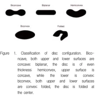
Figure 1. Classification of disc configuration. Bico- ncave, both upper and lower surfaces are concave; biplanar, the disc is of even thickness; hemiconvex, upper surface is concave, while the lower is convex;
biconvex, both upper and lower surfaces are convex; folded, the disc is folded at the center.
disc-condyle relationship in open-mouth position.
Finally, an anterior disc displacement without reduction group (ADWOR group) was defined by the disc always remaining anterior to the condyle in both the closed and open-mouth position.4,10)
To assess disc configuration, discs were categorized according to the shapes as shown in figure 1. The section where the deformation of the disc was most marked was used.11) The images were interpreted twice by one author and coincident results were selected.
The relative proportion of distribution of the type of the disc configuration in diagnostic subgroups was examined to see if there is any significant relationship between the progression of internal derangement and the degree of disc deformity. Each type of disc configuration was also correlated with TMJ pain and clicking. The presence of joint effusion was evaluated on the T2-weighted image and correlated with prevalence of joint pain.
Statistical Analysis
In analyzing the data, the chi-square test was used for categorical data and the test was done for the significance of group differences in proportion.
Ⅲ. RESULTS
One hundred sixty-nine temporomandibular joints in 90 patients were examined. Table 1 describes the diagnostic results from the MR imaging of these patients.
Results of the distribution of the disc configuration in the closed-mouth position in the anterior disc displacement with reduction group demonstrates that 40.00% of the total joint discs were biconcave, 22.35% were biplanar, 30.00% were convex, and 17.65% were folded, whereas, in the normal group, 91.67% of the discs were biconcave and 8.33% were biplanar, and in the anterior disc displacement without reduction group, 77.08% of the disc were folded, 20.83% were hemiconvex, and only 2.08%
were biconcave (Table 2).
In the open-mouth position, the disc configuration
Table 1. Number of subjects by diagnostic groups
Diagnosis Number
Normal
Anterior disc displacement with reduction Anterior disc displacement without reduction
48 85 36
Table 2. Percent distribution of the types of disc configuration in the closed- mouth position according to the diagnostic subgroups†
Group Disc Configuration Type
Ⅰ Ⅱ Ⅲ Ⅳ Ⅴ
Normal ADWR ADWOR
91.67 40.00 2.08
8.33 22.35 0.00
0.00 20.00 20.83
0.00 0.00 0.00
0.00 17.65 77.08 †; Statistically significant by chi-square test at level 0.05 (p = 0.001)
ADWR = Anterior disc displacement with reduction.
ADWOR = Anterior disc displacement without reduction Disc configuration type I = Biconcave; type II = Biplanar;
type III = Hemiconvex; type IV = Biconvex;
type V = Folded
of normal group was biconcave in 100% of the cases, whereas, in anterior disc displacement with reduction group, 92.94% of the discs were biconcave and 5.88%
were biplanar, 1.18% were folded, whereas, in the anterior disc displacement without reduction group, 93.75% of the disc were folded, 4.17% were biconcave and, 2.08% were biplanar (Table 3). There were statistically significant differences in the distribution of the types of disc configuration among groups in both the closed and open-mouth position (p<0.05).
In the normal group, all the pain free subjects have a disc of biconcave type, and 72.73% of the subjects with painful TMJ have a disc of biconcave type and the remaining 27.27% have a biplanar disc (Table 4).
In anterior disc displacement with reduction group, 60.53% of the click-free subjects have a biconcave disc, 21.05% of the same subjects have a biplanar disc, 10.53% have a hemiconvex disc, and only 7.89%
have a folded disc. However, the subjects with click
Table 3. Percent distribution of the types of disc configuration in open-mouth position according to the diagnostic subgroups†
Group Disc Configuration Type
Ⅰ Ⅱ Ⅲ Ⅳ Ⅴ
Normal ADWR ADWOR
100.00 92.94 4.17
0.00 5.88 2.08
0.00 0.00 0.00
0.00 0.00 0.00
0.00 1.18 93.75
†; Statistically significant by chi-square test at level 0.05 (p = 0.001)
Table 4. Percent distribution of disc configuration according to presence or absence of pain in patients with normal joint†
Group Disc Configuration Type
Ⅰ Ⅱ Ⅲ Ⅳ Ⅴ
Pain(+) Pain(-)
72.73 100.00
27.27 0.00
0.000 0.00
0.00 0.00
0.00 0.00
†; Statistically significant by chi-square test at level 0.05 (p = 0.006)
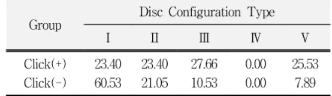
have a biconcave, biplanar, or hemiconvex disc with fairly even distributions ranging 23% to 28% (Table 7).
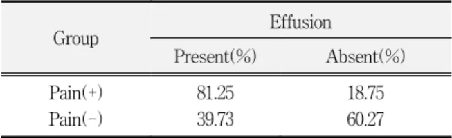
There was a strong relationship between the presence of joint effusion and TMJ pain, where the 81.25% of the subjects with evidence of joint effusion revealed joint pain (Table 10).
Table 5. Percent distribution of disc configuration according to presence or absence of clicking sound in patients with normal joint†
Group Disc Configuration Type
Ⅰ Ⅱ Ⅲ Ⅳ Ⅴ
Click(+) Click(-)
77.78 96.30
22.22 3.70
0.00 0.00
0.00 0.00
0.00 0.00
†; Statistically not significant by chi-square test at level 0.05 (p = 0.082)
Table 6. Percent distribution of the types of disc configuration according to presence or absence of pain in patients with ADD with reduction†
Group Disc Configuration Type
Ⅰ Ⅱ Ⅲ Ⅳ Ⅴ
Pain(+) Pain(-)
32.00 51.43
20.00 25.71
20.00 25.71
0.00 0.00
22.00 11.43
†; Statistically not significant by chi-square test at level 0.05 (p = 0.125)
Table 7. Percent distribution of disc configuration according to presence or absence of clicking sound in patients with ADD with reduction†
Group Disc Configuration Type
Ⅰ Ⅱ Ⅲ Ⅳ Ⅴ
Click(+) Click(-)
23.40 60.53
23.40 21.05
27.66 10.53
0.00 0.00
25.53 7.89
†; Statistically significant by chi-square test at level 0.05 (p = 0.003)
Table 8. Percent distribution of disc configuration according to presence or absence of pain in patients with ADD without reduction†
Group Disc Configuration Type
Ⅰ Ⅱ Ⅲ Ⅳ Ⅴ
Pain(+) Pain(-)
2.86 0.00
0.00 0.00
20.00 23.08
0.00 0.00
77.14 76.92
†; Statistically not significant by chi-square test at level 0.05 (p = 0.813)
Table 9. Percent distribution of disc configuration according to presence or absence of clicking sound in patients with ADD without reduction†
Group Disc Configuration Type
Ⅰ Ⅱ Ⅲ Ⅳ Ⅴ
Click(+) Click(-)
5.26 0.00
0.00 0.00
31.58 13.79
0.00 0.00
63.16 86.21
†; Statistically not significant by chi-square test at level 0.05 (p = 0.131)
Table 10. Prevalence of effusion by the presence or absence of pain†
Group Effusion
Present(%) Absent(%)
Pain(+) Pain(-)
81.25 39.73
18.75 60.27
†; Statistically significant by chi-square test at level 0.05 (p = 0.003)
Ⅳ. DISCUSSION
Morphologic alterations of the disc can be evaluated using double-space arthrography, but this requires an injection of contrast material into the upper and lower joint space compartments and additional radiation from multisection tomography.
However, MRI provides a noninvasive mechanism
for several aspects of discal pathology. Anterior displacements of the disc can be clearly visualized on MR images with an accuracy that is perhaps even greater than on arthrograms. The MR images can also give a good visualization for disc configuration.
According to the study of Tasaki and Westesson12), MR Imaging was 95% accurate in the assessment of disc position and disc form and 93% accurate in the assessment of osseous changes.
In this study, the disc position and shape was examined on T1- and T2-weighted images for every subject in each diagnostic subgroup of internal derangement. The disc shape was classified into 5 types according to the method of Murakami et al.11) They classified disc configuration on MR imaging into biconcave, biplanar, hemiconvex, biconvex, and folded type.
Disc deformation was rarely seen in the normal group. Most discs were biconcave in the normal group, whereas, in the anterior disc displacement with reduction group in closed-mouth position, less than half of them were biconcave and in the anterior disc displacement without reduction group, most of them were folded. Murakami et al.11) suggested that the disc may be distorted only after disc displacement, since few distorted discs were found in the normal disc position.
A few normally positioned discs and many of anteriorly displaced discs deviated from the biconcave configuration on the MRI when the mouth was closed, but was found to be biconcave on the MRI when the mouth was open. These findings are in an agreement with those reported by Murakami et. al.11) and DE Leeuw et. al.13) In some cases, folded discs in closed-mouth position were reduced into biconcave configuration during mouth opening.
Therefore, disc configuration and disc reduction must be examined in open-mouth position.13) Heffez and Jordan14) classified disc configuration into bow-tie, straight, funnel, bulge and Y by means of corrected lateral cephalometric arthrotomograms and histopathologic sagittal section. They found that the normal condyle-disc-fossa relationship and slight-to-moderate disc displacement occurred with
bow-tie shaped disc and severe disc displacement occurred predominantly with bulge shaped disc. But, there were certain problems innate to the development of a classification scheme based on the radiographs obtained with a corrected lateral hypocycloidal tomographic technique. First, the radiographic technique corrects for the condyle and not the disc in the closed position. Secondly, not all arthrotomograms show images of disc shapes that fit neatly into a category according to the established criteria.
Westesson et al.15) studied joint morphology, internal derangement, and joint function in randomly selected autopsy specimen using arthrography and cryosections. The result of their study showed that joints with superior disc position rarely demonstrated morphologic alterations and joints with completely anteriorly positioned discs showed disc deformation in 77% and irregularities of the articular surfaces in 65%.
In this study, 91.67% of the normal group showed biconcave disc, whereas, 40% of the ADWR group and only 2% of the ADWOR group showed it.
However, the folded disc was seen in 17.65% of the ADWR group and 77.08% of the ADWOR group.
There was not anyone who had a folded disc in the normal group. These results indicate that the degree of the disc displacement is closely related to the degree of the disc deformity, namely, the more the internal derangement progresses, the more the disc is deformed. Although it is still unclear which is the first change over the other, Katzberg and Westesson5) suggested the deformation of the disc appears to occur secondarily to the disc displacement.
Disc configuration appeared to be slightly associated with pain from the joint. In the normal group, all the pain-free subjects have biconcave discs, whereas, some subjects with joint pain have a biplanar disc. In the ADWR group, there is a tendency that the prevalence of joint pain was higher in the subjects with a hemiconvex or folded disc and it was lower in the subjects with a biconcave or biplanar disc although it was not statistically
significant.
However, the evidence of joint effusion in MR images appeared to be significantly associated with the subjective complaint of joint pain. Joint effusion can be easily demonstrated by the use of T2-weighted imaging sequences. Joint effusion is diagnosed when there is an area of high signal on the T2-weighted images, in the upper or lower joint spaces or both. It has been our experience that joint effusion is strongly associated with joint pain. This is also supported by another series of work, in which 88% of painful joints showed MR evidence of joint effusion. Also, Kim and Suh et al.16) described that MR findings of the amount of temporomandibular joint effusion correlated with joint pain and anterior disc displacement. In our study, 81.25% of painful joints showed joint effusion on MRI findings. The presence of fluid or inflammation in the joint without an apparent disc abnormality can be one of the determinants that can affect clinical management of the patient.
The etiology of pain related to the internal derangement is not completely understood. The pain has several possible origins, such as tearing and laceration of the joint capsule, compression of the posterior disc attachment, impingement of the inside of the capsule by the displaced disc, synovitis, expansion of the joint to peripheral adhesion and nerve entrapment. There is still a lot to be learned about the etiology of pain, its variation over time and its relationship to morphologic alterations in the joint.5)
Recent arthrographic and CT studies of patients who have TMJ pain and dysfunction have shown a close relationship between anterior disc displacement with reduction and painful clicking of the TMJ.
These investigations have shown that a loud audible clicking sound occurs as the displaced disc reduces.17) According to MRI finding in our study, in the anterior disc displacement without reduction group, deformed and displaced disc is more related with a clicking sound. Interestingly, it suggests that a significant percentage of patients with clicking TMJs do not have a reduction of the discs. These findings
suggest that the deviations in the surface contours of both the disc surface and condylar head could be a mechanism for the clicking sound. Stretching of ligaments with the sound arising from disc vibration as the condyle translates also is a possible factor of a clicking sound. Painful clicking of the TMJ need not always imply a reducing disc displacement.9) MRI finding can describe a clinically unrecognized abnormality of disc and its function associated with clicking, which can represent a more advanced stage of disease than would be suggested by clinical evaluation alone. Certain limitations must be considered in the present study. Medial or lateral displacement of the disc was not considered in MR Imaging. Coronal section is valuable for medial or lateral displacement of disc. It was cited that coronal images helped in avoiding a false-negative diagnosis in 13% of the joints by Tasaki and Westesson.12) Another limitation is that shape classification of disc on MR Imaging was determined by the same investigator.
In conclusion, there were a variety of disc configurations in the subjects with internal derangement and some clinical symptoms appeared to be significantly associated with the disc configuration. Therefore, in the clinical evaluation of patients with temporomandibualr disorders the examinations of soft tissue condition as well as hard tissue relationship of the joint should be included for successful management of the patients.
V. CONCLUSIONS
The aim of this study was to investigate the configuration and position of the disc on MRI and correlate them to clinical characteristics, such as pain, clicking and joing effusion.
Ninety patients (68 females, 22 males) with a mean age of 24.9 years (ranging from 13 to 63 years), who had been diagnosed with TMJ disorders, were included in the study.
The following conclusions were drawn.
1. The degree of the disc displacement was closely related to the degree of the disc deformity.
2. Disc configuration seems to be slightly associated with pain but it was not statistically significant.
3. MR evidence of joint effusion seems to correlate with joint pain. In our study, 81.25% of painful joints showed joint effusion.
4. The deviations in the surface contours of the disc and condylar head or disc vibration during condylar translation was suggested as a mechanism for the clicking sound.
5. For clinical evaluation of TMD, disc configuration should be examined and MRI is recommended as a excellent method for describing soft tissue condition.
REFERENCES
1. Jundh H, Westesson PL: Clinical signs of temporo- mandibular joint internal derangement in adults, Oral Surg Oral Med Oral Pathol., 72:637-641, 1991.
2. Okeson JP: Management of temporomandibular disor- ders and occlusion, 2nd ed. St. Louis The C.V. Mosby Co. 1985.
3. Kaplan PA, Tu HK, Williams SM, Lydiatt DD: The normal temporomandibular joint: MR and arthrographic correlation, Radiology, 165:177-178, 1987.
4. Drace JE, Enzmann DR: Defining the normal tempo- romandibular joint: Closed-, Partially open-, and Open-mouth MR Imaging of asymptomatic subjects, Radiology, 177: 67-71, 1990.
5. Katzberg RW, Westesson PL: Diagnosis of the tem- poromandibular joint, Philadelphia. W.B. Saunders Company, 1993.
6. Drace JE, Young SW, Enzmann DR: TMJ meniscus and bilaminar zone: MR Imaging of the substructure-Diagnostic landmarks and pitfalls of interpretation, Radiology, 177:73-76, 1990.
7. Bell WE: Clinical management of Temporomandibular joint disorders, Chicago, London, Year Book Medical Publishers, 1982.
8. Katzberg RW, Bessette RW, Tallents RH, Plewes DB, Manzione JV, Schenck JF, Foster TH, Hart HR:
Normal and abnormal temporomandibular joint: MR Imaging with surface coil, Radiology, 158:183-189, 1986.
9. Broods SL, Westesson PL: Temporomandibular Joint:
Value of coronal MR Imaging, Radiology, 188:317-321, 1993.
10. Hans MG, Jieberman J, Goldberg J, Rozencweig G, Bellon E: A comparison of clinical examination, history and magnetic resonance imaging for identifying orthodontic patients with temporomandibular joint disorders, Am. J. Orthod Dentofac, Orthop 101;54-9, 1992.
11. S. Murakami, A. Takahashi, H. Nishiyama, M.
Fujishita and H. Fuchihata: Magnetic resonance evaluation of the temporomandibular joint disc position and configuration, Dentomaxillofac. Radiol., 22: 205-207, 1993.
12. Tasaki MM, Westesson PL: Temporomandibular Joint: Diagnostic accuracy with sagittal and coronal MR Imaging, Radiology, 186:723-729, 1993.
13. DE Leeuw R, Boering G, Stegenga B and DE Bont LGM : TMJ articular disc position and configuration 30 years after initial diagnosis of internal derang- ement, J Oral Maxillofac Surg., 53:234-241, 1995.
14. Heffez L, Jordan S: A classification of temporo- mandibular joint disc morphology, Oral Surg Oral Med Oral Pathol., 67:11-9, 1989.
15. Westesson PL, Bronstein SL, Liedberg J: Internal derangement of the temporomandibular joint:
Morphologic description with correlation to joint function, Oral Surg. Oral Pathol., 59:323-331, 1985.
16. Kim KH, Suh KJ: A relationship between amount of joint effusion, disc displacement and presence of pain in the temporomandibular joint : MR Imaging, J.
Korean Radiol Soc., 36:149-153, 1997.
17. Miller TL, Katzberg RW, Tallents RH, Bessete RW, K. Hayakawa: Temporoman- dibular joint clicking with nonreducing anterior displacement of the meniscus, Oral Surg. Oral Med. Oral Pathol., 59:
323-331, 1985.
국문초록
악관절 내장증 환자에서 자기공명영상사진상의 관절원판 형태의 평가
경북대학교 치과대학 구강내과학교실†
대구보건대학 치위생과‡
김 귀 애†, 김 선 희‡, 기 우 천†, 최 재 갑†
이 연구의 목적은 악관절 내장증 환자에서 관절원판의 형태를 자기공명영상사진으로 평가하고 임상적 증상과 의 관계를 알아보고자 하는 것이다.
악관절 내장증의 증상을 가진 90명의 환자에서 169개의 관절을 자기공명영상사진으로 관절원판과 하악과두의 관계를 기준으로 정상군, 정복성 관절원판 변위군, 비정복성 관절원판 변위군으로 분류하고 관절원판의 형태를 5가지로 나누었다. 임상검사는 측두하악관절통과 관절음의 유무를 확인하여 그 상관관계를 알아보았다.
실험 결과에서 측두하악관절통은 각 군에서 관절원판의 형태와는 정상군을 제외하고는 통계적인 유의성이 없었 으며, 염증성 삼출액과 유의한 관계를 보였다. 관절음은 정복성 관절원판 변위환자에서 관절원판형태와 통계적으 로 유의성을 보였으며, 비정복성 관절원판 변위환자에서도 상당수 관절음이 발생하였음을 알 수 있었다. 이 결과 는 측두하악관절의 통증은 관절원판의 형태가 많이 변형될수록 통증이 심해지는 경우가 많았지만, 다른 여러 가 지 원인들이 존재할 수 있으며, 관절음은 정복성 관절원판 변위환자에서 관절원판의 변형이 심할수록 관절원판이 정복할 때 발생될 수 있다는 것을 의미한다. 따라서, 임상적인 검사와 더불어 자기공명영상사진으로 정확한 진단 을 함으로써 관절원판 변위의 적절한 치료가 행해질수 있으리라 생각된다.