https://doi.org/10.9721/KJFST.2020.52.4.342
342
©The Korean Society of Food Science and Technology
Antioxidant activities of flower, berry and leaf of Panax ginseng C. A. Meyer
Hee-Jeong Ryu1, Chul-Jong Jung1, and Gyung-Yun Beik1,*1Department of OkChundang Research institute
Abstract This study was conducted to investigate the applicability of the ground parts such as flower (GF), berry (GR), and leaf (GL) from Panax ginseng C. A. Meyer. The ground parts were extracted from hot water (WE) and 60% ethanol (EE). Total polyphenol and flavonoid contents were 15.02-32.74 and 21.60-484.05 mg GAE/g, respectively. Hot water extract of ginseng leaf (GLWE) and 60% ethanol extract of ginseng leaf (GLEE) showed higher total polyphenol and total flavonoid contents than other extracts. Crude saponin contents were found in the range of 15.30-37.27%. Antioxidant activity of these extracts from ginseng was also analyzed by DPPH, ABTS, H2O2 scavenging activity, reducing power, and inhibition effect on lipid peroxidation. We confirmed the results that hot water extract of ginseng leaf (GLWE), 60% ethanol extract of ginseng leaf (GLEE) has high anti-oxidative effects. According to the antioxidant activity results of each extract of ginseng flower, ginseng berry, and ginseng leaf, it is judged that their availability is very high, and if proper processing is performed, it can be used as a functional raw material.
Keywords: anti-oxidant, ginseng flower, ginseng berry, ginseng leaf, Panax ginseng
Introduction
Panax ginseng C. A. Meyer has been used as a natural healthy food of oriental based on scientific evidence and clinical for thousands of years. In recently, Saponins, ginsenosides possessed by ginseng, have been reported to have various effects on the human body such as anti-fatigue, anti-cancer, promoting liver function, preventing arteriosclerosis, preventing hypertension, anti-stress, anti-aging, promoting brain activity, and anti-inflammatory activity (Park, 1996).
In addition, ginseng mainly consumes only the root. but the content of saponin is contained in ginseng leaves and berries more than the root (Cho, 1977; Choi et al., 2009; Han et al., 2004; Hu et al., 2008; Shao et al., 1989; Shi et al., 2007; Yahara et al., 1979; Yahara et al., 1976a; Yahara et al., 1976b).
Ginseng flower, the ground part of ginseng, has more ginsenosides content than the root, and its kind is similar to a root, and has many Rb2, Rc, Rd, Re, Rg and total ginsenoside, especially Re component, which contains a large amount of ginseng (Choi et al., 2009). It is removed to promote, and ginseng berry is also produced only for more than 4 years of ginseng and is also removed in advance to increase the saponin content of the root. However, ginseng berry also has more than 2 times the content of saponin and more than 30 times the content of Re compared to the root of ginseng root (Wang et al., 2006). In 2000, cardiovascular disease improvement and anti-diabetes have been shown to be effective attracting attention.
Ginseng leaf also contains ginsenosides similar to ginseng roots (Chang, 2003; Cho, 1977; Choi et al., 2009; Seog et al., 2004). Ginseng leaf have the highest among ginseng sites, and have been reported to have a saponin content of about 4-5 times higher than ginseng roots and 9 times higher than stems (Jackson et al., 2003; Xie et al., 2004).
According to pharmacological studies, its effect is similar to that of ginseng root, and it is reported that it is effective against enhancing immunity, promoting metabolism, regulating the central nervous system, angina pectoral, and neurological dysfunction.
In addition, many studies have been conducted on pharmacological effects such as anti-cancer, blood glucose-lowering effects of polysaccharides (Kim et al., 1998), anti-viral and anti-fungal activity of the protein (Ng and Wang, 2001), the anti-lipolytic activity of oligopeptides (Kim et al., 1987), and antioxidant activity of phenolic compounds (Lee et al., 2000; Shin et al., 1990; Wee et al., 1989).
Most of the research on the ginseng’s ground part is the research on the saponin component of ginseng leaf (Hong et al., 1979; Kim and Staba, 1974) and research on utilization of Yang and Lee (1979), Kim et al. (1981), a study on the development of ginseng leaf tea a study on the extraction and nutritional value of ginseng leaf protein (Kim et al., 1989), whitening action, collagenase inhibitory activity for fermentation of ginseng flower, ginseng berry as a cosmetic ingredient (Jeon et al., 2011), a study on improving blood circulation and improving skin tone (Kim et al., 2010) has been reported.
It is rarely useful, and it is almost discarded to date. As such, studies on ginseng flowers, ginseng berry, and ginseng leaf which are by-products, are mostly related to changes in ginsenoside for each of them, and in particular, there have been few reports comparing and analyzing the antioxidant capacity of functional ingredients.
The purpose of this study was to investigate the antioxidant *Corresponding author: Gyung-Yun Beik, Okchundang Research
institute, Daegu 41059, Korea Tel: +82-53-950-0090 Fax: +82-53-965-9551 E-mail: [email protected]
Received April 8, 2020; revised May 11, 2020; accepted July 24, 2020
activity, crude saponin content of the above-ground parts of ginseng to find various ways to utilize ginseng flower, berry, and leaf and to provide basic data for the development of health functional food products.
Materials and Methods
Materials
The flower, berry, and leaf of ginseng used in this study were purchased from Yeongcheon branch of Okchundang (Okchundang Co., Daegu, Korea). The dried flower, berry, and leaf of 4-year-old ginseng were extract ed 80oC for more than 8 h (three times) in the water of decuple (sample: sol vent extraction=1:10, w/v) of the sample using a reflux extraction for the physiologically activities. And 60% of ethanol extraction was extracted for more than 8 hours at room temperatures. The reason for using 60% ethanol was report ed by Hwang et al. (2005) that crude polysaccharide content decreases rapidly when the concentration of the extraction solvent is less than 50% EtOH or more than 70% ethanol in the ginseng extraction method. The preliminary experiment showed the same tendency and extracted from an optimal concentration of 60% ethanol solvent.
The hot water extracts were expressed as GFWE, GRWE, GLWE (hot water extract of ginseng flower; GFWE, hot water extract of ginseng berry; GRWE and hot water extract of ginseng leaf; GLWE). And the ethanol extracts were extracted with 60% ethanol and expressed as GFEE, GREE, GLEE (60% ethanol extract of ginseng flower; GFEE, 60% ethanol extract of ginseng berry; GREE and 60% ethanol extract of ginseng leaf; GLEE). Each extract was filtered through filter paper (Whatman No. 2), concentrated using a rotary evaporator (Rotavapor R-100, Buchi Korea, Seoul, Korea) and lyophilized (TFD5505, ilShin BioBase, Gyeonggi, Korea).
Reagents
Folin-Ciocalteu reagent, DPPH (1,1-diphenyl-2-picrylhydrazyl), ABTS (2,2'-azino-bis(3-ethylbenzothiazoline-6-sulphonic acid), Hydrogen peroxide (H2O2), gallic acid, BHA (butylated hydroxy-anisole), ascorbic acid was purchased from Sigma-Aldrich Co. (St, Louis, MO, USA).
Determination of total polyphenol contents
Total phenolic contents were determined using a Folin-Ciocalteau reagent assay (Dewanto et al., 2002). Each extract was dissolved in methanol (0.1 mL) mixed with 2 mL of 2% sodium carbonate solution. After 0.1 mL of 50%, Folin-Ciocalteau reagent was added to the mixture. After 1 hour of standing, the extracts were measured at 720 nm. Gallic acid was used as a standard to construct the calibration curve and the total polyphenol contents of ginseng extracts were expressed in mg gallic acid equivalents (GAE)/g of extract.
Determination of total flavonoid contents
Total flavonoid content was determined based on the method described by Nieva Moreno et al. (2000) with minor modifications.
Each extract was dissolved in 80% ethanol (0.5 mL) and were mixture with 4.3 mL of 80% ethanol, 0.1 mL of 10% aluminum nitrate, 0.1 mL of 1 M potassium acetate. The mixture was then incubated at room temperature for 40 min. After, the extracts were measured at 415 nm. Gallic acid was used as a standard to construct the calibration curve and the total polyphenol contents of extracts were expressed in mg gallic acid equivalents (GAE)/g of extract.
Crude saponin content
Extraction and determination of crude saponin were carried out by modifying the method of Korean food standards codex. The samples (5 g) were placed into the reflux extractor and extracted with 50 mL water-saturated butanol at 70oC for 1 h. The extract was filtered through Whatman No. 2 filter paper and concentrated at 55oC using a rotary vacuum evaporator. The concentrate was dissolved in 60 mL of distilled water and washed two times in a separatory funnel with 60 mL diethyl ether. The aqueous layer was extracted three times with 60 mL water-saturated butanol. The butanol extracts were pooled and washed two times with 50 mL of distilled water to remove impurities. The resulting butanol layer was evaporated at 55oC using a rotary vacuum evaporator. The round-flask with the evaporated residue was dried at 105oC. After measuring the weight of the residue, the amount of crude saponin was calculated according to the formula of the Food public code.
DPPH radical scavenging activity
The DPPH radical scavenging capacity of samples was determined based on the method described by Blois (1958). A total of 1 mL of ethanolic DPPH radical (0.2 mM) was first mixed with 2 mL of extracts and stored in a dark environment at room temperature for 30 min. Subsequently, the absorbance values were determined at 517 nm using a microplate reader (EnSpire 2300, Perkin Elmer Co., Waltham, MA, USA). Ascorbic acid was used as a positive control. Inhibition rate of DPPH was calculated by the following formula:
DPPH radical scavenging activity (%) =1−
(
sample absorbance)
×100 control absorbanceABTS radical scavenging activity
The antioxidant activity capacities of the samples were evaluated by a method based on the decolonization of radical cation of ABTS (Pellegrin et al., 1999). The ABTS radical cation was prepared by the reaction of 7 mM ABTS with 140 mM potassium persulfate after the ABTS radical solution was allowed to stand in a dark environment at room temperature for 12-16 h. Prior to the assay, the ABTS solution was diluted with 50% ethanol to an absorbance of 0.70±0.02 at 734 nm. A total of 0.1 mL of ABTS radical solution was first mixed with 0.1 mL of extracts or gallic acid and stored in a dark environment at room temperature for 7 min. Subsequently, the absorbance values were determined at 517 nm using a Microplate reader (EnSpire 2300, Perkin Elmer Co.). The percentage of ABTS free radical scavenging activity was calculated using the following formula:
ABTS radical scavenging activity (%) =1−
(
sample absorbance)
×100 control absorbanceReducing power activity
The reducing power of samples determined according to the method of Oyaizu (1986). 0.25 mL of 200 mM sodium phosphate buffer (pH 6.6), 1 mL of sample, and 0.25 mL of 1% potassium ferricyanide were mixed, incubated at 50°C for 20 min. An aliquot of 0.25 mL trichloroacetic acid (10%, w/v) was added to the mixture, which was centrifuged at 3,000 rpm for 10 min. And then the supernatant (0.5 mL) was mixed with 0.5 mL of distilled water and 0.1 mL of 1% ferric chloride. The absorbance was measured at 700 nm.
Hydrogen peroxide (H2O2) scavenging activity
The H2O2 scavenging activity of samples determined according to the method of Yu et al (2017). An aliquot of 20µL of each sample was dispensed into a 96-well plate, followed by the addition of 100µL of PBS and 20 µL of 1 mM H2O2 and incubation at 37°C for 5 min. After that, 30µL of 1.25 mM ABTS and 30µL of 1 U/mL peroxidase were added and incubated at 37°C for 10 min. Subsequently, the absorbance values were determined at 405 nm using a microplate reader (EnSpire 2300, Perkin Elmer Co., Waltham, MA, USA). Ascorbic acid was used as a positive control. Inhibition percentage of H2O2 scavenging activity was calculated by the following formula:
H2O2 scavenging activity (%) =1−
(
sample absorbance)
×100 control absorbanceInhibition of linoleic acid peroxidation
The lipid peroxidation inhibitory activity of the lipids was measured in a thiobarbituric acid assay according to the method of Nakatan and Kikuzaki (1987). A mixture consisting of 5 mg/mL samples, 1 mL of 50 mM sodium phosphate buffer (pH 7.0), and 1 mL of linoleic acid in ethanol was incubated in a vial at 50oC for 24 h. The mixture (0.1 mL) was combined with 0.1 mL of 20% trichloroacetic acid (TCA, w/v) and 0.1 mL of 0.8% thiobabituric acid (TBA, w/v), and heated to 95oC for 20 min, and
the mixture was shaken. After centrifugation at 3,000 rpm for 15 min, the supernatant was isolated, and the absorbance was measured microplate reader at 532 nm.
Statistical analysis
Statistical significance of differences between groups was analyzed using a one-way ANOVA analysis of variance with Duncan’s multiple tests (SPSS, version 17.0, Chicago, IL, USA). The results are expressed as means with SE (n=3) for 3 experiments for each group unless otherwise indicated, and a p-value of less than 0.05 was considered statistically significant.
Results and Discussion
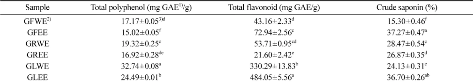
Determination of total polyphenol and total flavonoid contents Phenolic compounds widely distributed among the plant have various structures and molecular weights and their phenolic hydroxyl groups are known to bind to macromolecular proteins and have functions such as anti-bacterial, anti-cancer, blood pressure-enhancing and anti-oxidant functions (Park et al., 2003; Lee et al., 2006). Total polyphenol and total flavonoid contents of ground parts such as ginseng flower, berry, and leaf are shown in Table 1. The total polyphenol contents of GFWE and GFEE were 17.17±0.05, 15.02±0.05 mg GAE/g, respectively. GRWE and GREE were 19.32±0.25, 16.92±0.28 mg GAE/g, GLWE and GLEE were 32.74±0.08 and 24.49±0.05 mg GAE/g.
In the study by Lee et al. (2004), the total polyphenol content of regional ginseng was leaf 147-200 mg%, stem 110-153 mg% and root 61-86 mg%. Also, total phenol contents of the Allium hookeri were found to be 2.40-2.76 mg GAE/g in leaves, 0.65-0.82 mg/g in bulbs and 0.50-0.59 mg/g in roots, respectively (Hwang et al., 2015). It was confirmed that the ground parts had a significantly higher total phenol content than the roots, these results are similar to those of ginseng and contain higher polyphenols than root extracts.
The total flavonoids showed 330.29±13.83 mg GAE/g in GLWE and 484.05±5.56 mg GAE/g in GLEE. The other ground parts of ginseng (GFWE, GFEE, GRWE, GREE) were 43.16±2.33, 72.94±2.56, 53.71±0.95, and 21.60±2.42 mg GAE/g. As a result, it was confirmed that the hot water and ethanol extracts from ginseng leaf has the highest total flavonoid contents similar to the
Table 1. Total polyphenol, flavonoid and crud saponin content of hot water and 60% ethanol extracts of flower, berry, and leaf from Panax ginseng C. A. Meyer
Sample Total polyphenol (mg GAE1)/g) Total flavonoid (mg GAE/g) Crude saponin (%)
GFWE2) 017.17±0.053)d 43.16±2.33d 15.30±0.46f GFEE 15.02±0.05f 72.94±2.56c 37.27±0.47a GRWE 19.32±0.25c 053.71±0.95cd 28.47±0.54c GREE 016.92±0.28de 21.60±2.42e 26.87±0.35d GLWE 32.74±0.08a 330.29±13.83b 24.13±0.31e GLEE 24.49±0.01b 484.05±5.56a0 036.70±0.26ab
1)mg GAE/g: mg gallic acid equivalent/g
2)Abbreviation: GFWE; Hot water extract of ginseng flower, GFEE; 60% ethanol extract of ginseng flower, GRWE; Hot water extract of ginseng berry, GREE; 60% ethanol extract of ginseng berry, GLWE; Hot water extract of ginseng leaf, GLEE; 60% ethanol extract of ginseng leaf. 3)Values are mean±SE of triplicate determinations. Different superscript letters(a-e) within a same column are significantly different at p<0.05.
trend of total polyphenol contents. The ground parts of ginseng with a high physiologically actives substance such as total polyphenols and flavonoids show the possibility to be used as a material for health supplements.
Crude saponin content
As shown in Table 1, GFWE was the lowest in 15.30±0.46% among ground parts of ginseng, but the highest content of GFEE was 37.27±0.47%. The crude saponin content of GLEE was 36.70±0.26%, which is similar to GFEE content.
Kim et al. (1987) reported that the content of crude saponin was for 44.40±21.32% in ginseng leaves and 33.86±16.51% for ginseng peels. These results are as high as those of this experiment and it is considered that there is no difference as a raw material for obtaining the only saponin. According to Kamatusu et al. (1969) study on the chemical composition of ground parts of
ginseng, a considerable amount of saponin exists as various complexes about leaves. Saito et al. (1973) also reported that the total saponin content of ginseng leaves was higher than ginseng roots.
DPPH radical scavenging activity
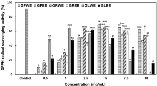
DPPH radicals are widely used to investigate the scavenging activities of many compounds. When DPPH radicals are scavenged the color of the reaction mixture changes from purple to yellow (Blois, 1958). The results shown in Fig. 2 indicate that the DPPH radical scavenging activity of GFWE, GFEE, GRWE, GREE, GLWE, GLEE was measured at the concentrations of 0.5, 1, 2.5, 5, 7.5, and 10 mg/mL. DPPH scavenging activity of the extracts from ground parts of ginseng was lower than that of positive control (1% ascorbic acid, 91.07%), but the DPPH radical scavenging activities tended to increase to a concentration-Fig. 1. DPPH radical scavenging activity of hot water and 60% ethanol extracts of flower, berry, and leaf from Panax ginseng C. A. Meyer. The positive control was used as ascorbic acid. Each value is mean±SE (n≥3) and different superscript letters (a-p) are significantly different at p<0.05 by Duncan’s multiple range test.
Fig. 2. ABTS radical scavenging activity of hot water and 60% ethanol extracts of flower, berry, and leaf from Panax ginseng C. A. Meyer. Positive control was used as gallic acid. Each value is mean±SE (n≥3) and different superscript letters (a-q) are significantly different at p<0.05 by Duncan’s multiple range test.
dependent manner at 0.5-5 mg/mL (p<0.05). The DPPH scavenging activity range of samples was GFWE (9.96-69.20%), GFEE (0.36-62.68%), GRWE (16.30-64.86%), GREE (8.88-63.22%), GLWE (−47.64-64.67%) and GLEE (15.22-61.78%), respectively. At the concentration of 1 mg/mL, these extracts showed activity in order of GLWE (64.67%) > GLEE (47.46%) > GRWE (30.98%) and the highest DPPH scavenging activity was observed in the GLWE. Also, as shown in Fig. 1., GFWE (69.20%) and GRWE (64.86%) showed high scavenging ability at 5 mg/mL concentration of each extracts also, GFWE and GRWE activities were higher than those at higher concentrations. These results have shown that the effective concentration of the different scavenging capacities was 5 mg/mL depending on the amount of each extract. However, the RC50 (Concentration required for 50% reduction of DPPH·) values showed that the GLWE (0.54 mg/mL), GLEE (1.69 mg/mL), and GRWE (1.90 mg/mL) has the best DPPH radical scavenging ability compared to the other samples. In general, ground parts of ginseng hot water extract have more scavenged activities than the 60% ethanol extracts and the leaf of the ground parts showed high DPPH radical scavenging activity similar to the trend of total polyphenol and flavonoid contents.
In another report, using a similar paper of Park et al.(1990), the DPPH radical scavenging activity of ground parts ginseng was judged to be involved in the physiologically active substance.
ABTS radical scavenging activity
ABTS radicals produced by mixing ABTS and potassium persulfate react with the antioxidants contained in the sample and the specific cyan color is expressed by the removal of the cation (Re et al., 1999). The results of the determination of antioxidant activity were demonstrated for Fig. 2. The ABTS radical scavenging activity of each extract from the ground parts of ginseng was significantly increased from 0.5 to 5 mg/mL with increasing concentration. At the 5 mg/mL, GLWE 89.20%, GRWE 88.21% and GFWE 85.68% shown the highest inhibition activity and these extracts were significantly different in the order of GLWE (86.33%), GLEE (73.91%), GRWE (67.13%), GFWE (67.13%) at 10 mg/
mL. The RC50 value of ABTS radical scavenging activity with 60% ethanol and hot water extract were GLWE (0.14 mg/mL), GLEE (1.80 mg/mL), GRWE (2.66 mg/mL), GFWE (2.71 mg/mL), GFEE (3.18 mg/mL) and GREE (3.58 mg/mL), respectively. Jeon et al. (2013) reported that ABTS radical scavenging activity of processed codonopsis lanceolata was 82.1 and 70.6% in 5 steaming with fermentation at hot water and 70% ethanol solvent extraction at a 10 mg/mL, these results were lower than that of GLWE.
Reducing power activity
Among the various mechanisms of antioxidant activity, the ability to release electrons to reactive oxygen species and free radicals is a reducing power, so it can be used as a means of measuring antioxidant activity (Yim et al., 2006). The reductive power of reducing the metal ions after adding the concentrations of extracts from the ground parts of ginseng to 0.5, 1, and 2 mg/ mL were measured as shown in Fig. 3.
Each extract significantly increased among them GLWE, GLEE, GRWE, and GFWE indicated higher reducing power in that order of the 2 mg/mL concentration. In the extracts of flower, berry, and leaf of ginseng, hot water extracts reconfirmed higher reducing power than ethanol extracts. In the previous scavenging activity of DPPH radicals and ABTS radicals, the hot water extracts judged higher antioxidant activity than the ethanol extracts. Especially, hot water extracts of leaf and berry from ginseng were the most active.
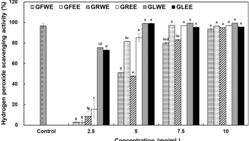
Hydrogen peroxide (H2O2) scavenging activity
H2O2 is very important because of its ability to penetrate biological membranes. The H2O2 is not reactive itself, but it can sometimes be toxic to cells because it may give rise to hydroxyl radicals in the cells (Ames et al., 1993). Thus, removing H2O2 is very important to the protection for food systems. The H2O2 scavenging activity of GFWE, GFEE, GRWE, GREE, GLWE and GLEE were measured at the concentrations of 2.5, 5, 7.5, 10 mg/ mL (Fig. 4). All extract from ground parts of ginseng showed dose-dependent activity, also the activity was similar to those of the positive controls at the high concentration. The extracts demonstrated the highest scavenging activity range (93.63-99.41%) at 10 mg/mL. The RC50 value of H2O2 scavenging activity was GLWE (1.32 mg/mL), GLEE (1.66 mg/mL), GREE (3.74 mg/mL), GFEE (4.00 mg/mL), GRWE (5.24 mg/mL) and GFWE (5.35 mg/ mL), respectively.
Inhibition of linoleic acid peroxidation
The most prominent and currently used assay as an index for lipid peroxidation products is the thiobarbituric acid assay (TBA assay). The TBA assay is used as an indicator of the degree of lipid peroxidation and complex formed by the reaction of malonaldehyde and TBA in lipid oxidation (Lee and Lee, 2016). In this experiment, the antioxidant effects were observed at 5 mg/ mL concentrations of flower, berry, and leaf extracts from ginseng using linoleic acid (an unsaturated fatty acid) as a substrate (Fig. 5). It was confirmed that the absorbance value was increased to 4 Fig. 3. Reducing power of hot water and 60% ethanol extracts of
flower, berry, and leaf from Panax ginseng C. A. Meyer. Each value is mean±SE (n≥3) and different superscript letters (a-j) are significantly different at p<0.05 by Duncan’s multiple range test.
days compared to the 0 days, and the lipid peroxide of the control group increased with time.
When the absorbance value of the negative control on the 4 days is taken as 100%. In the positive control 1% butylated hydroxytoluene (BHT), oxidation was suppressed to 93.02%, GREE 55.81%, GFEE 41.86%, GLEE 38.37%, GRWE 32.56%, GFWE 23.26% and GLWE 9.30%. Among them, ethanol extracts from flower, berry, and leaf from ginseng showed an excellent inhibitory effect on lipid peroxidation.
This antioxidant activity was thought to be different results depending on the measuring method however, generally similar results showed that leaf extracts from ginseng have outstanding antioxidant activity compared to flower and berry extract. In general, the higher amounts of physiological active component
such as total polyphenol and flavonoid exhibit stronger antioxidant activity, and the results are easily expected. Therefore, the antioxidant effects of flower, berry and leaf ginseng extracts were valuable research contribution, expanding the research evidence base on research and may be utilized in the high-value-added raw material.
Conclusion
The physiological active components and antioxidant activity of ginseng flower, ginseng berry, and ginseng leaf, which is the ground parts of ginseng, and obtained the following results. As a representative component of antioxidants, the total polyphenol contents were found to be the highest among GLWE, GLEE followed by GRWE. In a similar trend, the ginseng leaf (GLEE, GLWE) also had the highest total flavonoid. The contents of crude saponin were higher in 60% ethanol extracts as opposed to the results of total polyphenols and total flavonoids. Then, DPPH radical, ABTS radical, reducing power, H2O2 scavenging activity, and Inhibition activity of linoleic acid peroxidation was confirmed to evaluate antioxidant activity. These antioxidant activities were thought to be different results depending on the measuring. However, generally similar results showed that ginseng leaf extracts have the highest antioxidant activities compared to flower and berry extract at the DPPH, ABTS, H2O2 scavenging activity, and reducing power. In general, the higher the amounts of the physiological active component such as total polyphenol and flavonoid that exhibit strong antioxidant activities. Therefore, the antioxidant effects of the extract from the flower, berry, and leaf from ginseng were valuable research contribution to evidence-based research and maybe utilized of high-value-added raw materials.
Fig. 5. Inhibitory effects of hot water and 60% ethanol extracts of flower, berry, and leaf from Panax ginseng C. A. Meyer on linoleic acid peroxidation. The positive control was used as BHT. Each value is mean±SE (n≥3).
Fig. 4. H2O2 scavenging activity of hot water and 60% ethanol extracts of flower, berry, and leaf from Panax ginseng C. A. Meyer. The positive control was used as ascorbic acid. Each value is mean±SE (n≥3) and different superscript letters (a-g) are significantly different at p<0.05 by Duncan’s multiple range test.
Acknowledgments
We thank the anonymous referees for their useful suggestions.
References
Ames BM, Shigena MK, Hagen TM Oxidants, antioxidants, and the degenerative diseases of aging. Proc. Natl Acad Sci. 90: 7915-7922 (1993)
Blois MS. Antioxidant determinations by the use of a stable free rad-ical. Nature 181: 1199-1200 (1958)
Chang HK. Effect of processing methods on the saponin contents of Panax ginseng leaf-tea. Korean. J. Food Nutr. 16: 46-53 (2003) Cho SH. Saponins of Korean ginseng C. A. Meyer (Part II) The
saponins of the ground part of ginseng. J. Korean Agric. Chem. Soc. 20: 142-146 (1977)
Choi JE, Li X, Han YH, Lee KT. Changes of saponin contents of leaves, stems, and flower-buds of Panax ginseng C. A. Meyer by harvesting days. Korean J. Medicinal Crop. Sci. 17: 251-256 (2009)
Dewanto V, Xianzhang W, Liu RH. Processed sweet corn has higher antioxidant activity. J. Agric. Food Chem. 50: 4959-4964 (2002) Han JH, Park SJ, Ahn CN, Wee JJ, Kim KY, Park SH. Nutritional
composition ginsenoside content and fundamental safety evalua-tion with leaf and stem extract of Panax ginseng. J. Korean Soc. Food Sci. Nutr. 33: 778-784 (2004)
Hong SK, Park EK, Lee CY, Kim MU. High-performance liquid chromatographic determination of ginseng saponins. Yakhak Hoeji. 23: 181 (1979)
Hu JN, Lee JH, Shin JA, Choi JE. Lee KT. Determination of ginse-nosides content in korean ginseng seeds and roots by high perfor-mance liquid chromatography. Food Sci. Biotechnol. 17: 430-433 (2008)
Hwang JS, Lee BH, An XX, Jeong HR, Kim YE, Lee II, Kim DO. Total phenolics, total flavonoids, and antioxidant capacity in the leaves, bulbs, and roots of Allium hookeri. Korean J. Food Sci. Technol. 47: 261-266 (2015)
Hwang WI, Lee SD, Kim DC. An extraction method of physiologi-cally active in ginseng or red ginseng. Korea patent 10-0517128 (2005)
Jackson CJC, Dini JP, Lavandier C, Faulkner H, Rupasinghe HPV, John TA. Ginsenoside content of north american ginseng (Panax quinquefolius L. Araliaceae) in relation to plant development and growing locations. J. Ginseng Res. 6: 135-140 (2003)
Jeon JM, Choi SK, Kim YJ, Jang Sj, Cheon JW, Lee HS. Antioxi-dant and antiaging effect of ginseng berry extract fermented by lactic acid bactera. J. Soc. Cosmet. Scientissts Korea. 37: 75-81 (2011)
Jeon SM, Kim SY, Kim IH, Go JS, Kim HR. Antioxidant activities of processed Deoduck (Codonopsis lanceolata) extracts. J. Korean Soc. Food Sci. Nutr. 42: 924-932 (2013)
Kamatsu M, Tomomori T, Makiguchi S. Studies on the constituents of the herb of Panax ginseng C. A. Meyer. on the flavonoid con-stituents. Japanese J. Pharm. Sci. 89: 122-126 (1969)
Kim SD, Do JH, Oh HI, Lee SJ. Effects of processing methods on the quality of ginseng leaf tea. Korean J. Food Sci. Technol. 13: 267-272 (1981)
Kim JH, Lee MS, Nam CW. Protein concentrate from ginseng leaf and its nutritive value. Korean J. Food Sci. Technol. 21: 441-445 (1989)
Kim JK, Kim BS, Park CW, Seo DB, Yoo HR, Lee SJ. Effect of ginseng-berry extract on the improvement of blood microcircula-tion and skin brightness. J. Physiol & Pathol. 24: 85-90 (2010) Kim MW, Ko SR, Choi KJ, Kim SC. Distribution of saponin in
vari-ous sections of Panax ginseng root and changes of its contents according to root age. Korean J. Ginseng Sci. 11: 10-16 (1987) Kim KH, Lee YS, Jung IS, Park SY, Chung HY, Lee IR, Yun YS.
Acidic polysaccharide from Panax ginseng, ginsan, induces Th1 cell and macrophage cytokines and generates LAK cells in syn-ergy with r -2. Planta Med. 64: 110-115 (1998)
Kim KH, Na JY, Jo DH, Lee CY. Extraction and purification of gin-seng oligopeptides with antilipolytic activities. Korean Agric. Chem. Soc. 30: 88-94 (1987)
Kim JY, Staba EJ. Proceedings of International Ginseng Symposium. The Central Reserch Institute, Office of Monopoly, Seoul, Korea. pp. 77 (1974)
Lee SH, Lee SO. Polyphenol contents and antioxidant activities of lentil extracts from different cultivars. J. Korean Soc. Food Sci. Nutr. 45: 973-979 (2016)
Lee SE, Lee SW, Bang JK, Yu YJ, Seong NS. Antioxidant activities of leaf, stem, and root of Panax ginseng C. A. Meyer. Korean J. Medicinal Crop. Sci. 12: 237-242 (2004)
Lee SJ, Park DW, Jang HGm Kim CY, Park YS, Kim TC, Heo BG. Total phenol electron donating ability and tyrosinase inhibition activity of pear cut branch extract. Kor. J. Hort. Sci. Technol. 24: 338-342 (2006)
Lee JW, Sohn HO, Do JH. Function of the water-soluble browning reaction products isolated from korean red ginseng 2. Linoleic acid, Ox-brain antioxidant and Fe2+ ADP/NAD system. J. Gin-seng Res. 24: 35-40 (2000)
Ng TB, Wang H. Panaxagin, a new protein from chinese ginseng possesses antifungal, anti-viral, translation-inhibiting and ribonu-clease activities. Life Science 68: 739-749 (2001)
Nieva Moreno MI, Isla MI, Sampietro AR, Vattuone MA. Compari-son of the free radical-scavenging activity of propolis from sev-eral regions of Argentina. J. Ethnopharmacol. 71: 109-114 (2000) Nakatani N, Kikuzaki H. A new antioxidative glucoside isolated
from oregano (Origanum vulgare L.). Agric. Biol. Chem. 51: 2727-2732 (1987)
Oyaizu M. Studies on product of browning reaction prepared from glucosamine. Jap. J. Nutr. 44: 307-315 (1986)
Park CK, Jeon BS, Yang JW. The chemical components of korean ginseng. Korean J. Food Industry Nutr. 8: 10-23 (2003)
Park JD. Recent studies on the chemical constituents of korean gin-seng. Korean J. Ginseng Sci. 20: 389-415 (1996)
Pellegrin N, Ke R, Yang M, Rice-Evans C. Screening of dietary car-otenoids and carotenoid-rich fruit extract for antioxidant activities applying 2.2’-azinobis (3-ethylenbenzothiazoline-6-sulfonic acid) radical cation decolorization assay. Methods Enzymol. 299: 379-389 (1999)
Park SN, Choi SW, Boo YC. Effects of flavonoids of ginseng leaves on erythrocyte membranes against singlet oxygen caused damage. Korean J. Ginseng Sci. 14: 191-199 (1990)
Re R, Pellegrini N, Proteggente A, Panala A, Yang M, Rice-Evans C. Antioxidant activity applying an improved ABTS radical cat-ion decolorizatcat-ion assay. Free Radic. Biol. Med. 26: 1231-1237 (1999)
Saito H, Morita M, Takagi K. Pharmacological studies of Panax gin-seng leaves. Jap. J. Pharmacol. 23: 43-56 (1973)
Seog HM, Jung CH, Choi IW, Choi HD. Changes in contents of gin-senosides and phenolic compounds in wild ginseng leaves during tea proccesing. Food Sci. Biotechnol. 13: 516-518 (2004)
Shao CJ, Xu JD, Kasai R, Tanaka O. Saponins from flower-buds of Panax ginseng cultivated at Jilin. Chem Pharm Bull. 37: 1934-1935 (1989)
Shi W, Wang Y, Li J, Zhang H, Ding I. Investigation of ginsenosides in different parts and ages of Panax ginseng. Food Chem. 102:664-668 (2007)
Shin JG, Park JW, Pyo JK, Chung MH. Protective effects of a gin-seng component, maltol (2-methyl-3-hydroxyl-4-pyrone) against tissue damages induced by oxygen radicals. Korean J. Ginseng Sci. 14: 187-190 (1990)
Wang CZ, Wu JA, Mcentee E, Yuan CS. Saponins composition in American ginseng leaf and berry assayed by high-performance liquid chromatography. J. Agric. Food Chem. 54: 2261-2266 (2006)
Wee JJ, Park JD, Kim MW. Identification of phenolic antioxidants components isolated from Panax ginseng. J. Korean Agric. Chem. Soc. 32: 50-56 (1989)
Xie JT, Wu JA, Lin E, Wang CZ, Yuan CS. Constituents and effects of ginseng leaf. Orient. Pharm. Exp. Med. 4: 1-8 (2004)
saponins of roots, leaves. flower-buds, and fruits of Panax gin-seng C.A. Meyer. Chem. Pharm. Bull. 271: 88-92 (1979)
Yahara S, Matsuura k, Kasai R, Tanaka O. Saponins of buds and flowers of Panax ginseng C. A. Meyer. (1). Isolation of ginsenos-ide-Rd, -Re, and -Rg1. Chem. Pharm. Bull. 24: 3212-3213 (1976a)
Yahara S, Tanaka O, Komori T. Saponins of the leaves of Panax gin-seng C.A. Meyer. Chem. Pharm. Bull. 24: 2204-2208 (1976b)
Yang HC, Lee SY. A study on the preparation of ginseng-leaf tea. J. Korean Agri. Chem. Soc. 22: 51-57 (1979)
Yim MH, Hong TG, Lee JH. Antioxidant and antimicrobial activity of fermentation and ethanol extracts of pine needles (Pinus densi-flora). Food Sci. Biotechnol. 15: 582-588 (2006)
Yu MH, Lee HS, Cho HR, Lee SO. Enzymatic preparation and anti-oxidant activities of protein hydrolysates from tenebrio molitor larvae. J. Korean Soc. Food Sci. Nutr. 46: 435-441 (2017)