INTRODUCTION
Atopic dermatitis (AD) is a chronic inflammatory skin disease with impaired cell-mediated immune function and compromised skin barrier function. The incidence of AD is generally considered to be increasing worldwide (1, 2).
The presence of a defect in cutaneous permeability barrier function is generally viewed as a downstream consequence of inflammation (‘‘inside-outside’’ hypothesis) (1). Yet, alter- natively, it has been hypothesized that the xerosis and per- meability barrier abnormality, or both could drive disease activity in AD and other inflammatory dermatoses (‘‘outside- inside’’ hypothesis) (3). The primary cytokines, such as, IL- 1aand IL-1binitiate the cytokine cascade in AD (4). IL-4 inhibits ceramide synthesis, and exogenous applications of the IL-4 impede permeability barrier recovery after acute perturbations (5, 6). Two members of human AMPs, hBD2 and hCAP18 (LL-37) are down-regulated in a Th2-depen- dent fashion in AD. They are co-localized with lipids with-
in epidermal lamellar bodies before their secretion, hence some hypothesized that these two functions could be linked, and explored the relationship between the cutaneous perme- ability and antimicrobial barriers (7-9).
Recently, several studies have shown that permeability bar- rier function and antimicrobial defense are not discrete but rather regulated in parallel (7, 10, 11). However, there are no reports confirming that the mechanism is working in patients with AD. Here, we investigated whether restoration of per- meability barrier is accompanied by the concomitant improve- ment of antimicrobial defense in patients with AD.
MATERIALS AND METHODS Subjects
The diagnostic criteria for AD were established according to Hanifin and Rajka (12). The study protocol was approved
766
Kui Young Park1, Dong Ha Kim1, Mi Sook Jeong2, Kapsok Li1, and Seong Jun Seo1
Department of Dermatology1and Chung-Ang Medical Research Center2, Chung-Ang University College of Medicine, Seoul, Korea
Address for Correspondence Seong Jun Seo, M.D.
Department of Dermatology, College of Medicine, Chung-Ang University Hospital, 29 Heukseong-no, Dongjak-gu, Seoul 156-755, Korea
Tel : +82.2-6299-1525, Fax : +82.2-823-1049 E-mail : [email protected]
This study was supported by a grant of the Korea Healthcare technology R&D Project, Ministry for Health, Welfare & Family Affairs, Republic of Korea (A091121).
Changes of Antimicrobial Peptides and Transepidermal Water Loss After Topical Application of Tacrolimus and Ceramide-dominant Emollient in Patients with Atopic Dermatitis
Increased transepidermal water loss (TEWL) and downregulated antimicrobial pep- tides (AMPs) are observed in patients with atopic dermatitis (AD). Tacrolimus and ceramide-dominant emollients are effective in the treatment of AD by preventing the production of inflammatory cytokines and by correcting skin barrier dysfunctions, respectively. Present study was designed to investigate the relationship between antimicrobial and barrier factors by measuring the changes of AMPs and TEWL after topical application of tacrolimus and ceramide-dominant emollient in the patients with AD. A total of three patients with AD were treated with tacrolimus in one lesion and ceramide-dominant emollient in another lesion for 4 weeks. RT-PCR and west- ern blotting revealed that the mRNA and protein expression levels of hBD-2 and LL- 37 were increased on the both study sites. Immunohistochemical analysis showed significant increase of AMPs and IL-1a, while, IL-4 was decreased on the both study sites. The mean changes of TEWL and AMPs showed no statistical difference bet- ween both sites. Tacrolimus and ceramide-dominant emollient influence on both TEWL and AMPs expression in patients with AD, namely they have similar effects on both of the two. This study shows that restoration of permeability barrier func- tion is accompanied by the concomitant improvement of antimicrobial defense in patients with AD.
Key Words : Antimicrobial peptide; Dermatitis, Atopic; Ceramides; Permeability Barrier; Tacrolimus
Received : 23 March 2009 Accepted : 23 October 2009
ⓒ 2010 The Korean Academy of Medical Sciences.
This is an Open Access article distributed under the terms of the Creative Commons Attribution Non-Commercial License (http://creativecommons.org/licenses/by-nc/3.0) which permits unrestricted non-commercial use, distribution, and reproduction in any medium, provided the original work is properly cited.
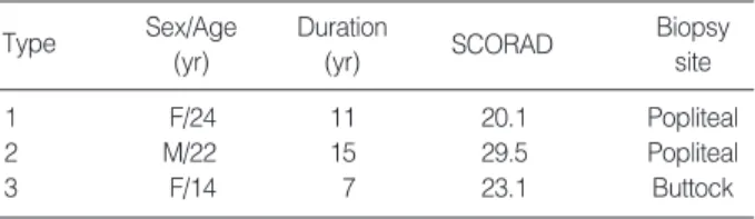
by the Ethical Committee of Chung-Ang University Hos- pital Institute Review Board (I2007014). Informed consents were obtained from all subjects after they were fully informed about the details and the potential risk of the study. The char- acteristics of subjects are summarized in Table 1. Two, clini- cally similar and symmetric lesions of the patients were select- ed, after measuring transepidermal water loss (TEWL), punch biopsies were performed on the involved skin of 3 patients with AD. Thin layers of tacrolimus ointment 0.1% (Protopic) and ceramide-dominant emollient (TriCeram) were applied twice daily for 4 weeks, respectively.
TEWL assessment
To assess epidermal permeability barrier function, we per- formed measurements of TEWL, which have been utilized to provide information about status of permeability barrier under either normal, experimentally perturbed, or diseased conditions.
The published guidelines for the measurement of TEWL were followed (13). In detail, all subjects’ conditions were first stabilized for 15 to 20 min in a climate- and humidity-con- trolled room. Ambient temperature ranged between 21℃ and 24℃, with a mean relative humidity of 45%. TEWL was measured with a Tewameter TM 210 (Courage & Khaz- aka, Cologne, Germany) and estimated over 2 representative involved sites, tacrolimus-applied skin and emollient-applied skin, respectively.
Reverse transcription-polymerase chain reaction (RT-PCR)
The tissues from punch biopsy were cut by scissor. Total RNA was isolated from skin using TRIZol reagent (Invit- rogen, Carlsbad, CA, USA) after adding 1 mL of TRIZol reagent. And the tissue was homogenized by homogenizer.
After 5 min at room temperature, 0.2 mL of chloroform per 1mL of TRIZol reagent was added. Tubes were shaken vig- orously. The mixtures were centrifuged with 12,000 rpm at 4℃for 20 min, the upper aqueous phase were transferred to a fresh tube, and the same amount of 2-propanol was added.
After the mixture was incubated at 4℃for 15 min, it was centrifuged with 12,000 rpm for 15 min. The supernatant was removed, then the RNA pellet was washed with 70%
ethanol and centrifuged with 12,000 rpm at 4℃for 15 min, and briefly dried. The purified RNA was dissolved in DEPC-
DW. Total cellular RNA was reverse transcribed at 42℃for 30 min in containing reverse trancriptase (TaKaRa, Shiga, Japan), 10×buffer, 10 mM dNTP (dNTP mix), oligo dT primer, RNase inhibitor, and 25 mM MgCl2.
RT-PCR analysis of LL-37 and hBD-2 mRNA using spe- cific primers were performed.
We synthesized the PCR primer on the basis of GenBank data. Primers were chemically synthesized by DNA synthe- sizer (Pharmacia, Biogatan, Uppsala, Sweden). Their sequences were as follows:
hBD-2 (128 bp),
5′-ATC TCC TCT TCT CGT TCC TC-3′(sense) 5′-ACC TTCTAG GGC AAA AGA CT-3′(anti-sense);
LL-37 (208 bp),
5′-CTG ATG CCT CTT CCA GGT GT-3′(sense) 5′-GAG GGA GCC CTT TCT GAA TC-3′(anti-sense);
GAPDH (593 bp),
5′-CCA CCC ATG GCA AAT TCC ATG GCA-3′(sense), 5′-GGT GCT GCT TGT TAG GAG GTC AAG TAA
AGG GC-3′(anti-sense).
Two mL of each cDNA sample from the RT-PCR was am- plified by PCR in containing Taq polymerase (TaKaRa), 10
×buffer, 25 mM MgCl2and 10 pM primer.
Electrophoresis
The products were run in 1.5% agarose gel contain 1 mg ethidium bromide per millimeter. Twenty mL of reaction mix- ture was mixed with loading buffer, separated by electrophore- sis for 15 min at 100 volts and visualized by UV transillu- mination. PCR products of hBD-2 and LL-37 were normal- ized with GAPDH by using densitometer (volume of hBD- 2/volume of GAPDH×100, volume of LL-37/volume of GAPDH×100).
Western blotting
The tissues were cut by scissor. Skin were lysed in a buffer containing 50 mM Tris-Cl (pH 8.0), 150 mM NaCl, 0.02%
sodium azide, 100 mg/mL phenylmethanesulfonyl fluoride, 1 mg/mL aprotinin, 1% Triton×100, the tissue was homog- enized by homogenizer. After centrifuging with 12,000 rpm at 4℃for 30 min, the supernatant was transferred into new tube, 30 mg of soluble protein were loaded in 15% sodium dodecyl sulfate polyacrylamide gel electrophoresis (SDS-PA- GE) with sample buffer containing 1 M Tris and 50% glyc- erol. Samples were heated at 95℃for 5 min prior to gel load- ing. For hBD-2 detection, separated protein on gel electro- phoresis was transferred to nitrocellulose membrane (Osmon- ics, Minnesota, MN, USA) at 0.16 A for 1 hr. The membrane was washed 3 times with Tris-buffered saline Tween 20 (TBST), and blocked with 5% skim milk for 1 hr at room tempera- ture. Following this, the membrane was incubated overnight
SCORAD, Severity Scoring of Atopic Dermatitis.
Type Sex/Age (yr)
Duration
(yr) SCORAD Biopsy
site
1 F/24 11 20.1 Popliteal
2 M/22 15 29.5 Popliteal
3 F/14 7 23.1 Buttock
Table 1. Characteristics of subjects at study entry
at 4℃with goat anti-hBD-2 polyclonal antibody (1:1,500 in 5% bovine serum albumin, SantaCruz, Delaware, CA, USA) and goat anti-LL-37 polyclonal antibody (1:1,500 in 5% bovine serum albumin, SantaCruz, Delaware, CA) and then washed 3 times with TBST. The secondary mouse anti- goat peroxidase conjugated antibody (1:2,000 in blocking solution, SantaCruz) was incubated for 1 hr at room temper- ature. After washing with ECL solution (SantaCruz) for 3 min,
the membrane was then exposed to radiography film (Roche, Indianapolis, IN, USA).
Immunohistochemistry (IHC)
IHC was carried out using tissue section of involved sites of atopic patients. In brief, 4 mm thick sections were deparaf- finized in xylene 2 times for 15 min each, and epitopes were retrieved by autoclaving (60℃) for 1 hr in citrate-buffered saline (pH 6.0). After 40 min of cooling at room temperature, the activity of endogenous peroxidase was quenched by treat- ment with 3% H2O2in TBST for 10 min. The sections were blocked with normal goat serum for 1 hr, and incubated with mouse anti-human LL-37, hBD-2, IL-1aand IL-4 polyclonal antibodies, respectively. After 5 washes with PBS, the sec- tions were incubated with peroxidase conjugated goat anti- mouse secondary antibody, FITC anti-mouse secondary anti- body and color was developed with diaminobenzidine. Two independent blind observers evaluated serial sections. For quantitative analysis, the stained cells were counted in three consecutive microscopic fields (×400).
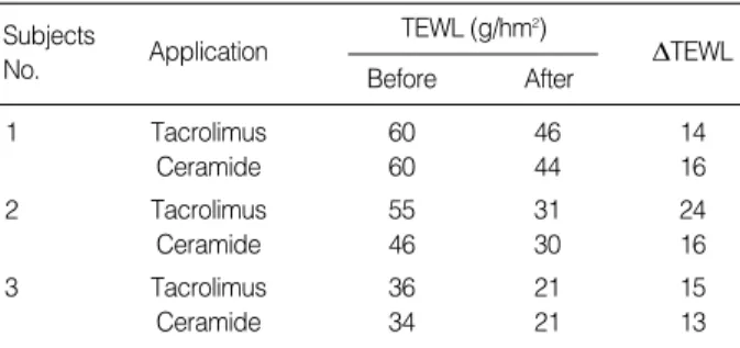
DTEWL, mean changes of TEWL before and after application.
Subjects
No. Application
Before After TEWL (g/hm2)
DTEWL
1 Tacrolimus 60 46 14
Ceramide 60 44 16
2 Tacrolimus 55 31 24
Ceramide 46 30 16
3 Tacrolimus 36 21 15
Ceramide 34 21 13
Table 2. Comparison of mean TEWL levels between tacrolimus- applied and emollient-applied lesion in AD
hBD-2/GAPDH ×100
0.5 0.4 0.3 0.2 0.1 0.0
GAPDH 598 bp
* *
*
* *
*
hBD-2
Pt.1 Pt.2 Pt.3 Pt.1 Pt.2 Pt.3
Tacrolimus
128 bp
Ceramide A
LL-37/GAPDH ×100
0.4
0.3
0.2
0.1
0.0
GAPDH 598 bp
*
* * *
*
*
LL-37
Pt.1 Pt.2 Pt.3 Pt.1 Pt.2 Pt.3
Tacrolimus
208 bp
Ceramide B
C
DhDB-2
0.25
0.2
0.15
0.1
0.05
0
Pt.1 Pt.2 Pt.3
Tacrolimus Ceramide
Tacrolimus Ceramide
D
DLL-37
0.25
0.2
0.15
0.1
0.05
0
Pt.1 Pt.2 Pt.3
Fig. 1. RT-PCR for expression of hBD-2 and LL-37 before and after application of tacrolimus and ceramide-dominant emollient. (A) Increased expression of hBD after application of tacrolimus and ceramide-dominant emollient. (B) Increased expression of LL-37 after application of tacrolimus and ceramide-dominant emollient (*P<0.05). (C) No statistical difference of hBD-2 between tacrolimus-applied and emollient- applied skin. (D) No statistical difference of LL-37 between tacrolimus-applied and emollient-applied skin. Pt., patient.
Statistical analysis
Statistical analysis was conducted using Wilkoxon T test.
Statistical differences in mRNA expression or protein stain- ing of hBD-2 and LL-37 between groups were determined, with significant differences conferred when P≤0.05.
RESULTS
TEWL before and after application of tacrolimus and ceramide-dominant emollient
For comparison of TEWL levels between tacrolimus-ap- plied and emollient-applied skin in atopic patient, measure- ments were performed 5 times for each site, repeatedly. The mean TEWL levels decreased on the both sites. The mean changes of TEWL were 17.7 g/hm2(14-24 g/hm2) for tac- rolimus-applied lesions and 15 g/hm2 (13-16 g/hm2) for emollient-applied lesions, but there is no statistical differ- ence between tacrolimus-applied and emollient-applied skin (Table 2).
Expression of LL-37 and hBD-2 before and after application of tacrolimus and ceramide-dominant emollient using RT-PCR method
The expressions of hBD-2 and LL-37 mRNA were increas- ed on the both sites after application of either tacrolimus or ceramide-dominant emollient. The mean changes of AMPs, however, showed no statistical difference between tacrolimus- applied and emollient-applied skin (Fig. 1).
Expression of LL-37 and hBD-2 before and after application of tacrolimus and ceramide-dominant emollient using Western blotting
The expressions of hBD-2 and LL-37 proteins were increased on the both sites after application either tacrolimus or cera- mide-dominant emollient. The mean changes of AMPs, how- ever, showed no statistical difference between tacrolimus- applied and emollient-applied skin (Fig. 2).
Actin 42
kDa
hBD-2 7
kDa
Pt.1 Pt.2 Pt.3 Pt.1 Pt.2 Pt.3
Tacrolimus Ceramide A
Actin 42
kDa
LL-37 18
kDa
Pt.1 Pt.2 Pt.3 Pt.1 Pt.2 Pt.3
Tacrolimus Ceramide B
Fig. 2. Western blotting for expression of hBD-2 and LL-37 before and after application of tacrolimus and ceramide-dominant emol- lient. (A) Increased expression of hBD after application of tacro- limus and ceramide-dominant emollient. (B) Increased expres- sion of LL-37 after application of tacrolimus and ceramide-domi- nant emollient.
Fig. 3. Immunohistochemical staining for hBD-2 and LL-37 in section from patients with atopic dermatitis. Expression of hBD-2, LL-37, IL-1a and IL-4 receptors before and after tacrolimus and ceramide treatments as assessed by immunohistochemistry. Y axis: numbers of stained cells in microscopic fields (×400).
10 8 6 4 2
0 Pt.1 Pt.2 Pt.3
Tacrolimus
hBD-2 LL-37
10 8 6 4 2
0 Pt.1 Pt.2 Pt.3
Ceramide
10 8 6 4 2
0 Pt.1 Pt.2 Pt.3
Tacrolimus
10 8 6 4 2
0 Pt.1 Pt.2 Pt.3
Ceramide
10 8 6 4 2
0 Pt.1 Pt.2 Pt.3
Tacrolimus
IL-1α IL-4
10 8 6 4 2
0 Pt.1 Pt.2 Pt.3
Ceramide
10 8 6 4 2
0 Pt.1 Pt.2 Pt.3
Tacrolimus
10 8 6 4 2
0 Pt.1 Pt.2 Pt.3
Ceramide
Before After
IHC for LL-37, hBD-2, IL-1a, and IL-4 before and after application of tacrolimus and ceramide-dominant emollient
The intensity of staining of LL-37, hBD-2 were significant- ly stronger in the epidermis after application on the both sites.
And the intensity of staining of IL-4 was slightly weaker in the papillary dermis after application (Fig. 3).
DISCUSSION
Although both patients with AD and psoriasis have altered skin barrier function, patients with psoriasis are more resis- tant to skin infection; about 30 percent of patients with AD have bacterial or viral infections of the skin, as compared with only 7 percent of patients with psoriasis (14, 15). In psoriatic skin, keratinocytes are rapidly proliferating and differentiat- ing, making epidermis thickened. So, quantitatively, abun- dant antimicrobial peptides are expressed in the psoriatic epi- dermis (15, 16). A deficiency in the expressions of AMPs in AD may account for the susceptibility of AD patients to skin infection, such as Staphylococcus aureus (9). The inability to increase AMPs may be due to suppression by Th2 cytokines that are elevated in AD. In fact, in vitro keratinocytes in cul- ture could be shown to lose that ability to increase hBD-2 expression when exposed to IL-4 or IL-13 (17, 18). Enhanced Th2 cell activity is a hallmark of acute AD (17). Increased production of IL-4, IL-5, and IL-13 by Th2 cells, in turn, inhib- it Th1 cytokine production, including IFN-gand IL-18, which are two beneficial mediators of AMPs. IL-4, IL-13 have also direct inhibitory effects for AMPs expression (19, 20).
When emollients or moisturizers are used in the treatment of AD and other inflammatory dermatoses, the intended aim is limited to an improvement in skin hydration and mitiga- tion of the xerosis. No reduction in inflammatory disease activ- ity is expected, and such activity has been documented only infrequently (21). However, Chamlin et al. (22) reported that ceramide-dominant, barrier repair lipids alleviate childhood atopic dermatitis, and attribute the improvement seen in their patients to a normalization of barrier function. The normal- ized barrier function, however, dampened the cytokine cas- cade that initiates and sustains AD. It implied that emollients are helpful in not only skin hydration but also ceasing to cyto- kine cascade leading to inflammation through normalization of permeability barrier function.
Various immunological parameters are affected in patients with AD: the relative and absolute numbers of circulating T cells are reduced; lymphocyte response to mitogens are im- paired; the cytotoxic potential of several cell types, such as monocytes and natural killer cells, is decreased; and chemotat- ic responsiveness is reduced (17, 20). There are few published data on the effect of tacrolimus ointment on these parame- ters, although Nghiem et al. (23) have shown that topical calcineurin inhibitors specifically target key immunological
mechanisms that have been implicated in the pathogenesis of AD, and act primarily on T cells in lesional skin. Failure of AMPs to up-regulate appropriately in AD is attributed currently to excess Th2 cytokines (9). Tacrolimus suppresses Th2 activity and increases AMPs in AD. Although Kis et al.
(24) showed no effect on the IL-1a, IL-8, and TNF-aproduc- tion by tacrolimus in normal human keratinocytes, it is regard- ed as being fundamental immunologic differences between normal and atopic conditions.
While levels of the IL-1agenerally increase in inflamma- tory dermatoses, they showed paradoxical decline in AD epi- dermis (25). Recently, Elias et al. (4) proposed the hypothe- sis that increase in serine protease activity would generate active forms of IL-1, which is a primary contributor to inflam- mation in AD. However, the results in this study support the opposite conclusion about the cytokine milieu of AD;
elevated skin production of Th2 cytokines and low levels of proinflammatory cytokines, such as, TNF-a, IFN-g, and IL- 1. And it is also reported that IL-13 and IL-4 inhibited TNF-a and IFN-g-induced hBD-3 production (20).
The antimicrobial barrier is intimately linked to the per- meability barrier, and as with water egress, pathogen ingress occurs through the extracellular domains (7). We investigat- ed whether improvement of antimicrobial defense by tacro- limus could simultaneously restored permeability barrier, and the study yielded the expected results. There are several reports supporting the hypothesis that the permeability and antimicrobial barriers of the skin may not be discrete, but rather co-regulated and even inter-dependent (7, 11). The intimate link between these two functions is likely originat- ed from the co-assembly of AMP with barrier lipid precur- sors within nascent lamellar body (7, 8). The metabolic res- ponse of AMP parallels the metabolic response of the lipid to acute disruption of permeability barrier. When the per- meability barrier is perturbed by solvent treatment or tape stripping, a sequential, lipid-synthetic response ensues in the underlying epidermis that rapidly restores normal per- meability barrier homeostasis (26). Permeability disruption not only removes lipid, but it also initiates a concomitant and rapid loss of AMP from stratum corneum (4). Accord- ing to the ‘outside-inside’ hypothesis, the barrier abnormal- ity first provokes and sustains AD through activation of an epidermis-initiated cytokine cascade, followed by recruit- ment of the specific immunologic response (3).
This study showed that ceramide-replacement improved not only permeability barrier function but also antimicro- bial barrier in AD; meanwhile tacrolimus-application im- proved permeability barrier in addition to antimicrobial bar- rier. In summary, the restoration of permeability barrier leads the recovery of antimicrobial barrier, and the recovery of anti- microbial barrier leads the restoration of permeability barri- er. Through the present study, we could support the theory about interrelationship between antimicrobial barrier and permeability barrier in AD patient. Yet, further studies about
the intermediate process linking antimicrobial barrier to per- meability barrier (e.g., inflammatory cells, cytokines, skin pH, filaggrin, serine protease) are required.
REFERENCES
1. Leung DY. Atopic dermatitis: new insights and opportunities for ther- apeutic intervention. J Allergy Clin Immunol 2000; 105: 860-76.
2. Williams HC. Is the prevalence of atopic dermatitis increasing? Clin Exp Dermatol 1992; 17: 385-91.
3. Elias PM, Wood LC, Feingold KR. Epidermal pathogenesis of inflam- matory dermatoses. Am J Contact Dermat 1999; 10: 119-26.
4. Elias PM, Steinhoff M. ‘‘Outside-to-inside’’(and now back to ‘‘out- side’’) pathogenic mechanisms in atopic dermatitis. J Invest Derma- tol 2008; 128: 1067-70.
5. Kurahashi R, Hatano Y, Katagiri K. IL-4 suppresses the recovery of cutaneous permeability barrier functions in vivo. J Invest Dermatol 2008; 128: 1329-31.
6. Hatano Y, Terashi H, Arakawa S, Katagiri K. Interleukin-4 suppress- es the enhancement of ceramide synthesis and cutaneous permeabili- ty barrier functions induced by tumor necrosis factor-alpha and inter- feron-gamma in human epidermis. J Invest Dermatol 2005; 124:
786-92.
7. Elias PM. Stratum corneum defensive functions: an integrated view.
J Invest Dermatol 2005; 125: 183-200.
8. Oren A, Ganz T, Liu L, Meerloo T. In human epidermis, beta-defen- sin 2 is packaged in lamellar bodies. Exp Mol Pathol 2003; 74: 180-2.
9. Ong PY, Ohtake T, Brandt C, Strickland I, Boguniewicz M, Ganz T, Gallo RL, Leung DY. Endogenous antimicrobial peptides and skin infections in atopic dermatitis. N Engl J Med 2002; 347: 1151-60.
10. Aberg KM, Man MQ, Gallo RL, Ganz T, Crumrine D, Brown BE, Choi EH, Kim DK, Schröder JM, Feingold KR, Elias PM. Co-regu- lation and interdependence of the mammalian epidermal permeabil- ity and antimicrobial barriers. J Invest Dermatol 2008; 128: 917-25.
11. Elias PM, Choi EH. Interactions among stratum corneum defensive functions. Exp Dermatol 2005; 14: 719-26.
12. Hanifin JM, Rajka G. Diagnostic features of atopic dermatitis. Acta Derm Venereol Suppl (Stockh) 1980; 92: 44-7.
13. Pinnagoda J, Tupker RA, Agner T, Serup J. Guidelines for transepi- dermal water loss (TEWL) measurement. A report from the Standard- ization Group of the European Society of Contact Dermatitis. Con- tact Dermatitis 1990; 22: 164-78.
14. Howell MD, Jones JF, Kisich KO, Streib JE, Gallo RL, Leung DY.
Selective killing of vaccine virus by LL-37: implications for eczema vaccinatum. J Immunol 2004; 172: 1763-7.
15. Kim JE, Kim BJ, Jeong MS, Seo SJ, Kim MN, Hong CK, Ro BI.
Expression and modulation of LL-37 in normal human keratinocytes, HaCaT cells, and inflammatory skin diseases. J Korean Med Sci 2005; 20: 649-54.
16. Rieg S, Steffen H, Seeber S, Humeny A, Kalbacher H, Dietz K, Garbe C, Schittek B. Deficiency of dermcidin-derived antimicrobial pep- tides in sweat of patients with atopic dermatitis correlates with an impaired innate defense of human skin in vivo. J Immunol 2005; 74:
8003-10.
17. Boguniewicz M. Update on atopic dermatitis: insights into patho- genesis and new treatment paradigms. Allergy Asthma Proc 2004;
25: 279-82.
18. Raychaudhuri SP, Jiang WY, Raychaudhuri SK, Krensky AM. Lesion- al T cells and dermal dendrocytes in psoriasis plaque express in- creased levels of granulysin. J Am Acad Dermatol 2004; 51: 1006-8.
19. Fellermann K, Wehkamp J, Stange EF. Antimicrobial peptides in the skin. N Engl J Med 2003; 348: 361-3.
20. Nomura I, Goleva E, Howell MD, Hamid QA, Ong PY, Hall CF, Darst MA, Gao B, Boguniewicz M, Travers JB, Leung DY. Cytokine milieu of atopic dermatitis, as compared to psoriasis, skin prevents induction of innate immune response genes. J Immunol 2003; 171:
3262-9.
21. Loden M, Andersson AC, Lindberg M. Improvement in skin barrier function in patients with atopic dermatitis after treatment with a mois- turizing cream (Canoderm). Br J Dermatol 1999; 140: 264-7.
22. Chamlin SL, Frieden IJ, Fowler A, Williams M, Kao J, Sheu M, Elias PM. Ceramide-dominant, barrier-repair lipids improve childhood atopic dermatitis. Arch Dermatol 2001; 137: 1110-2.
23. Nghiem P, Pearson G, Langley RG. Tacrolimus and pimecrolimus:
from clever prokaryotes to inhibiting calcineurin and treating atopic dermatitis. J Am Acad Dermatol 2002; 46: 228-41.
24. Kis K, Bodai L, Polyanka H, Eder K, Pivarcsi A, Duda E, Soos G, Bata-Csorgo Z, Kemeny L. Budesonide, but not tacrolimus, affects the immune functions of normal human keratinocytes. Int Immuno- pharmacol 2006; 6: 358-68.
25. Reilly DM, Parslew R, Sharpe GR, Powell S, Green MR. Inflam- matory mediators in normal, sensitive and diseased skin types. Acta Derm Venereol 2000; 80: 171-4.
26. Feingold KR. The regulation and role of epidermal lipid synthesis.
Adv Lipid Res 1991; 24: 57-82.