INTRODUCTION
Isolated amyloid deposition has been reported in almost every organ systems including the genitourinary tract (1).
Localized amyloidosis in the seminal vesicle is an unusual finding and most of the cases have been described in autopsy series with its incidence being about 9-16% (1-3). The inci- dence of localized amyloidosis of the seminal vesicles increases with age, representing a form of senile amyloidosis (3). Some authors document its incidental detection in the surgical specimens and the incidence rate is also very low (4, 5). Amy- loid deposition tends to be nodular and subepithelial. Involve- ment of vessel walls is characteristically not seen in localized form.
In cases associated with genitourinary carcinomas, the amyloid deposition in the seminal vesicle can be misdiag- nosed as tumor invasion on magnetic resonance imaging (MRI) findings (6, 7). Our cases were also suggested exten- sion of the prostatic adenocarcinoma both to seminal vesicles and vasa deferentia on the MRI study. However, these sus- picious lesions in both seminal vesicles and vasa deferentia turned out to be localized amyloid deposition in these organs.
We reported these cases because amyloid deposition in vas
deferens is extremely rare and amyloid deposition in the vas deferens simulating tumor invasion has not been reported.
Electron microscopic analysis was performed on these cases and we made a review of the literature.
CASE REPORT Case 1
A 60-yr-old male patient who has been taken medicines for treatment of hypertension and benign prostatic hyper- plasia came to our hospital, because of the recent elevation of serum prostate-specific antigen (PSA) level (5.4 ng/mL).
At that time, serum free PSA level was 0.76 ng/mL (14.1%
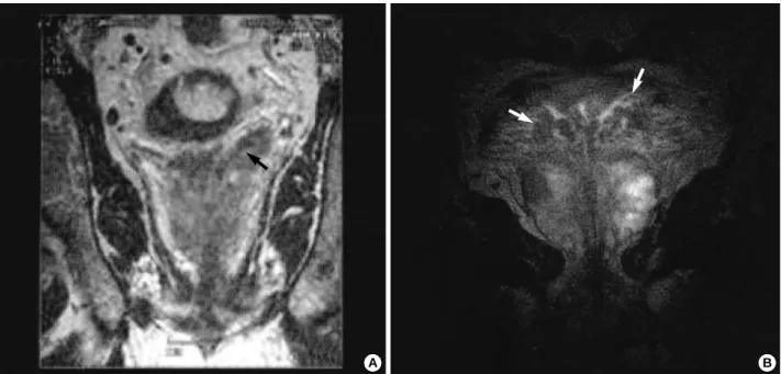
of total PSA level). On digital rectal examination of the prostate, a poorly-defined firm nodule was palpable in the right side. MRI was performed at an outside hospital and revealed multiple irregular foci of low signal intensity in the prostate as well as both seminal vesicles and vasa defer- entia on T2-weighted imaging, suggesting of prostatic car- cinoma extension both to seminal vesicles and vasa deferen- tia (Fig. 1A). Based on the clinical diagnosis of T3b prostatic
*
Department of Pathology and Diagnostic Radiology*, University of Ulsan College of Medicine, Asan Medical Center, Seoul, Korea
Jae Y. Ro, M.D.
Department of Pathology, University of Ulsan College of Medicine, Asan Medical Center, 388-1 Pungnap-dong, Songpa-gu, Seoul 138-736, Korea Te : +82.2-3010-4550, Fax : +82.2-472-7898 E-mail : [email protected]
447
We reported localized amyloidosis involving seminal vesicles and vasa deferen- tia, which was found in two patients with prostatic adenocarcinoma. A 60-yr-old (Case 1) and a 59-yr-old (Case 2) man came to our hospital with elevation of serum prostate-specific antigen (PSA) and biopsy proven carcinoma, respec- tively. MRI revealed multiple irregular foci of low signal intensity in the prostates as well as in both seminal vesicles and vasa deferentia on T2-weighted imaging, suggesting prostatic carcinoma with extension to both seminal vesicles and vasa deferentia in both cases. Under the clinical diagnosis of stage III prostatic adeno- carcinoma, a radical prostatectomy was performed in both patients. Microscopi- cally, Gleason score 7 adenocarcinoma was observed in both patients. In addi- tion, isolated amyloidosis of both seminal vesicles and vasa deferentia was found without carcinoma involvement. Localized amyloidosis in the seminal vesicles, which is considered as senile process, has been occasionally reported in the autop- sy and in the surgical specimens. Amyloid deposition in the vas deferens has also been reported in the literature, however, the deposition mimicking extension of carcinoma has not been reported. In this report, two cases of isolated amyloidosis of the seminal vesicles and vasa deferentia are described with electron microscopic study and literature review.
Key Words : Amyloidosis, Seminal Vesicles, Vas Deferens, Prostatic Neoplasms
Received : 11 June 2002 Accepted : 12 August 2002
carcinoma, a radical prostatectomy was performed.
On gross examination of the radical prostatectomy speci- men, the prostate was slightly enlarged (5.0 4.5 3.0 cm) and weighed 35.5 g. Two well-defined ovoid masses (1.5 0.9 0.7 cm and 1 0.6 0.3 cm) were present in the right and left sides of the prostate, abutting the prostatic
capsule. The tumor occupied less than 10% of the total pro- state. The cut surfaces of the masses were yellowish tan and firm. Neither hemorrhage nor necrosis was seen. On the serial sections of both seminal vesicles and vasa deferentia, they were focally thickened and firm with yellowish tan dis- coloration, suggesting tumor extension (Fig. 2A). Micro-
Fig. 1.Multiple irregular foci of low signal intensity are observed in the prostate as well as in the area of both seminal vesicles and vasa deferentia (arrow) on the T2-weighted MR imaging, suggesting tumor extension of prostatic carcinoma to the seminal vesicles and vasa deferentia. (A) Case 1. (B) Case 2.
A B
Fig. 2.(A) There are multiple foci of yellowish tan and firm nodular lesions, both in seminal vesicles. These nodular lesions are better illus- trated on the right side of seminal vesicles (short arrow). In this picture, a tip of prostatic carcinoma mass is seen in the left lower corner (long arrow). The remaining prostatic parenchyma shows benign nodular hyperplasia. (B) Microscopically, prostatic masses are con- firmed to be adenocarcinoma of Gleason score 7 (4+3) (H&E, 40).
A B
scopically, two prostatic masses were diagnosed as Gleason score 7 (4+3) adenocarcinoma (Fig. 2B). The tumor was confined within the prostate with no extraprostatic exten- sion. In both vasa deferentia and seminal vesicles, nodular deposits of amorphous eosinophilic material were seen beneath the epithelial layer with no evidence of prostatic carcinoma involvement (Fig. 3A, B). Pelvic lymph nodes were free of tumor. The final TNM stage of the prostatic carcinoma was T2bN0M0 (stage II). He is alive and well 6 months after surgery. His most recent serum PSA is less than 0.2 ng/mL.
Case 2
A 59-yr-old male patient has been taken medicines for symptomatic treatment of right flank pain and gross hema- turia originated from a known renal multicystic disease for 4 yrs. However, the patient did not have an evidence of renal failure nor receive a hemodialysis treatment. Past history was noncontributory with right ureterolithotomy 20 yrs ago and the resection of unknown brain tumor 9 yrs ago. He was admitted in an outside hospital due to the recent aggrava- tion of the low urinary tract symptoms, including urinary
Fig. 3.(A, B) In both vasa deferentia, amorphous eosinophilic materials are markedly deposited beneath the epithelial layer of the vas deferens (H&E, A, 40; B, 400). (C) The deposits exhibit apple green birefringence under a polarized microscope ( 40). (D) Ultra- structurally, these deposits are fine, rigid, non-branching filaments of variable length with haphazard arrangement ( 10,000).
A B
C D
level), respectively. On digital rectal examination of the pro- state, no palpable nodule was present. MRI of the prostate, both seminal vesicles and vasa deferentia showed similar findings with those of Case 1 (Fig. 1B), except a focus of high signal intensity both in T1- and T2-weighted imaging in this case, suggesting of intraprostatic hemorrhage. No change of the renal cysts was observed. Like Case 1, a radical prostatectomy was performed, based on the clinical diagno- sis of T3b prostatic carcinoma.
On gross examination of the radical prostatectomy speci- men, the prostate was slightly enlarged (5.0 4.8 3.5 cm) and weighed 49 g. Two well-defined ovoid masses (1.2 1.2 1.1 cm and 0.7 0.4 0.3 cm) were present both in the left and right side of the prostate, abutting the prostatic capsule. The tumor occupied less than 10% of the total pro- state. The cut surfaces of the prostatic masses and both semi- nal vesicles and vasa deferentia were similar to those of Case 1. Microscopically, two prostatic masses were diagnosed as Gleason score 7 (3+4) adenocarcinoma. The tumor was con- fined within the prostate with no extraprostatic extension and pelvic lymph node metastasis. In this case, nodular de- posits of amorphous eosinophilic material were seen beneath the epithelial layer in the ejaculatory ducts as well as both vasa deferentia and seminal vesicles with no evidence of pro- static carcinoma involvement. The final TNM stage of the prostatic carcinoma was also T2bN0M0 (stage II) like Case 1. This patient is recently discharged from the hospital after operation with uneventable outcome.
The deposits of both Cases 1 and 2 exhibited Mahogany red color with Congo red amyloid staining and apple green birefringence under a polarized microscope. By electron microscopic examination, these deposits were composed of fine, 7.5 to 10 nm diameter, rigid, and non-branching fila- ments of variable length with haphazard arrangement (Fig.
3C, D), indicating amyloid fibers. This amyloid material was observed in the subepithelial connective tissue without vascular wall deposition.
DISCUSSION
Localized amyloidosis in the seminal vesicle has been un- commonly reported as an incidental finding in the autopsy (1-3). This process is considered as a senile change because of the increase incidence with age. Some authors rarely doc- ument the incidental amyloid deposition in the seminal vesicle in the surgical specimens (4, 5). Localized amyloido- sis in the seminal vesicle occurs bilaterally and the deposits tend to be nodular and subepithelial with no vascular involve-
cases of localized amyloidosis both in seminal vesicles and vasa deferentia (2). All these cases were incidentally detected.
Our cases will be the fourth and fifth reported cases of local- ized amyloidosis involving vas deferens.
It has been suggested that this type of amyloid is derived from a secretory exocrine product of the normal seminal vesicle epithelium (9). Unger et al. (5) demonstrated that the prior luteinizing hormone-releasing hormone (LHRH) treatment may act as a seminal vesicle epithelial stimulant for elaboration of amyloid. One of our patients (Case 1) has been taken unknown medicines for benign prostatic hyper- plasia during the last 3 yrs at an outside hospital and this medication may be related with the amyloid deposition.
Various organs, including lung, larynx, skin, tongue, eye and genitourinary tracts have been reported to be the sites of localized amyloidosis which amyloid involves subepithe- lial connective tissue without involving vascular wall. Unlike systemic amyloid deposition, which is mostly composed of AL (lambda chain) type, the amyloid of the seminal vesicle was negative for the known amyloidogenic substances, such as AA protein, AL (kappa and lamda chains), transthyretin (prealbumin), 2-microglobulin, -amyloid protein, cys- tatin C, calcitonin, and amylin by immunohistochemical stainings (10-12). Tsutsumi et al. (10) proposed that the lacto- ferrin was the major constituent in localized senile amyloi- dosis of seminal vesicle.
Some authors (6, 7) reported that the localized amyloido- sis of the seminal vesicle was clinically and radiologically similar to direct tumor invasion from bladder or prostate cancer, because these deposited areas also displayed low sig- nal intensity on T2-weighted imaging of MRI. To differen- tiate amyloid deposition from tumor invasion of seminal vesicle, an administration of the gadopentetate dimeglumine may be useful since amyloid deposition shows no enhance- ment unlike tumor involvement (6).
We report very unusual cases of the localized amyloidosis in both vasa deferentia as well as seminal vesicles, which were found in the radical prostatectomy specimens. Our cases were originally interpretated as the extension of prostatic adenocarcinoma to seminal vesicles and vasa deferentia based on the preoperative radiologic studies. Therefore, a possible seminal vesicle and/or vas deferens involvement of prostatic or bladder cancer by image analysis is not necessarily true involvement of tumor on pathologic examination. Therefore, a possibility of localized amyloidosis of the seminal vesicle and/or vas deferens should be considered in prostatic cancer cases, which showed abnormal MR imagings in the seminal vesicles and vasa deferentia.
REFERENCES
1. Coyne JD, Kealy WF. Seminal vesicle amyloidosis: morphological, histochemical and immunohistochemical observations. Histopathol- ogy 1993; 22: 173-6.
2. Goldman H. Amyloidosis of seminal vesicles and vas deferens. Pri- mary localized cases. Arch Pathol 1963; 75: 94-8.
3. Pitkanen P, Westermark P, Cornwell GG, Murdoch W. Amyloid of the seminal vesicles. Am J Pathol 1983; 110: 64-9.
4. Seidman JD, Shmookler BM, Connolly B, Lack EE. Localized amyloidosis of seminal vesicles: report of three cases in surgically obtained material. Mod Pathol 1989; 2: 671-5.
5. Unger PD, Wang Q, Gordon RE, Stock R, Stone N. Localized amy- loidosis of the seminal vesicle. Possible association with hormonal- ly treated prostatic adenocarcinoma. Arch Pathol Lab Med 1997;
121: 1265-8.
6. Jager GJ, Ruijter ET, de la Rossette JJ, van de Kaa CA. Amyloidosis of the seminal vesicles simulating tumor invasion of prostatic carci- noma on endorectal MR images. Eur Radiol 1997; 7: 552-4.
7. Kaji Y, Sugimura K, Nagaoka S, Ishida T. Amyloid deposition in seminal vesicles mimicking tumor invasion from bladder cancer:
MR findings. J Comput Assist Tomogr 1992; 16: 989-91.
8. Krane RJ, Klugo RC, Olsson CA. Seminal vesicle amyloidosis. Urolo- gy 1973; 2: 70-2.
9. Cornwell GG, Westermark GT, Pitkanen P, Westermark P. Seminal vesicle amyloid: The first example of exocrine cell origin of an amy- loid fibril precursor. J Pathol 1992; 167: 297-303.
10. Tsutsumi Y, Serizawa A, Hori S. Localized amyloidosis of the semi- nal vesicle: identification of lactoferrin immunoreactivity in the amy- loid material. Pathol Int 1996; 46: 491-7.
11. Suess K, Moch H, Epper R, Koller A, Durmuller U, Mihatsch MJ.
Heterogeneity of seminal vesicle amyloid. Immunohistochemical detection of lactoferrin and amyloid of the prealbumin-transthyretin type. Pathologe 1998; 19: 115-9.
12. Esslimani M, Serre I, Granier M, Robert M, Baldet P, Costes V. Uro- genital amyloidosis: clinico-pathological study of 8 cases. Ann Pathol 1999; 19: 487-91.