ISSN 0378-6471 (Print)⋅ISSN 2092-9374 (Online)
http://dx.doi.org/10.3341/jkos.2016.57.2.310
Case Report
상층각막성형술을 받은 환자에서 시행한 백내장 수술 1예
A Case of Cataract Surgery in an Epikeratophakia Patient
전수지⋅조양경
Soo Ji Jeon, MD, Yang Kyung Cho, MD, PhD
가톨릭대학교 의과대학 안과 및 시과학교실
Department of Ophthalmology and Visual Science, College of Medicine, The Catholic University of Korea, Seoul, Korea
Purpose: To report a case of cataract surgery in an epikeratophakia patient.
Case summary: A 59-year-old female with a history of epikeratophakic surgery 20 years ago complained of decreased visual acuity of both eyes for several months. She had nucleosclerotic and posterior subcapsular types of cataracts. Phacoemulsification and posterior capsule intraocular lens implantation were performed in both eyes. During surgery, corneal edema was especially prominent at the cornea with epikeratophakic lenticules in both eyes. In the left eye, severe corneal edema after one day of sur- gery was observed; however, after one week, corneal edema had subsided and visual acuity of both eyes had improved.
Conclusions: When it necessary that cataract surgery is performed in patients with epikeratophakic lenticules, it is important to anticipate the corneal edema intraoperatively and postoperatively. Moreover, the surgeon should consider the acute calculation of the target refraction of intraocular lens in an epikeratophakia patient.
J Korean Ophthalmol Soc 2016;57(2):310-315
Keywords: Cataract surgery, Corneal edema, Epikeratophakia
■Received: 2015. 7. 17. ■ Revised: 2015. 9. 2.
■Accepted: 2015. 11. 11.
■Address reprint requests to Yang Kyung Cho, MD, PhD Department of Ophthalmology, The Catholic University of Korea St. Vincent’s Hospital, #93 Jungbu-daero, Paldal-gu, Suwon 16247, Korea
Tel: 82-31-240-7340, Fax: 82-31-251-6225 E-mail: yangkyeung@hanmail.net
ⓒ2016 The Korean Ophthalmological Society
This is an Open Access article distributed under the terms of the Creative Commons Attribution Non-Commercial License (http://creativecommons.org/licenses/by-nc/3.0/) which permits unrestricted non-commercial use, distribution, and reproduction in any medium, provided the original work is properly cited.
상층각막성형술은 고도근시나 무수정체안의 시력교정을 위해 예전에 널리 시행되었던 굴절 교정수술로, 각막의 앞면 에 다른 수여각막을 붙여주는 술기이다. 1980년 Kaufman1이 무수정체증의 교정에 상층각막성형술을 사용한 이래 원추 각막, 고도근시 환자 등에서 각막의 굴절력을 변화시켜 굴 절이상을 교정하기 위하여 시행되었다.2-5 이 수술은 가역적 인 수술로서 오래 전 이 수술을 시행 받은 환자 중에는 상 층의 각막절편을 제거하고 다른 굴절 수술을 추가로 받은 환자도 많이 있다.6
국내에서는 상층각막성형술의 시행이 많지 않았고, 수술 결과에 불만족했던 환자의 경우 상층각막을 제거하는 복원 수술을 이미 시행 받았기 때문에 상층각막성형술을 한 환 자들의 백내장 수술에 대한 보고가 불충분하다. 저자들은 상층각막성형술을 시행 받은 환자에서 양안 백내장 수술을 시행하여 양호한 수술 후 시력 결과를 보였으나, 수술 중과 수술 후 각막부종이 쉽게 오고, 또 빠르게 가라앉는 것을 경험하였다. 이러한 예가 국내에는 아직 보고된 바가 없기 에 이를 보고하는 바이다.
증례보고
59세 여자 환자가 수개월 전부터 시작된 양안의 시력저 하를 주소로 내원하였다. 과거력상 환자는 내원 20년 전 국 내에서 근시교정을 위해 양안의 상층각막성형술을 시행 받 았다. 내원 당시 나안시력은 우안은 0.06, 좌안은 0.1이었고
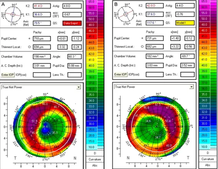
Figure 1. Pentacam® examination of both eyes before cataract surgery. (A) Right eye: Pentacam® shows a flattened curvature of the
right cornea due to epikeratophakic lenticule. The location of epikeratophakic lenticule is slightly nasal to the center of the cornea and the keratometric value (true net power) at the center of the right cornea is 31.1 diopter. (B) Left eye: Pentacam® shows a flat- tened curvature of the left cornea due to epikeratophakic lenticule. The location of epikeratophakic lenticule is slightly nasal to the center of the cornea and the keratometric value (true net power) at the center of the left cornea is 36.2 diopter.최대교정시력은 우안 0.2, 좌안 0.15였다. 현성굴절검사상 우안은 -5.50 Dsph=-3.50 Dcyl×150°였으며 좌안은 -6.50 Dsph=-4.00 Dcyl×140°였다.
세극등 현미경 검사에서 양안의 상층각막절개술로 인한 반흔이 관찰되었다. 양안은 중증도의 핵경화성 백내장 및 후낭하 백내장을 보였으며 좌안의 핵경화 정도가 더 심했 다. A-scan 초음파(Compact II device, Quantel Medical, Eyecubed, Ellex, Adelaide, Australia)에서 환자는 우안 안 축장 27.21 mm, 좌안 안축장 27.25 mm의 축성 근시를 보 였고, 경면현미경(NSP-9900®, Konan, Japan) 검사에서 각막 내피세포밀도는 우안 2,531 cells/mm2, 좌안 1,976 cells/mm2 로 측정되었다. 각막두께는 상층각막성형술에 의해 두꺼워 져서 우안 755 µm, 좌안 763 µm였다. 우안과 좌안의 초음 파 수정체 유화술 및 인공수정체 삽입술을 하루 간격으로
시행하였다. 상층각막성형술에 대한 기록을 얻을 수 없어 서 Pentacam® (Oculus, OptikgereGmBH, Wetzlar, Germany) 을 이용하여 측정한 K값과 Sanders-Retzlaff-Kraff (SRK-T) 공식을 사용하여 -1.0D의 굴절력을 목표로 인공수정체 도 수를 결정하였다. Pentacam®에서 조직렌즈이식에 의해 각 막이 편평해졌음을 알 수 있었다(Fig. 1).
수술은 lidocaine과 bupivacaine을 이용한 구후 마취 및 nadbath 마취하에 시행되었다. 상층각막성형술을 받은 각 막의 전반적인 부종과 조직절편의 테두리 부분의 굴곡으로 인해 전낭절개술 및 초음파유화술 전반에서 다른 수술에 비해 시야에 지장을 주었다(Fig. 2). 핵쪼개기 방법은 stop and chop 방법을 사용하였다. 초음파 유화술 도중에 상층 각막성형술을 받은 부위의 부종이 심해져서 시야의 흐림 증상으로 수술실 조명을 끄고 수술을 마저 시행하였다. 수술 시
A B
A-1 B-1
A-2 B-2
A-3 B-3
A-4 B-4
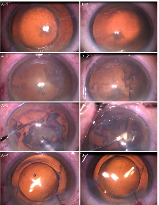
Figure 2. Intraoperative microscopic view of the both eyes during cataract surgery. (A) Right eye. (B) Left eye. (A-1, B-1)
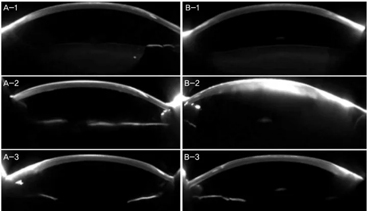
Immediately before corneal incision: the white arrows indicate 12 o’clock position of the cornea at the intraoperative microscopic view. In each right and left eye, the epikeratophakic lenticule is located slightly nasal to the center of the cornea. (A-2, B-2) Immediately after hydrodissection: the margin of the epikeratophakic lenticule caused blurring of the image due to scattering of light, thus made it difficult to distinguish the capsulorrhexis margin before and after hydrodissection. (A-3, B-3) During phacoe- mulsificaiton: the corneal edema increased during phacoemulsification, in which the surgeon needed to decrease the illumination of the operating room to improve the microscopic view. (A-4, B-4) At the end of operation.Figure 3. Pentacam® Scheimpflug images of both eyes. (A) Right eye. (B) Left eye. (A-1, B-1) Preoperative images: the epiker-
atophakic lenticules are well attached to the corneal stroma in both eyes. (A-2, B-2) Postoperative 1 day: (A-2) In the right eye, there is mild corneal edema in both epikeratophakic lenticule and stromal bed. (B-2) In left eye, there was severe corneal edema in both epikeratophakic lenticule and stromal bed. (A-3, B-3) Postoperative 1 week: the corneal edema subsided in both eyes.사용한 초음파 양은 우안 18%, 30.9 seconds, 좌안 15%, 42.3 sec- onds였다. 양안 모두 인공수정체(HOYA YA60BBR, HOYA, Tokyo, Japan) 삽입 후 수술은 안정적으로 종료되었다.
우안은 술 후 1일째 나안시력 0.5, 굴절력 +1.25 Dsph=
-4.50 Dcyl×90°로 측정되었고 좌안은 술 후 1일째 나안 시력 안전수지 30 cm, 굴절력은 각막부종으로 찍히지 않았다.
수술 전과 비교했을 때(Fig. 3A-1, B-1), 수술 후 1병일 때 우안은 경미한 각막의 부종을 보였고, 좌안은 심한 각막 부 종을 보였다. Pentacam®의 샤임플러그 이미지에서 좌안은 각막 부종이 심하고 상층 각막도 부어 있는 것이 확인되었 다(Fig. 3A-2, B-2).
술 후 7일째 우안은 나안 시력 0.5, 굴절력 +0.50 Dsph=
-0.50 Dcyl×60°, 좌안은 나안 시력 0.5, 굴절력 +0.75 Dsph=
-2.75 Dcyl×130°로 측정되었다. 술 후 Pentacam®의 샤임플 러그 이미지에서 각막부종은 모두 없어지고 술 전 각막두 께로 돌아온 것이 확인되었다(Fig. 3A-3, B-3).
술 후 6주째 우안의 나안 시력은 0.5, 굴절력은 0.00 Dsph=0.00 Dcyl×180°였고, 좌안의 나안 시력은 0.4, 교정 시력은 0.5로 측정되었으며 굴절력은 +0.25 Dsph=-1.75 Dcyl×130°였다. 이후 술 후 4개월까지 특이 변화 없이 유 지되었다.
고 찰
상층각막성형술 이후 시행하는 백내장 수술에서 고려해 야 할 사항으로는 크게 두 가지를 생각해 볼 수 있다. 첫 번째는 인공수정체 도수 계산의 불확실성이고, 두 번째는 각막 부종이 쉽게 생긴다는 점이다.7,8 먼저 인공수정체 도 수 계산의 측면에서 본 증례를 고찰하였을 때, 조직렌즈의 이식부위가 각막의 중심에서 벗어나 있었음에도 불구하고, Pentacam®에 의한 목표 굴절력 계산이 성공적이었음을 알 수 있었다. 우안의 수술 후 1병일 때 보인 원시는 경한 각 막부종에 의한 것이라 생각된다.
본 증례에서 경험했던 것처럼, 상층각막성형술을 받은 환자의 경우 백내장 수술 도중에 각막 부종에 의한 시야의 흐림이 올 수 있고, 상층각막성형술을 한 테두리 부분의 심 한 굴곡은 술자의 시야확보에 지장을 줄 수 있다. 또한 상 층각막(조직렌즈)의 중심이 정확히 동공중심이나 각막중심 과 일치하지 않았기 때문에 조명에 의한 반사나, 눈의 위치 에 따라 각막혼탁에 의한 시야확보 가능 정도의 변이가 심 했다.
백내장 수술 중 및 수술 후 발생하는 각막 부종의 대부분 의 원인은 각막내피 손상이다. 우안의 경우 수술 전 각막
A-1 B-1
A-2 B-2
A-3 B-3
내피세포밀도는 2,531 cells/mm2이고 좌안의 수술 전 내피 세포밀도는 1,976 cells/mm2였다. 술 전 내피세포가 낮은 밀도를 보이고, 균일하지 못한 모양을 보인 것은 상층각막 성형술로 인하여 두꺼워진 각막에 대한 내피세포의 변화로 생각된다.9
우안의 수술 1주 후 내피세포 밀도는 1,980 cells/mm2로 수술 전에 비해 약 21.77%의 내피세포의 감소가 있었고, 좌안은 수술 1주 후 내피세포 밀도는 1,420 cells/mm2로 수 술 전에 비해 약 28.13%의 내피세포 감소가 있었다. 이는 양안의 백내장 수술 도중 사용한 초음파 양에 의한 내피세 포 손상의 차이로 보인다(우안: 18%, 30.9 seconds, 좌안:
15%, 42.3 seconds).
좌안의 경우 우안에 비해 수술 중 사용한 초음파 에너지 가 더 많았고, 이와 연관되어 수술 후 내피세포의 손실이 더 많았으나, 수술 직후 발생한 좌안 각막의 심한 부종을 설명할 수 있는 원인으로 충분하지 않았다. 실제 좌안에서 수술 후 1병일 때 보인 각막의 부종은 일반적인 백내장 수 술 직후에 보이는 각막 부종에 비해 현저하게 심한 정도였 다. 즉 수술 직후에 보였던 좌안의 심한 각막부종은 사용한 초음파 양이나 이로 인한 각막내피세포의 손상 이외에 다 른 부가적인 원인도 생각해야 할 것으로 보인다.
좌안의 심한 각막부종을 설명할 수 있는 요인을 고찰해 보면, 먼저 좌안의 수술 전 내피세포가 낮은 밀도 및 다형 성, 다면성을 보인 것을 생각할 수 있고, 두 번째로 수술 중 사용한 초음파에너지에 의한 손상을 고려할 수 있으며, 마 지막으로 상층각막성형술을 한 경우 조직렌즈의 존재가 각 막부종에 대한 endothelial pump에 의한 보상 기전에 방해 가 될 수 있음을 시사한다.
수술 직후 심한 각막부종을 보인 좌안의 샤임플러그 이 미지를 관찰하여 수동으로 각막두께를 측정해 보면 수술 전 조직렌즈의 두께는 205 μm, 수여각막의 두께는 545 μm 였으며, 술 후 1병일 때 조직렌즈의 부종은 310 μm, 남아 있는 수여각막 부분의 부종은 820 μm였다. 이 수치들은 각 각 수술 전과 비교했을 때 비슷한 증가율을 보인다. 술 후 1주일 때 조직렌즈의 두께는 201 μm, 수여각막의 두께는 547 μm로 측정되어 수술 전 상태로 거의 회복되었다. Fan et al9에 의하면 조직렌즈와 수여각막 사이에는 틈이 벌어 질 수 있고 그 사이로 ghost vessel이 자라나는 경우도 있다 고 보고되어 있다. 본 증례에서 만약 조직렌즈가 수여각막 과 유착되지 않았다면 각막부종에 의해 조직절편과 수여각 막 사이의 틈새 벌어짐이나 부종이 샤임플러그 이미지에서 관찰이 가능했을 것으로 보이나, 본 증례의 양안 모두에서
틈새 벌어짐은 관찰되지 않았다. 이는 수술한 지 20여년이 지난 상태이므로 조직렌즈와 수여 각막의 유착이 잘 이루 어져 있음을 알 수 있는 간접적인 소견으로 보인다.
상층각막성형술은 현재는 근시의 교정술로 거의 시행되 지 않는 수술이고, 무수정체안이나 심한 각막의 얇아짐을 동반한 원추각막 등에서는 고려해 볼 수 있는 수술이다. 국 내에서 행해진 상층각막이식술 중 많은 경우에서는 이 수 술이 가역적인 수술이라는 장점을 이용하여, 불만족스럽거 나 불규칙적인 굴절교정 효과를 보인 환자에서는 상층각막 을 다시 제거하는 수술이 많이 행해졌다.10,11 그 영향으로 국내에서는 아직까지 상층각막성형술을 시행한 환자에서 백내장 수술을 시행한 경우에 대한 보고가 없다.
저자들은 본 증례의 환자를 경험하여 상층 조직렌즈를 그대로 유지한 채 백내장이 생긴 환자의 경우에 정밀한 인 공 수정체의 도수 계산 및 수술 중 각막 부종이나 각막의 굴곡에 의한 시야확보의 어려움, 그리고 수술 후 각막 부종 을 예상하고 수술에 임해야 할 것으로 생각되어 이를 보고 하는 바이다.
REFERENCES
1) Kaufman HE. The correction of aphakia. XXXVI Edward Jackson Memorial Lecture. Am J Ophthalmol 1980;89:1-10.
2) Halliday BL. Epikeratophakia for aphakia, keratoconus, and myopia. Br J Ophthalmol 1990;74:67-72.
3) Kaufman HE, Werblin TP. Epikeratophakia for the treatment of keratoconus. Am J Ophthalmol 1982;93:342-7.
4) McDonald MB, Kaufman HE, Aquavella JV, et al. The nationwide study of epikeratophakia for aphakia in adults. Am J Ophthalmol 1987;103(3 Pt 2):358-65.
5) Panda A, Gupta AK, Sharma N, et al. Anatomical and functional graft survival, 10 years after epikeratoplasty in keratoconus. Indian J Ophthalmol 2013;61:18-22.
6) Lee KW, Choi TH, Lee HB. The effects of epikeratoplasty & laser in situ keratomileusis(LASIK) for correction of high myopia. J Korean Ophthalmol Soc 1998;39:1707-15.
7) Labor PK, Ignacio T, Johnson M, Janku-Lestock L. Accommodating intraocular lens implantation in an epikeratophakia patient. J Cataract Refract Surg 2010;36:347-50.
8) Seitz B, Langenbucher A. Intraocular lens power calculation in eyes after corneal refractive surgery. J Refract Surg 2000;16:349-61.
9) Fan JC, Patel DV, McGhee CN. Long-term microstructural changes following epikeratophakia: in vivo confocal microscopy study. J Cataract Refract Surg 2008;34:1793-8.
10) Shin YJ, Park WC, Lee JH. Clear lens extraction and epiker- atophakic lenticule removal in complicated epikeratophakic patients. J Korean Ophthalmol Soc 2002;43:1397-401.
11) Greenbaum A, Kaiserman I, Avni I. Long-term reversibility of epikeratophakia. Cornea 2007;26:1210-2.
= 국문초록 =
상층각막성형술을 받은 환자에서 시행한 백내장 수술 1예
목적: 상층각막성형술을 받은 환자에서 백내장수술을 시행한 1예를 경험하여 아직까지 국내에 보고된 바가 없어 이를 보고하고자 한다.
증례요약: 59세 여자 환자가 수개월 전부터 발생한 양안 시력저하를 주소로 내원하였다. 환자는 20년 전 양안에 상층각막성형술을 시행 받았으며, 양안에 중증도의 핵경화성 백내장 및 후낭하 백내장을 보였다. 초음파유화술 및 인공수정체 삽입술을 시행하였다.
백내장 수술 중 상층각막성형술을 시행 받은 각막 부분에서 현저한 각막부종이 발생하고, 조직렌즈 부착부위의 굴곡으로 수술시야에 장애를 주었다. 수술 후 1병일 때 좌안의 현저한 각막 부종이 관찰되었으나 수술 1주일 후 각막 부종은 수술 전 상태로 회복되었다.
술 후 6주째 우안 시력은 0.5, 굴절력은 0.00 Dsph=0.00 Dcyl×180o, 좌안 시력은 0.5로 측정되었고 굴절력은 +0.25 Dsph=-1.75 Dcyl×130o였으며, 술 후 4개월까지 유지되었다.
결론: 본 증례에서는 상층각막성형술을 시행 받은 환자에서 조직렌즈를 그대로 유지한 채 백내장 수술을 시행하여 만족스러운 수술 결과를 얻었으나, 술 중 각막부종이나 각막의 굴곡에 의한 시야확보의 어려움을 경험하였고 수술 직후 심한 각막부종을 경험하였다.
상층각막성형술 후 시행하는 백내장 수술에서는 정밀한 인공수정체 도수의 계산과 더불어 시야확보의 어려움 및 수술 중과 수술 후 각막의 부종 가능성을 예상하고 수술에 임해야 할 것으로 생각된다.
<대한안과학회지 2016;57(2):310-315>