72
Mutational and Expressional Analysis of ATG5 Gene in Non-Small Cell Lung Cancers
Purpose: Several lines of evidence have indicated that perturbations of autophagy are involved in the development of many human diseases, including cancer. The autophagy-related genes (ATG) encode proteins that play important roles in autophagic processes. The aim of this study was to see whether alterations of the ATG5 protein expression and somatic mutations of the ATG5 gene are present in human non-small cell lung cancers (NSCLCs).
Materials and Methods: We analyzed the ATG5 somatic mutations in 45 NSCLCs by performing single-strand conformation polymorphism (SSCP). We examined the ATG5 protein expression in 45 NSCLCs by performing immunohistochemistry. Results: The SSCP analysis revealed no evidence of somatic mutation in the DNA sequences encoding the ATG5 gene in the 45 NSCLCs. On the immunohistochemistry, ATG5 protein was expressed in the normal bronchial epithelial cells, while it was lost in 9 (20%) of the NSCLCs.
Conclusion: Our data indicates that ATG5 is altered in NSCLC at the expressional level, but not at the mutational level. The data also suggests that the loss of expression of ATG5 might play a role in the pathogenesis of NSCLC by altering autophagic and apoptotic cell death. (J Lung Cancer 2010;9(2):72 76)
Key Words: Non-small cell lung cancer, ATG5, Autophagy, Mutation
Min Sung Kim, M.S.
Nam Jin Yoo, M.D. and Sug Hyung Lee, M.D.
Department of Pathology, The Catholic University of Korea College of Medi- cine, Seoul, Korea
Received: September 10, 2010 Revised: September 28, 2010 Accepted: September 29, 2010 Address for correspondence Sug Hyung Lee, M.D.
Department of Pathology, The Catholic University of Korea College of Medi- cine, 505, Banpo-dong, Seocho-gu, Seoul 137-701, Korea
Tel: 82-2-2258-7311 Fax: 82-2-537-6586
E-mail: [email protected] This work was supported by a grant from Ministry for Health, Welfare and Family Affairs (A100098).
서 론
Autophagy는 오래된 단백질과 손상된 세포 내 소기관을 라이소좀(lysosome)에 의해 제거하는 자기소화 과정이다 (1-3). 이 과정은 진화적으로 잘 보존된 막이동성(membrane trafficking) 과정으로 autophagosome의 형성과 함께 시작한 다(1). Autophagy 과정의 조절에는 많은 유전자가 관여하지 만, 이 중 가장 중요한 유전자는 autophagy-related gene (ATG)이라고 명명된 단백질 집단이다(4). 현재까지 31종의 ATG 유전자가 효모(yeast)에서 발굴되었고, 이 중 16종이 사람에서도 확인되었다(1-4). Apoptosis는 type I programmed cell death (PCD)로 분류되며 autophagy는 type II PCD로 분 류되지만, apoptosis와 autophagy는 상호연관성 및 연결성이 존재한다. 하지만, apoptosis와는 달리 autophagy는 세포생존 (cell survival)에도 중요한 역할을 한다(1-3).
Apoptosis의 이상이 많은 질환에 연관되어 있는 것이 밝
혀져 있다. 유사하게 autophagy의 이상이 질환의 발생에 중 요하다는 근거가 퇴행성 질환, 미생물 감염, 암 등에서 점차 밝혀지고 있다(1-3). 가장 잘 알려진 autophagy 관련 유전자 인 BECN1 (ATG6)은 다양한 암에서 결손되며, BECN+/− 돌 연변이 마우스는 높은 빈도의 암 발생이 나타난다(5). 이는 autophagy 유발성 유전자의 억제가 암 발생을 촉진하며 이 들이 종양억제 유전자임을 제시하는 결과이다. 일부의 ATG 유전자의 돌연변이가 다양한 암에서 확인되었다(6-8).
비소세포성폐암에서 BECN1 유전자의 돌연변이가 연구되 었지만 아주 낮은 빈도로 나타났으며(1/124, 0.8%) (6), BECN1 이외의 ATG 유전자에 대한 연구가 비소세포성폐 암에서 보고된 바가 없는 실정이다.
세포사멸 기전에 관여하는 유전자의 이상이 발암과정에 중요한 역할을 한다는 증거들이 제시되고 있다. 예를 들면 caspase-3, Fas 등 apoptosis 유전자의 돌연변이가 비소세포 폐암을 비롯한 여러 암에서 보고된 바 있다(9-14). ATG5는 autophagy 과정의 초기 autophagosome 형성에 관여하는 유
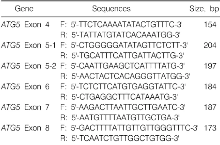
Table 1. Primer Sequences of ATG5 Gene Used in This Study
Gene Sequences Size, bp
ATG5 Exon 4 F: 5'-TTCTCAAAATATACTGTTTC-3' 154 R: 5'-TATTATGTATCACAAATGG-3'
ATG5 Exon 5-1 F: 5'-CTGGGGGATATAGTTCTCTT-3' 204 R: 5'-TGCATTTCATTGATTACTTG-3'
ATG5 Exon 5-2 F: 5'-CAATTGAAGCTCATTTTATG-3' 197 R: 5'-AACTACTCACAGGGTTATGG-3' ATG5 Exon 6 F: 5'-TCTCTTCATGTGAGGTATTC-3' 184
R: 5'-CTGAGGCTTTCATAAATG-3'
ATG5 Exon 7 F: 5'-AAGACTTAATTGCTTGAATC-3' 187 R: 5'-AATGTTTTAATGTTGCTGA-3'
ATG5 Exon 8 F: 5'-GACTTTTATTGTTGTTGGGTTTC-3' 173 R: 5'-TCAATCTGTTGGCTGTGG-3'
전자이며 ATG5와 ATG12는 함께 autophagosome 형성을 촉 진한다(1-3). 또한, ATG5는 autophagy뿐 아니라 apoptosis의 진행에도 중요한 역할을 하는 것으로 알려져 있다(15,16).
ATG5가 세포사멸에서 중요한 기능을 담당하지만, 현재까 지 이 유전자의 돌연변이가 암에서 보고된 바는 없다. 이에 저자들은 본 연구에서 ATG5 유전자의 돌연변이가 비소세 포성폐암의 세포사멸 이상에 관여하는지를 알아보기 위해, 비소세포성폐암 조직을 대상으로 돌연변이 및 발현 연구를 시행하였다.
대상 및 방법 1) 연구 대상
1999년 이후 근치적 폐 절제술을 받고 진단된 45명의 비 소세포성폐암 환자의 폐조직을 대상으로 하였다. 폐조직은 메타칸에 고정되고 파라핀에 포매되었다. 폐암환자의 파라 핀 포매 조직을 5μm 두께로 박절하여 hematoxylin & eosin (H&E)염색을 실시한 후 2명의 진단병리 의사가 독립적으 로 국제보건기구(World Health Organization, WHO) 분류에 따라 분류하였으며, 이들은 편평상피암 22예, 샘암 23예였 다. 36∼79세의 연령분포를 보였으며 평균연령은 57세였 다.
2) 돌연변이 조사
H&E 염색된 조직에서 미세절제술(microdissection)을 이 용하여 암세포 및 정상세포를 각각 분리 수집한 후, proteinase K를 처리하여 DNA를 얻었다. ATG5 유전자를 이 루고 있는 genomic DNA는 7개의 엑손(exon) (엑손 2∼8)으 로 이루어지며 275개의 염기를 번역한다. 과거의 연구를 통 해 아미노 말단(1∼70)은 세포사멸 유도기능이 없고 다른 부분(아미노산 서열 71∼275)은 세포사멸 유도기능이 있는 것이 밝혀진 바 있다(16). 따라서, 본 연구는 아미노산 79∼
275를 코딩하는 엑손 4∼8의 돌연변이를 조사하였다. 이를 위해 이 부위를 증폭할 수 있는 시발체(primer) 6쌍을 제작 하였다(Table 1). 방사성 동위원소인([32P]dCTP)를 중합효소 연쇄반응에 포함시켜서 자기방사법(autoradiogram)으로 중 합효소연쇄반응 산물을 분석할 수 있게 하였다. 중합효소 연쇄반응은 혼합액을 94oC에서 10분간 변성시킨 후 94oC에 서 30초, 53∼62oC에서 40초와 72oC에서 40초씩 각각 35회 반복하였으며 72oC에서 5분간 연장반응을 실시하였다. 중 합효소연쇄반응 산물을 single-strand conformation polymor- phism (SSCP) 분석을 위해 non-denaturing gel에 running 후 건조하고 관찰하여, 정상 DNA에서 관찰되는 야생형(wild-
type) band 이외의 band가 나타난 경우, 2회 이상 반복하여 확인하고, aberrant band를 잘라서 cyclic sequencing으로 DNA 염기서열을 분석하였다. SSCP, DNA 염기서열 분석에 관한 내용은 이전의 논문에 자세히 기술되어 있다(9).
3) 면역화학염색
박절된 45예의 폐암 조직절편을 이용하여 ATG5 단백질 에 대한 면역조직화학법 검사를 시행하였다. 사람의 ATG5 단백질에 대한 토끼의 항체(dilution 1/800; Abcam, Cambridge, UK)가 일차 항체로 사용되었다. 면역조직화학검사의 신호 를 증폭시키기 위하여 전자레인지를 이용한 항원 복원 (antigen retrieval)이 시행되었으며 자세한 방법은 이전의 논 문에 자세히 기술되었다(9). 면역염색은 일차항체를 4oC에 서 15시간 처리한 후 DAKO REAL EnVision System (DAKO, Glostrup, Denmark)을 사용하여 시행하였다. 이후 diamino- benzidine으로 반응산물을 현상하고 hematoxylin으로 염색 하였다. 결과는 30% 이상의 세포가 양성이면 양성으로 판 정하였다. 양성을 발현 강도에 따라서 −, +로 나누었다.
결과는 2명의 진단병리 의사가 독립적으로 판독하였다. 통 계처리는 Fisher’s exact test를 이용하여 발현의 차이를 분석 하였다.
결 과 1) ATG5 돌연변이
미세절제를 통해서 암 및 정상세포를 비소세포성폐암 조 직에서 선택적으로 분리할 수 있었고, 추출된 DNA를 이용 하여 6쌍의 시발체로 증폭하고 SSCP로 분석하였다. 중합효 소연쇄반응 산물은 SSCP에서 잘 관찰되어서, 미세절제를 통한 DNA의 획득 및 중합효소연쇄반응에 이상이 없음을
Fig. 1. Representative single-strand conformation polymor- phism (SSCP) of ATG5 gene in non-small cell lung cancers.
Exon 5 of ATG5 gene was amplified by PCR using a specific primer set. The PCR products from three representative cases of non-small cell lung cancers were visualized on SSCP. SSCP of DNA from the non-small cell lung cancers (T) shows no aberrant bands as compared to SSCPs from the normal tissues (N).
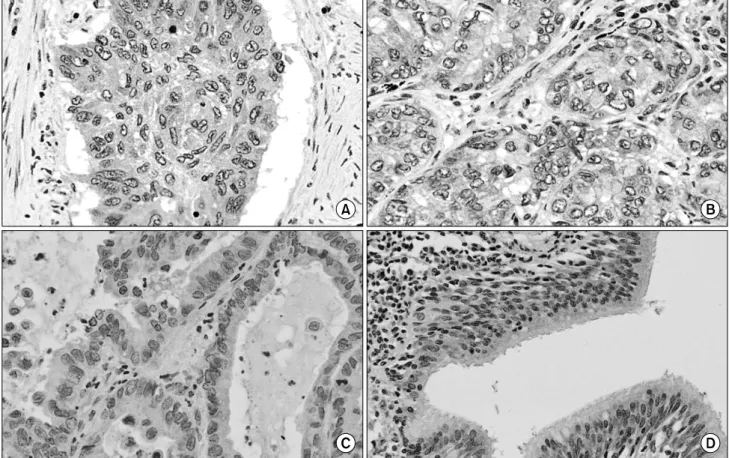
Fig. 2. Visualization of ATG5 expression in non-small cell lung cancer tissues by immunohistochemistry. (A) A squamous cell carcinoma shows ATG5 immunostaining in the cancer cells. (B) A adenocarcinoma shows ATG5 immunostaining in the cancer cells. (C) In another adenocarcinoma, the cancer cells are negative for ATG5 immunostaining. (D) Normal bronchial epithelial cells are positive for ATG5 immunostaining (original magnification A∼C, ×200; D, ×150).
알 수 있었다(Fig. 1). SSCP를 분석한 결과 중합효소연쇄반 응의 결과물은 정상조직의 DNA와 같이 야생형의 band로만
나타났으며, 돌연변이에서 보이는 이상 band는 관찰할 수 없었다(Fig. 1). 이들은 염기서열 분석 결과 돌연변이가 없 는 정상 염기서열을 가지고 있었다(data not shown). SSCP 결과는 해당 ATG5 유전자의 돌연변이가 분석한 45예의 폐 암에서 나타나지 않음을 의미했다. 이 실험은 미세절제, 중 합효소연쇄반응, SSCP 및 염기서열분석을 2회 반복하였으 며, 결과는 2회 모두 일치하였다.
2) ATG5 단백질 발현
조사한 45예의 폐암에서 ATG5 단백질의 발현은 36예 (80%)에서 양성이었고 9예에서는 음성이었다(Table 2, Fig.
2). 22예의 편평상피암에서는 16예(73%)가 양성이었고, 6예 에서는 발현이 나타나지 않았다. 23예의 샘암에서는 20예 (87%)가 ATG5 발현이 양성이었고, 3예에서는 발현이 나타 나지 않았다. ATG5 발현은 편평상피암 및 샘암에서 발현의 통계학적 차이는 없었다(Fisher’s exact test, two tails p>
0.05). ATG5의 면역조직화학 염색은 양성의 경우에는 세포 질 및 핵에 나타났다(Fig. 2). 정상 기관지 혹은 세기관지 상
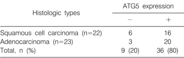
Table 2. Summary of ATG5 Expression in the Lung Cancers
Histologic types ATG5 expression
− +
Squamous cell carcinoma (n=22) 6 16
Adenocarcinoma (n=23) 3 20
Total, n (%) 9 (20) 36 (80)
피는 ATG5 양성을 나타냈다(Fig. 2). 일차항체를 처리하지 않은 음성 대조군은 ATG5 면역염색에 대해 음성을 보여 면역염색이 특이적인 것을 나타냈다. ATG5의 발현은 환자 의 성별, 나이, 암의 전이, 암의 진행단계 등에 의해 차이가 없었다(Fisher’s exact test, two tails p>0.05).
고안 및 결론
세포사멸 관련 단백질을 코딩하는 유전자에 흔히 암 돌 연변이가 발생하고 기능적으로 돌연변이가 세포사멸을 저 하시켜 암세포의 증식을 유도한다는 선행연구(9-14)에 따라 본 연구는 세포사멸의 일종인 autophagy를 유발시키는 유전 자인 ATG5에 관한 연구를 비소세포성폐암을 대상으로 시 행하였다. 요약하면, 본 연구의 목적은 ATG5 유전자의 돌 연변이와 발현을 함께 비소세포성폐암 조직에서 조사하여 비소세포폐암의 발생과 autophagy와의 관련성을 규명하는 것이었다. ATG5는 정상 폐기관지 상피세포에서는 잘 발현 되었지만 20%의 비소세포성폐암에서는 발현되지 않았다.
그러나 ATG5 유전자가 45예의 비소세포성폐암 조직에서 돌연변이가 되지 않았다는 것을 확인하였다. 이를 통해서 본 연구자들은 비소세포폐암의 병 발생기전에서 ATG 단백 질의 발현 감소가 모종의 역할을 할 가능성을 제시하였다.
하지만, 이 유전자의 돌연변이는 중요한 역할을 담당하지 않으리라는 것을 알 수 있었다.
세포사멸 불활성화에 의한 세포의 증식 및 생존 증가가 암의 시작 및 진행에 중요한 단계라는 것이 잘 알려져 있다 (17). 모든 세포사멸 저해 유전자는 암유전자이며, 모든 세 포사멸 유발 유전자는 암 억제 유전자로 간주된다(17). 암 에서 autophagy의 세포사멸에 대한 논란이 존재한다(1-4).
일부의 연구는 autophagy가 암의 발생을 억제한다고 밝힌 반면, 다른 연구들은 autophagy가 암의 발생을 촉진하고 암 세포의 세포사멸을 억제한다고 주장한다(1-4). ATG5의 autophagy 촉진자로의 역할은 확실하지만 실제 암 발생에서 ATG5가 어떤 역할을 하는지는 좀 더 밝혀져야 할 것이다.
하지만 흥미롭게도 ATG5는 다른 autophagy 관련 유전자에
비해 apoptosis를 유발하는 기능이 강력한 것으로 알려져 있 다. Apoptosis는 ATG5를 분절시키고 분절된 ATG5는 미토 콘드리아(mitochondria)로 이동하여 caspase를 활성화한다.
또한, ATG5는 암세포를 항암제 치료에 잘 반응하게 하는 것으로 알려져 있다(15,16). 이는 적어도 ATG5의 발현 저하 가 암 발생을 촉진할 가능성이 많은 것으로 해석될 수 있는 근거이며, ATG5가 비소세포성폐암의 발생에 암 억제유전 자의 역할을 수행할 가능성이 높은 것을 제시한다.
본 연구가 비소세포성폐암의 ATG5 소실을 밝혔지만, 빈 도(20%)는 높지 않은 편이다. 단백질 발현의 소실은 다양한 기전에 의해 나타난다. 대표적인 autophagy 단백질인 BECN1 의 발현 소실이 유방암에서 흔히 나타나는데 이 경우 단백 질 발현 소실은 BECN1 유전자의 과메칠화(hypermethyla- tion)에 의한다(18). ATG5 단백질의 발현이 과메칠화에 의 해 조절되는지에 대한 연구는 아직 없으므로 ATG5 과메칠 화가 폐암에서 나타날 가능성에 대한 조사가 필요한 실정 이다. 세포사멸 기전은 apoptosis, autophagy를 포함한 많은 경로의 종합으로 이루어지는 복잡한 기전이다. 한 가지 유 전자의 발현 이상으로 세포사멸을 결정하기는 힘든 것으로 알려져 있다(19). 따라서, 한 유전자의 이상과 임상 데이터 의 관련성은 잘 나타나기가 어려운 구조이며, 본 연구의 ATG5의 발현 감소가 비소세포성폐암 환자의 임상정보와 연관성이 없는 것과도 관련이 높다고 하겠다. 이제까지 autophagy와 관련된 유전자의 이상이 다수 보고된 바 있지 만, 비소세포성폐암에서는 BECN1 유전자 돌연변이 정도가 연구된 실정이다. 다른 암에서 밝혀진 ATG 유전자, UVRAG 유전자의 돌연변이 및 발현 연구가 향후의 연구를 통해 비소세포성폐암에서 분석될 필요가 있으며, 이를 기 존의 apoptosis 데이터 및 임상 데이터와 동시에 연구한다면 비소세포성폐암의 암 발병기전 및 진단, 치료 관련성을 규 명할 가능성이 높을 것이다.
REFERENCES
1. Baehrecke EH. Autophagy: dual roles in life and death? Nat Rev Mol Cell Biol 2005;6:505-510.
2. Edinger AL, Thompson CB. Death by design: apoptosis, necrosis and autophagy. Curr Opin Cell Biol 2004;16:663-669.
3. Kondo Y, Kanzawa T, Sawaya R, Kondo S. The role of auto- phagy in cancer development and response to therapy. Nat Rev Cancer 2005;5:726-734.
4. Klionsky DJ. Autophagy: from phenomenology to molecular understanding in less than a decade. Nat Rev Mol Cell Biol 2007;8:931-937.
5. Aita VM, Christiano AM, Gilliam TC. Mapping complex traits
in diseases of the hair and skin. Exp Dermatol 1999;8:439-452.
6. Lee JW, Jeong EG, Lee SH, Yoo NJ, Lee SH. Somatic muta- tions of BECN1, an autophagy-related gene, in human cancers.
APMIS 2007;115:750-756.
7. Kang MR, Kim MS, Oh JE, et al. Frameshift mutations of autophagy-related genes ATG2B, ATG5, ATG9B and ATG12 in gastric and colorectal cancers with microsatellite instability.
J Pathol 2009;217:702-706.
8. Kim MS, Jeong EG, Ahn CH, Kim SS, Lee SH, Yoo NJ.
Frameshift mutation of UVRAG, an autophagy-related gene, in gastric carcinomas with microsatellite instability. Hum Pathol 2008;39:1059-1063.
9. Lee SH, Shin MS, Park WS, et al. Alterations of Fas (Apo-1/
CD95) gene in non-small cell lung cancer. Oncogene 1999;18:
3754-3760.
10. Kim HS, Lee JW, Soung YH, et al. Inactivating mutations of caspase-8 gene in colorectal carcinomas. Gastroenterology 2003;125:708-715.
11. Lee JW, Soung YH, Kim SY, et al. Inactivating mutations of proapoptotic Bad gene in human colon cancers. Carcinogenesis 2004;25:1371-1376.
12. Shin MS, Kim HS, Kang CS, et al. Inactivating mutations of
CASP10 gene in non-Hodgkin lymphomas. Blood 2002;99:
4094-4099.
13. Soung YH, Lee JW, Kim SY, et al. Somatic mutations of CASP3 gene in human cancers. Hum Genet 2004;115:112-115.
14. Lee SH, Shin MS, Kim HS, et al. Alterations of the DR5/
TRAIL receptor 2 gene in non-small cell lung cancers. Cancer Res 1999;59:5683-5686.
15. Yousefi S, Perozzo R, Schmid I, et al. Calpain-mediated cleavage of Atg5 switches autophagy to apoptosis. Nat Cell Biol 2006;8:1124-1132.
16. Pyo JO, Jang MH, Kwon YK, et al. Essential roles of Atg5 and FADD in autophagic cell death: dissection of autophagic cell death into vacuole formation and cell death. J Biol Chem 2005;280:20722-20729.
17. Hanahan D, Weinberg RA. The hallmarks of cancer. Cell 2000;100:57-70.
18. Li Z, Chen B, Wu Y, Jin F, Xia Y, Liu X. Genetic and epige- netic silencing of the beclin 1 gene in sporadic breast tumors.
BMC Cancer 2010;10:98.
19. Reed JC. Mechanisms of apoptosis. Am J Pathol 2000;157:
1415-1430.