Low-grade osteosarcoma is a rare tumor that occurs mainly on the long bones such as the distal femur and proximal tibia. It may also develop in flat bones, such as ribs and craniofacial bones (1, 2). It is an unusual variant of an osteosarcoma, with a significantly better prognosis than a conventional osteosarcoma. It is characterized by slow growth, a low metastatic rate and prolonged sur- vival after treatment. However, to the best of our knowledge there have been no reports on MR and positron emission tomography (PET) computed tomog- raphy (CT) findings for a low-grade osteosarcoma of the spine. We will describe the detailed radiologic findings for a low-grade osteosarcoma that is rarely encountered in spine imaging.
Case Report
A 48 year-old male complained of severe low back
pain that radiated to both lower extremities for a 5- month duration. A physical examination did not reveal any tenderness or other symptoms associated with this complaint. The alkaline phosphatase level (87 IU/L) was within the normal limit. All levels of serum tumor mark- ers, including alfa feto protein (AFP), carcinoembryonic antigen (CEA), carbohydrate antigen 125 (CA 125) and carbohydrate antigen 19-9 (CA19-9) were within nor- mal limits. Plain radiography of the lumbar spine did not reveal a bone lesion. CT showed a lytic bone lesion with sclerotic margins. The lesion involved the right pedicle and extended to the right transverse process and posterior vertebral body. The vertebral cortex invasion was visible (Fig. 1). A PET scan revealed a focus of FDG- tracer uptake along the L1 vertebra. MRI revealed an enhancing mass in the right posterior portion of the L1 vertebral body that involved the right pedicle and the transverse process (Fig. 2). The lesion showed low signal on sagittal T1-weighted images and strong enhancement on sagittal and axial T1-weighted post-contrast images.
On sagittal T2-weighted images, the lesion exhibited predominantly low signal, but some areas of the posteri- or portion did exhibit some high signals. Following imaging assessment, the patient underwent a pediculec- tomy for tissue confirmation. The patient underwent
J Korean Radiol Soc 2007;56:575-578
─ 575 ─
Low-grade Osteosarcoma of the Spine: A Case Report1
Young Chul Kim, M.D., Jin-Suck Suh, M.D., Myung In Kim, M.D., Hye-Jung Choo, M.D., Yong-Min Huh, M.D.
1Department of Diagnostic Radiology, Severance Hospital, Research Institute of Radiological Science, Yonsei University College of Medicine, Seoul, South Korea.
Received January 2, 2007 ; Accepted February 12, 2007
Address reprint requests to : Young Chul Kim, M.D., Department of Diagnostic Radiology, Severance Hospital, Seodaemun-gu, Shinchon- dong 134, Seoul 120-752, South Korea
Tel. 82-2-2228-7400 Fax. 82-2-393-3035 E-mail: [email protected]
Low-grade osteosarcoma is not typically found in the long bone and pelvis. Most pri- mary osteosarcomas that arise in the spine are high-grade malignancies. A low-grade osteosarcoma arising in the spine has not been previously described. We report here the clinical, radiological, and histological findings of a case of low-grade osteosarcoma that arose in the spine.
Index words :Osteosarcoma Spine
Positron emission tomography (PET)
regular follow-up without further treatment.
The histology of the tumor was described as spindle cell proliferation with woven and lamellar bone forma- tion, resembling a fibrous dysplasia (Fig. 3). The tumor
was subsequently diagnosed as a low-grade osteosarco- ma via microscopic correlation with the radiological findings.
Young Chul Kim, et al: Low-grade Osteosarcoma of the Spine
─ 576 ─
A B
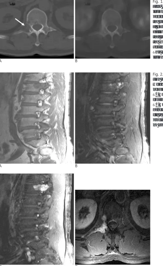
Fig. 2. MRI images (A--D). (A) The le- sion shows intermediate signals on T1- weighted images and (B) heteroge- neously mixed high and low signal on T2-weighted images. (C) On a post- contrast, sagittal (C) and (D) an axial T1-weighted image an intense en- hanced mass centered in the L1 pedi- cle is seen, extending to the transverse process body and lamina. There was no protruding soft tissue mass.
C D
A B
Fig. 1. CT (A), PET-CT fusion (B) im- ages. (A) The CT scan shows the lytic lesion (white arrow) involving the pedicle and posterior body, as well as the right transverse process. The later- al cortex disruption is clearly seen. (B) Fusion images of PET and CT show that the focus of uptake corresponds to the bone destructive lesion in the L1 vertebra and Rt. transverse process.
This indicates metabolic activity at the lesion.
Discussion
Low-grade osteosarcoma is a rare tumor, the inci- dence ranging from 1 to 4 % of all osteosarcomas (3, 4).
It shows a slight female predominance and occurs in the third or fourth decade of life. A low-grade osteosarcoma is typically located in the long bones, with a predilection for the distal femur and proximal tibia. These same loca- tions are commonly affected for conventional high- grade osteosarcomas. Other bone involvement includes the other long bones (fibula, radius, humerus and ulna), short tubular bones (metatarsal bone, phalanx and clav- icle) and flat bones (pelvis, rib and scapula) in rare cases (2, 3). However, no vertebral lesions have been reported previously (5). Of all primary malignant spinal tumors, osteosarcomas account for about 5% of cases.
Furthermore, most primary osteosarcomas that arise in the spine are high-grade malignancies (6).
Pathologists occasionally experience difficulty in dis- tinguishing low-grade osteosarcomas from fibrous dys- plasia because their histological features overlap (7, 8).
The histological features of this tumor were character- ized by bundles of spindled cells with minimal cytologi- cal atypia, rare mitotic figures and a variable osteoid (7).
This tumor may be frequently misdiagnosed as a fibrous dysplasia due to its minimal cytological atypia and rare mitotic figures (9). It is important to distinguish a low- grade osteosarcoma from a benign lesion, as although this tumor has a good prognosis, it poses a rare chance of recurrence when not properly eradicated by proper surgical measures (8).
Radiological findings of these tumors are variable and include sclerotic or heavily trabeculated matrix, intra- medullary involvement, localized cortical destruction, poor margins, and lack of new periosteal bone forma- tion (5, 10). Recently, low-grade central osteosarcomas have been classified into four patterns in descending or-
J Korean Radiol Soc 2007;56:575-578
─ 577 ─
A B
C
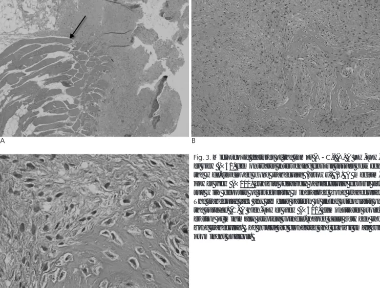
Fig. 3. Microscopic features of the tumor (A--C). (A) A low-pow- er view (×40) demonstrates intervening fibrous tissues between the well-developed bone trabeculae (arrows). (B) A medium- power view (×100) exhibits relatively paucicellular fibrous tis- sue with deposits of irregularly mineralized bone trabeculae.
The trabeculae lack any lamellar pattern or lining osteoblasts on the surface. (C) A high-power view (×400) demonstrates prolif- eration of minimally atypical spindle-shaped cells between the bone trabeculae. The nuclei are elongated and exhibit small but prominent nucleoli.
der of frequency of radiological patterns: 1. lytic with varying amounts of coarse trabeculation; 2. predomi- nantly lytic; 3. densely sclerotic; 4. mixed lytic and scle- rotic without significant coarse trabeculation (5).
Most conventional osteosarcomas in the spine arise within the vertebral body, often eccentrically, and fre- quently extend into the posterior elements (11).
However, the lesion in our case was centered at the right pedicle of the L1 vertebra, extending to the posteri- or body and the transverse process. CT and MRI are useful for evaluating cortical interruption and soft tissue mass formation that is suggestive of a malignancy rather than a benign lesion. Regardless of the presence of sub- tle mitotic figures and atypia, the features of cortical dis- ruption or soft tissue mass seen on CT and MRI can help lead to a correct histological diagnosis. Despite the ab- sence of a soft mass formation in this patient, a cortical interruption and increase of FDG uptake suggested a malignancy.
The differential diagnosis of this tumor includes os- teoblastoma, giant cell tumor as well as fibrous dyspla- sia. The osteoblastoma may involve the posterior verte- bral elements and frequently extends into the vertebral body. An edematous reaction within the adjacent bone marrow is seen for a osteoblastoma more frequently than for a osteosarcoma. Most of the giant cell tumors are found in the sacrum and third decades. Cystic areas as well as areas of previous hemorrhage with hemo- siderin are also seen (6).
In conclusion, if aggressive radiographic findings often are subtle and the histological finding shows features of
fibrous dysplasia or another benign lesion, the possibili- ty of low-grade osteosarcoma should be considered.
References
1. Vlychou M, Ostlere SJ, Kerr R, Athanasou NA. Low-grade os- teosarcoma of the ethmoid sinus. Skeletal Radiol In press 2006 2. Yamaguchi T, Shimizu K, Koguchi Y, Saotome K, Ueda Y. Low-
grade central osteosarcoma of the rib. Skeletal Radial 2005;34:490- 493
3. Bugnone AN, Temple HT, Pitcher JD. Low-grade central osteosar- coma of the foot and ankle: radiographic and pathologic features in two patients: case report and literature review. Foot Ankle Int 2005;26:494-500
4. Kenan S, Ginat DT, Steiner GC. Dedifferentiated high-grade os- teosarcoma originating from low-grade central osteosarcoma of the fibula. Skeletal Radiol In press 2006
5. Andresen KJ, Sundaram M, Unni KK, Sim FH. Imaging features of low-grade central osteosarcoma of the long bones and pelvis.
Skeletal Radiol 2004; 33:373-379
6. Erlemann R. Imaging and differential diagnosis of primary bone tumors and tumor-like lesions of the spine. Eur J Radiol 2006;58:
48-67
7. Fukunaga M. Low-grade central osteosarcoma of the skull. Pathol Res Pract 2005;201:131-135
8. Ogose A, Hotta T, Emura I, Imaizumi S, Takeda M, Yamamura S.
Repeated dedifferentiation of low-grade intraosseous osteosarco- ma. Hum Pathol 2000;31:615-618
9. Franceschina MJ, Hankin RC, Irwin RB. Low-grade central os- teosarcoma resembling fibrous dysplasia. A report of two cases.
Am J Orthop 1997;26:432-440
10. Ellis JH, Siegel CL, Martel W, Weatherbee L, Dorfman H.
Radiologic features of well-differentiated osteosarcoma. AJR Am J Roentgenol 1988;151:739-742
11. Drevelegas A, Chourmouzi D, Boulogianni G, Sofroniadis I.
Imaging of primary bone tumors of the spine. Eur Radiol 2003;
13:1859-1871
Young Chul Kim, et al: Low-grade Osteosarcoma of the Spine
─ 578 ─
대한영상의학회지 2007;56:575-578
척추에서 발생한 저등급 골육종: 증례 보고1
1연세의대 세브란스병원 영상의학과
김영철・서진석・김명인・추혜정・허용민
저등급 골육종은 장골(long bone)과 골반에서 발생한다. 척추에서 발생하는 대부분의 골육종은 고등급 악성종양 이다. 이제까지 보고된 증례가 없는 것으로 알려진 척추에 생긴 저등급 골육종의 임상적, 방사선학적 그리고 병리 학적 소견을 보고자 한다.