A sinus of Valsalva aneurysm (SVA) is a relatively rare cardiac anomaly (1). SVA most commonly originates in the right coronary sinus of Valsalva (75-90%), the non- coronary sinus (10-25%), and rarely, in the left sinus (1, 2). Unruptured SVA usually remains asymptomatic, but it can sometimes cause cardiac arrhythmia, myocardial ischemia, or systemic embolic events (1-3).
Multidetector computed tomography (MDCT) is a use- ful modality for examining the anatomy of the heart, but there have been few radiologic reports of sinus of Valsalva aneurysms diagnosed by MDCT. We report here a case of an unruptured left- and non-coronary si- nus of Valsalva aneurysms detected by MDCT.
Case Report
A 51-year-old woman presented with chest discomfort
for four days and was taking medication to control her high blood pressure. She denied any history of previous surgery or other cardiac diseases.
An electrocardiography (ECG) performed on admis- sion revealed atrial fibrillation. Her blood biochemistry findings were unremarkable. The chest radiograph showed cardiomegaly and right pleural effusion. A transthoracic echocardiography (TTE) showed the di- latation of the non-coronary sinus and left coronary si- nus (Fig. 1), and the left atrium (LA) was compressed by the dilated left coronary sinus. TTE also revealed a mod- erate to severe degree of aortic regurgitation, a mild to moderate degree of tricuspid regurgitation and a mild degree of mitral regurgitation.
Chest CT examinations were performed with a 64 channel MDCT (Aquilion, Toshiba, Tokyo, Japan). The chest CT scans were obtained from the level of the tho- racic inlet to the level of the diaphragm. The images were obtained with a 2 mm slice thickness in the cranio- caudal direction for the axial scan, and the coronal and sagittal images were reconstructed automatically on the console. The contrast enhanced images were routinely obtained 40 seconds after the initiating the injection of contrast medium (a total of 130 mL) at a rate of 3-4 mL/s. In addition to traditional images, multiplanar re-
J Korean Soc Radiol 2010;63:509-512
─ 509 ─
MDCT Findings of Sinus of Valsalva Aneurysms Involving Two Coronary Sinuses: Case Report1
Eun Mi Kim, M.D., Su Young Kim, M.D., Sung Yoon Lee, M.D.2, Woo-Ik Chang, M.D.3, Chang Young Kim, M.D.3, Gham Hur, M.D.
1Department of Radiology, Inje University Ilsan-Paik Hospital
2Department of Internal Medicine, Inje University Ilsan-Paik Hospital
3Department of Cardiothoracic Surgery, Inje University Ilsan-Paik Hospital
Received May 17, 2010 ; Accepted August 12, 2010
Address reprint requests to : Su Young Kim, M.D., Department of Radiology, Ilsan Paik Hospital, Inje University School of Medinine, 2240, Daehwa-dong, Ilsanseo-gu, Goyang-si, Gyeonggi-do 411-706, Korea.
Tel. 82-31-910-7397 Fax. 82-31-910-7369 E-mail: [email protected]
A sinus of Valsalva aneurysm is relatively rare and usually involves a single sinus.
We describe here the multidetector computed tomography features of a case of an un- ruptured sinus of Valsalva aneurysms that affected the left and noncoronary sinuses in a 51-year-old woman.
Index words :Sinus of Valsalva Aneurysm
Tomography, X-Ray Computed
constructions (MPR) and 3-dimensional volume-render- ing images were made by an image processing worksta- tion (Rapidia; Infinitt, Seoul, Korea) to clearly depict the coronary sinuses. On the MPR images (Fig. 2), a 4.7 × 4.3 cm sized left coronary sinus of Valsalva aneurysm (LC-SVA) without thrombosis and a 3.0 × 3.4 cm sized non-coronary sinus of Valsalva aneurysm (NC-SVA) were observed. The left main coronary artery (LM) was compressed by the LC-SVA (Fig. 3) and about 50%
stenosis was suggested.
A cardiopulmonary bypass with moderate systemic hypothermia and cold blood cardioplegia was per-
formed. The operative findings confirmed the huge aneurysms of the left coronary sinus and the non-coro- nary sinus of Valsalva. The leaflets of the aortic valve and ascending aorta were morphologically normal. An aortic valve sparing procedure with a 24 mm Vaxcutek graft was attempted, but failed. As a result, a Bentall op- eration was performed.
Discussion
The most common cause of SVA is due to a congenital problem. SVA accounts for 0.1-3.5% of congenital heart disease and 0.14% of all open heart surgical procedures (1, 2). The incidence of SVA appears to be higher in countries of the Far East and the male-female ratio is 4:1 (2, 4). The aneurysm is thought to arise from the incom- plete fusion of the distal bulbar septum that divides the aorta and pulmonary artery and attaches to the annulus fibrosus of the aortic valve. As a result, the right coro- nary or noncoronary sinus of Valsalva is usually affect- ed. Because the left sinus of Valsalva does not arise from the bulbar septum, a left sinus of Valsalva aneurysm is rare and it is commonly acquired rather than being con- genital (2). Aneurysms affecting both sinuses of Valsalva are extremely rare. We found only three such cases in the English medical literature (5-7).
SVAs usually remain asymptomatic unless rupture oc- curs. The incidence of unruptured SVAs has been re- ported to be 20%, based on previous reports of necropsy and cardiac surgical findings (1). Unruptured SVAs
Eun Mi Kim, et al: MDCT Findings of Sinus of Valsalva Aneurysms Involving Two Coronary Sinuses
─ 510 ─ Fig. 1. A transthoracic echocardiography shows the dilatation of the left coronary (LC) and non-coronary (NC) sinuses. The left atrium (LA) is compressed by the dilated left coronary si- nus.
A B C
Fig. 2. 64 slice-MDCT scans of a 51-year-old woman presenting with chest discomfort for four days.
A, B. Two axial images show the unruptured left coronary sinus of Valsalva aneurysm (A, arrow) and the noncoronary sinus of Valsalva aneurysm (B, arrow head).
C. The MPR image shows the dilation of the left coronary (LC) and noncoronary (NC) sinus of Valsalva and the normal right coro- nary sinus (RC).
sometimes cause symptoms by mechanical obstruction.
Expanding SVAs can obstruct the ventricular outflow tract and compress the coronary arteries or conducting system, and this can induce an acute ischemic attack, complete heart block, or arrhythmia. Furthermore, sys- temic embolic events may come about from thrombus formation within a SVA (1). Because of its location, dis- tortion or deformation of the pulmonary or aortic valve leaflets can occur and this can cause aortic regurgitation or pulmonary insufficiency. Congenital SVAs are often associated with a ventricular septal defect (2, 3).
Clinically, the diagnosis of unruptured sinus of Valsalva aneurysms may be difficult because most cases are asymptomatic (8). TTE with Doppler ultrasonogra- phy is highly useful for diagnosing sinus of Valsalva aneurysms. TTE shows the wall of the SVAs or a throm- bus within the SVAs, as well as show valve movement or cardiac wall motion for evaluating the cardiac func- tion. Yet obesity and accompanying pulmonary diseases can limit the use of TTE. Transesophageal echocardiog- raphy offers the potential for a more accurate characteri- zation of an aneurysm, but this procedure is moderately invasive (2, 4).
In the past, CT could not show the shape of the sinus of Valsalva or the cardiac valves because of rapid car- diac movement (2, 9). Because of the great advances in
CT technology, MDCT has recently provided images of the cross-sectional anatomy of the heart with excellent spatial and temporal resolution (4). MDCT is useful for delineating a sinus of Valsalva aneurysm and its rup- ture, especially in patients whose echocardiography ex- amination is suboptimal (2). MDCT can accurately as- sess the aneurysm size, the sinus of origin, and aortic valve involvement (9).
In our case, MDCT accurately showed the unruptured left and noncoronary sinus of Valsalva aneurysms and the patient was successfully treated by surgical correc- tion. MDCT can provide superior anatomic orientation between the sinus of Valsalva aneurysm and the sur- rounding structures (8). This case demonstrates the use- fulness of MDCT for the diagnosis of unruptured aneurysms of both the left coronary and non-coronary sinuses of Valsalva.
References
1. Kim KH, Yang TH, Han YC, Cho HJ, Um SJ, Seol SH, et al. Huge aneurysm of the sinus of valsalva compressing the left atrium. J Cardiovasc Ultrasound 2008;16:140-142
2. White CS, Plotnick GD. Case 33: sinus of valsalva aneurysm.
Radiology 2001;219:82-85
3. Thankachen R, Gnanamuthu R, Doshi H, Shukla V, Korula RJ.
Unruptured aneurysm of the sinus of valsalva presenting with right ventricular outflow obstruction. Tex Heart Inst J 2003;30:152- J Korean Soc Radiol 2010;63:509-512
─ 511 ─
A B
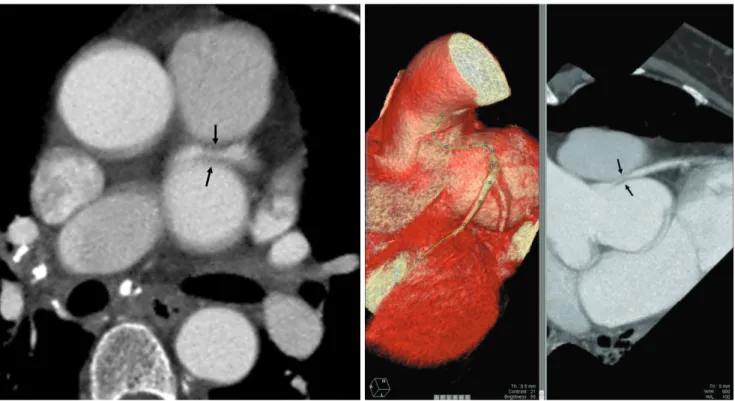
Fig. 3. A, B. The MPR image (A) and 3-dimensional volume-rendering images (B) show the compression of the left main coronary artery (arrows) by the LC-SVA.
154
4. Kantarci M, Doganay S, Gundogdu F, Unlu Y. A case with non- coronary sinus of valsalva aneurysm: multidetector computed to- mography findings. Heart Surg Forum 2008;11:E372-E374 5. Tami LF, Turi ZG, Arbulu A. Sinus of valsalva aneurysms involv-
ing both coronary ostia. Cathet Cardiovasc Diagn 1993;29:304-308 6. Zannis K, Tzvetkov B, Deux JF, Kirsch EW. Unruptured congeni-
tal aneurisms of the right and left sinuses of valsalva. Eur Heart J 2007;28:1565
7. Vijayalakshmi IB, Devananda NS, Chitra N. A patient with aneurysms of both aortic coronary sinuses of valsalva obstructing both ventricular outflow tracts. Cardiol Young 2009;19:537-539 8. Hwang SH, Kim TH, Kim SJ, Kwon HM, Yu KJ. Multidetector-
row computed tomography of a valsalva sinus aneurysm in a pa- tient with behcet disease. J Thorac Imaging 2006;21:300-302 9. Matteucci ML, Rescigno G, Capestro F, Torracca L. Syncope trig-
gered by a giant unruptured sinus of valsalva aneurysm. Interact Cardiovasc Thorac Surg 2009;9:1047-1048
Eun Mi Kim, et al: MDCT Findings of Sinus of Valsalva Aneurysms Involving Two Coronary Sinuses
─ 512 ─
대한영상의학회지 2010;63:509-512
MDCT로 진단한 두 개의 Coronary Sinus를 침범한 발살바동 동맥류 증례 보고1
1인제대학교 일산백병원 영상의학과
2인제대학교 일산백병원 내과
3인제대학교 일산백병원 흉부외과
김은미∙김수영∙이성윤2∙장우익3∙김창영3∙허 감
발살바동에서 발생한 동맥류는 비교적 드물며 대개 한 개의 발살바동을 침범한다. 저자는 51세 여자 환자에서 발 생한 좌관상동과 비관상동을 침범하는 동맥류의 다검출기전산화단층촬영 소견 1예를 보고하고자 한다.