Introduction
Round-shoulder posture (RSP) is typified by a protracted, anterior tipped, and downwardly rotated scapular position (Lee et al, 2015; Magee, 2008;
Thigpen et al, 2010; Wong et al, 2010). There are various factors that contribute to RSP, such as
tightness or shortness of the pectoralis minor, greater thoracic kyphosis, and the scapular anatomical struc- ture itself (Ekstrom et al, 2003; Lee et al, 2015;
Ludewig et al, 2004; Sahrmann, 2002; Thigpen et al, 2010). In addition, weakness or insufficient activities of the lower trapezius (LT) and serratus anterior (SA) are common causal factors of RSP (Lee et al,
Corresponding author: Chung-hwi Yi [email protected]
This research was supported by Basic Science Research Program through the National Research Foundation of Korea (NRF) funded by the Ministry of Education (2015R1D1A1A01057620).
Selective Muscle Activation With Visual Electromyographic Biofeedback During Scapular Posterior Tilt Exercise in Subjects With
Round-Shoulder Posture
Jae-ik Son1, BPT, PT, One-bin Lim1, MSc, PT, Hae-rim Han1, BPT, PT, Heon-seock Cynn2,3, PhD, PT, Chung-hwi Yi2,3, PhD, PT
1Dept. of Physical Therapy, The Graduate School, Yonsei University
2Dept. of Physical Therapy, College of Health Science, Yonsei University
3Dept. of Ergonomic Therapy, The Graduate School of Health Science, Yonsei University
Abstract
1)The purpose of this study was to investigate the effects of visual electromyography (EMG) biofeedback on the EMG activity of the lower trapezius (LT), serratus anterior (SA), and upper trapezius (UT) muscles, the LT/UT and SA/UT EMG activity ratios, and the scapular upward rotation angle during scapular posterior tilting exercise (SPTE). Twenty-four subjects with round-shoulder posture participated in this study. The EMG activities of the LT, SA, and UT were collected during SPTE both without and with visual EMG biofeedback. The scapular upward rotation angle was measured at the baseline, after SPTE without visual EMG biofeedback, and after SPTE with visual EMG biofeedback. The LT, SA, and UT EMG activities, and the LT/UT and SA/UT EMG activity ratios were analyzed by paired t-test. The scapular upward rotation angle was statistically analyzed using one-way repeated analysis of variance. If a significant difference was found, a Bonferroni correction was performed (p=.05/3=.017). The EMG activities of LT and SA significantly increased, and the EMG activity of UT significantly decreased during SPTE with visual EMG biofeedback compared to SPTE without visual EMG biofeedback (p<.05).
In addition, the LT/UT and SA/UT EMG activity ratios significantly increased during SPTE with visual EMG biofeedback compared to SPTE without visual EMG biofeedback (p<.05). Significant increases were found in the scapular upward rotation angle after SPTE without and with visual EMG biofeedback compared to baseline (p<.017), and no significant differences were observed in the scapular upward rotation angle between SPTE without and with visual EMG biofeedback. In conclusion, SPTE using visual EMG biofeedback may be an effective method for increasing LT and SA activities while reducing UT activity.
Key Words: Biofeedback; Electromyography; Round-shoulder posture; Scapular posterior tilting exercise.
2015; Sahrmann, 2002; Thigpen et al, 2010). The LT and SA are prime movers for scapular posterior tilt and upward rotation, which are required for widening the subacromial space during overhead activities to prevent impingement of the subacromial tissues (Ha et al 2012; Ludewig and Braman, 2011; Ludewig and Cook, 2000). In the case of insufficient activation of the LT and SA, individuals with RSP could be vulner- able to subacromial impingement or other problems that could be caused by altered shoulder kinematics (Ludewig and Cook, 2000; Thigpen et al, 2010).
Therefore, activation of the LT and SA is important during RSP intervention programs for people with RSP.
There are many exercises to increase activity of LT and SA, such as the scapular posterior tilting exercise in quadruped position (SPTE), wall facing arm lift, and prone arm lifting (Arlotta et al, 2011;
Ekstrom et al, 2003; Ha et al, 2012). Ha et al (2012) suggested that SPTE is highly efficient for activation of the LT and SA by stabilizing the scapula to the thoracic wall. However, Page et al (2010) suggested that individuals with RSP tend to use the upper tra- pezius (UT) more than the LT and SA during over- head activity involving the shoulder. The reduced activation of the UT has been emphasized in many previous studies for rehabilitation of patients with impingement syndrome (Cools et al, 2007a; Cools et al, 2007b; Huang et al, 2013; Ludewig and Braman, 2011; Ludewig and Cook, 2000). Since the scapular characteristics of movement in people with RSP dur- ing overhead tasks include greater scapular anterior tilting and internal rotation, individuals with RSP would experience negative effects such as impinge- ment syndrome (Ludewig and Braman, 2011; Ludewig and Cook, 2000; Thigpen et al, 2010). In addition, Ludewig and Braman (2011) suggested that excessive activation of the UT could result in excess clavicular elevation and scapular anterior tilting, both of which cause a decrease in scapular posterior tilt. Therefore, selective activation for effective exercise is required to increase the activity of the LT and SA and de- crease activity of the UT for individuals with RSP.
In many previous studies, visual electromyographic (EMG) biofeedback, which is the recommended ap- proach for learning functional motor control in re- habilitation settings (Holtermann et al, 2009;
Holtermann et al, 2010) has been used to selectively activate or inhibit specific muscles. Lim et al (2014) investigated selective activation between the infra- spinatus and posterior deltoid using visual EMG bio- feedback and there was a positive effect on the re- ducing activity of the posterior deltoid. In addition, Huang et al (2013) reported that the activities of the LT and SA increased while the UT activity de- creased during shoulder forward flexion using visual EMG biofeedback for individuals without and with impingement syndrome.
However, there are few previous studies doc- umenting selective activation with visual EMG bio- feedback for individuals with RSP during exercises to activating of the LT and SA. Therefore, the pur- pose of this study was to investigate LT, SA, and UT muscle activity, LT/UT and SA/UT ratio, and the scapular upward rotation angle during SPTE without and with visual EMG biofeedback in sub- jects with RSP. We hypothesized that SPTE with visual EMG biofeedback would increase LT and SA activities while reducing UT activity. And the LT/UT and SA/UT activity ratios would increase during STPE with visual EMG biofeedback.
Furthermore, as a result of increased LT and SA activities, the scapular upward rotation angle would increase after SPTE with visual EMG biofeedback.
Methods
Subjects
The G-power software ver. 3.1.2 was used for power analyses (Franz Faul, University of Kiel, Kiel, Germany). The sample size was calculated from data to achieve a power of .80 and an effect size of .66 with an alpha level of .05. The estimated sample size was 16 and 24 subjects (15 males and 9 females)
with RSP participated in this study. The general characteristics of the subjects are presented in Table 1. Inclusion criteria were as follows: 1) the measure- ment between the subject’s posterior border of acro- mion and the treatment table in the supine position as ≥2.5 ㎝ for the dominant side [interclass correla- tion coefficient (ICC)=.88∼.94] (Nijs et al, 2005;
Wong et al, 2010); 2) the subjects could perform full flexion in the sagittal plane, full abduction in the frontal plane and full scaption in the scapular plane, and they were asymptomatic (Ha et al, 2012).
Subjects were excluded if they had: 1) current shoulder pain or problems; 2) a history of neuro- logical, musculoskeletal, or cardiopulmonary disease that could interrupt shoulder motion (Ha et al, 2012).
As the subjects’ dominant sides were satisfied the inclusion criteria, the dominant arm was used in all tests. The dominant arm was determined by the pre- ferred arm used to eat and write (Yoshizaki et al, 2009) and all subjects’ right arms were dominant.
Prior to experimentation, the examiner explained the study and subjects wrote an informed consent. The study was approved by the Yonsei University Wonju Institutional Review Board (approval number:
1041849-201510-BM-054-02).
Instrumentation and electrode placement Surface EMG (Noraxon TeleMyo DTS, Noraxon Inc., AZ, USA) was used to investigate activities in the LT, SA and UT. Prior to electrode placement,
the skin was shaved and swabbed with alcohol.
Disposable, self-adhesive Ag/AgCl surface electrodes were used and placed at locations 2 ㎝ apart on the muscle belly of the LT, SA, and UT. The LT elec- trodes were attached obliquely upward and laterally along the line between the intersection of the spine of the scapula and the 7th thoracic spinal process (Criswell, 2011). The SA electrodes were attached anterior to the latissimus dorsi and posterior to the pectoralis major (Huang et al, 2013). The UT elec- trodes were attached midway between the spinous process of the 7th cervical vertebra and the posterior tip of the acromion process (Criswell, 2011). During the investigation, EMG signals were collected at 1,000 ㎐; the raw signal was filtered using a band- pass filter (Lancosh FIR) between 10 and 450 ㎐, and 60 ㎐ and 120 ㎐ notch filters were used to re- duce noise. EMG data were processed into root mean square values, which was calculated from 300 ㎳ data points of windows.
Scapular posterior tilting exercise (SPTE) In the quadruped position, the examiner instructed the subjects to rock backward slowly until the but- tocks touched both heels. The subjects’ non-domi- nant hand was placed under the forehead and the examiner passively abducted the subjects’ dominant arm until 145°. In this position, the subjects was in- structed to lift the dominant arm with elbow ex- tended until the radial border of the wrist slightly touched the target bar and maintained position to re- cord EMG activities (Ha et al, 2012; Lee et al, 2015) (Figure 1). The target bar was set at the level of the subjects’ earlobe. The subjects were given 12-sec to complete one movement cycle: the initial 3-sec were spent moving to the target position;
the middle 6-sec holding the target position; and last 3-sec were spent moving to the starting position. A metronome was used to guide the sub- jects to perform SPTE at a standard speeds. The EMG activities of the LT, SA, and UT were col- lected for 6-sec each trial during isometric con-
Parameters Mean±SDa
Age (year) 23.2±2.2
Height (㎝) 170.0±8.1
Weight (㎏) 66.4±12.9
BMIb (㎏/㎡) 22.8±3.0
Degree of RSPc (㎝) 3.7±.8
amean±standard deviation, bbody mass index, cdegree of round-shoulder posture: distance between the subject’s posterior border of acromion and the treatment table.
Table 1. General characteristics of the subjects (N=24)
traction period and the EMG activities at 2∼5 sec were used (Ha et al, 2012). Subjects practiced the SPTE prior to data collection for 5 min. After practice, the subjects were given a 5-min rest period. The subjects performed three trials with 2-min rest periods between trials to prevent muscle fatigue (Ha et al, 2012).
Scapular upward rotation angle measurement A previous study found that healthy subjects’
average maximum arm elevation angle for the domi- nant arm was approximately 135° (Yano et al, 2010).
Therefore, the measurement of the scapular upward rotation angle was at 135° shoulder abduction in co- ronal plane while standing. Two inclinometers were used to measure the scapular upward rotation angle.
One inclinometer was used to measure the shoulder abduction angle and at the same time, the other one was used to directly measure the scapular upward rotation angle by manually positioning it along the scapular spine. (Figure 2). Watson et al (2005) re- ported that the ICC was .88. The examiner meas- ured the angle three times at each conditions (baseline, after performing SPTE without and with visual EMG biofeedback), and the mean values of the angle were used for comparison. The scapular upward rotation after performing SPTE without and with visual EMG biofeedback was measured imme- diately after SPTE.
Visual EMG biofeedback
The visual EMG biofeedback used the EMG bio- feedback measurement option of MyoResearch Master Edition 1.07 XP software (Noraxon Inc., AZ, USA).
The biofeedback information was displayed on a computer screen under the treatment table (Figure 3).
In the biofeedback measurement option, we used a moving bar graph, which indicated the EMG activity (Lim et al, 2014; Jeon et al, 2011). For example, if the activity of a muscle increased or decreased, the bar graph was increased or decreased, accordingly.
We set the EMG activity threshold to give feedback
Figure 2. Scapular upward rotation angle measurement with two inclinometers.
Figure 1. The scapular posterior tilting exercise (turn off the monitor screen:
without visual electromyographic biofeedback, turn on the monitor screen: with visual electromyographic biofeedback).
1. LT, uV 2. SA, uV 3. UT, uV
Figure 3. Display of the visual electromyographic biofeedback (LT: lower trapezius, SA: serratus anterior, UT: upper trapezius).
for the LT, SA and UT. The mean value of the tar- get muscle EMG activities during SPTE without visual EMG biofeedback was determined as a threshold, respectively. In previous studies that aimed to selectively reduce muscle activities (posterior del- toid and pectoralis major), the thresholds of EMG activity were set based on 10% of the maximal vol- untary isometric contraction (MVIC) (Jeon et al, 2011; Lim et al, 2014). However, the results of the pilot study in the current study suggested that it was difficult to reduce the UT activity below 10% of the MVIC. Although the study by Huang et al (2013) that used visual EMG biofeedback to reduce the UT activity did not set an EMG activity thresh- old, the current study needed an EMG activity threshold to simultaneously control the LT, SA and UT. Thus, current study used the mean values of the EMG activities of LT, SA, and UT in SPTE without visual EMG biofeedback to define the EMG activity thresholds. The examiner instructed the sub- jects to decrease the UT EMG activity below the threshold and increase the LT and SA EMG activ- ities above the threshold during the SPTE by watching the computer display screen (Huang et al, 2013; Jeon et al, 2011; Lim et al, 2014). There was a 10-min session to familiarize the subjects with the use of visual EMG biofeedback, and after a 5-min rest period, the data for SPTE with visual EMG bi- ofeedback were collected.
Procedures
The experimental process for this study was as follows: 1) the examiner measured the subject’s scap- ular upward rotation angle as a baseline; 2) the sub- ject performed SPTE without visual EMG biofeedback while recording EMG data of SPTE without visual EMG biofeedback; 3) the examiner immediately measured the subject’s scapular upward rotation an- gle after the subject performed SPTE without visual EMG biofeedback; 4) the subject familiarized with the use of visual EMG biofeedback for 10 min (Holtermann et al, 2009; Lim et al, 2014); 5) after the
familiarizing session, the subject performed SPTE with visual EMG biofeedback while recording EMG data of SPTE with visual EMG biofeedback; 6) after performing the SPTE with visual EMG biofeedback, the examiner immediately measured the subject’s scapular upward rotation angle. The experimental process was not randomized to prevent learning effect.
EMG data processing
The MVIC was used to normalize a basis for EMG signal amplitude. The MVIC of the LT, SA and UT were respectively collected period of 5-sec.
The standard methods using gravity and manual re- sistance were used to collect MVIC value (Kendall et al, 2005). The MVIC value for the LT was collected with the subject in the prone position. The subject place their arm diagonally overhead, in line with the lower fibers of the trapezius muscle during external rotation, while resistance was applied distal to the elbow. The SA MVIC value was collected with the subject in the sitting position. The subject rotated internally and abducted to 125° their shoulder in the scapular plane; the examiner applied resistance above the elbow. The UT MVIC value was also collected with the subject in the sitting position. The subject’s elevated the acromial end of the clavicle and scapula, and posterolaterally extended the neck bringing the occiput toward the elevated shoulder with the face turned in the opposite direction. Resistance was ap- plied against the shoulder, in the direction of depres- sion, and against the head, in the direction of flexion anterolaterally. EMG activities collected between 2∼4 sec were used to define the mean MVIC amplitude, and the normalized EMG activity was presented as a percentage of MVIC (%MVIC).
Statistical analysis
A one-sample Kolmogorov-Smirnov test was used to define the normality of distribution. The variables (EMG values and scapular upward rotation angles) satisfied the normal distribution. Therefore, para- metric tests were used to analyze the variables. The
paired t-test was used to analyze the EMG activities (LT, SA, and UT) and the EMG activity ratios (LT/UT and SA/UT). One-way repeated measures analysis of variance was used to analyze scapular up- ward rotation angles for the three conditions (baseline and after performing SPTE without and with visual EMG biofeedback), and the Bonferroni correction was used for clarification of the differences among the three conditions (.05/3=.017). SPSS ver. 21.0 (SPSS Inc., Chicago, IL, USA) was used to analyze statistical significance, the significance level was set α=.05.
Results
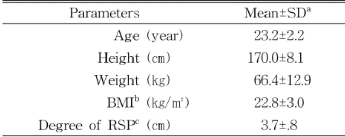
EMG activity and EMG activity ratio The EMG activities of LT and SA significantly increased with visual EMG biofeedback compared to without visual EMG biofeedback (LT without visual EMG biofeedback: 55.55±16.38 %MVIC; LT with vis- ual EMG biofeedback: 58.90±19.66 %MVIC; SA with- out visual EMG biofeedback: 24.77±13.34 %MVIC;
SA with visual EMG biofeedback: 38.01±18.85
%MVIC; p<.05). The EMG activity of UT sig- nificantly decreased with visual EMG biofeedback compared to without visual EMG biofeedback (UT without visual EMG biofeedback: 29.84±14.26
%MVIC; UT with visual EMG biofeedback:
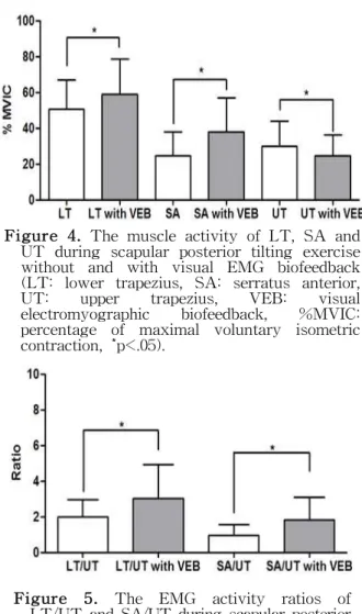
24.50±11.97 %MVIC; p<.05) (Figure 4). The LT/UT and SA/UT EMG activity ratios significantly in- creased with visual EMG biofeedback compared to without visual EMG biofeedback (LT/UT without visual EMG biofeedback: 2.00±.97 %MVIC; LT/UT with visual EMG biofeedback: 3.02±1.90 %MVIC;
SA/UT without visual EMG biofeedback: .95±.62
%MVIC; SA/UT with visual EMG biofeedback:
1.84±1.25 %MVIC; p<.05) (Figure 5).
Scapular upward rotation angle
There were significant differences in the scapular upward rotation angle among the baseline, after per- forming SPTE without and with visual EMG bio-
feedback (F2,22=30.965, p<.05). The scapular upward rotation angle significantly increased after performing SPTE without and with visual EMG biofeedback compared to the baseline (p<.017). There were no significant differences between scapular upward rota- tion angles after performing SPTE without and with visual EMG biofeedback (p>.017) (Table 2).
Discussion
This study investigated whether visual EMG bio- Figure 5. The EMG activity ratios of
LT/UT and SA/UT during scapular posterior tilting exercise without and with visual EMG biofeedback (LT: lower trapezius, SA:
serratus anterior, UT: upper trapezius, VEB:
visual electromyographic biofeedback,
*p<.05).
Figure 4. The muscle activity of LT, SA and UT during scapular posterior tilting exercise without and with visual EMG biofeedback (LT: lower trapezius, SA: serratus anterior, UT: upper trapezius, VEB: visual electromyographic biofeedback, %MVIC:
percentage of maximal voluntary isometric contraction, *p<.05).
feedback could increase the LT and SA activities while reducing UT activity during SPTE and changes in scapular upward rotation angles in sub- jects with RSP. The results of this study partially supported the hypotheses. The LT and SA activities significantly increased while the UT activity reduced during SPTE with visual EMG biofeedback compared to SPTE without visual EMG biofeedback. EMG ac- tivity ratios (LT/UT and SA/UT) significantly in- creased in SPTE with visual EMG biofeedback com- pared to SPTE without visual EMG biofeedback.
However, the scapular upward rotation angle was not significantly different between the measurements without and with visual EMG biofeedback.
The current study found that LT and SA activ- ities were significantly increased during SPTE with visual EMG biofeedback compared to those per- formed without visual EMG biofeedback. Previous studies reported that visual EMG biofeedback train- ing could help subjects learn how to use their mus- cles (Holtermann et al, 2009; Holtermann et al, 2010;
Huang et al, 2013; Lim et al, 2014). Although the current study investigated immediate effects, the re- sults suggest that the use of visual EMG biofeed- back training would support the selective control of the lower part and upper part within the trapezius muscle as well as the SA. The SPTE was designed to raise subjects’ arm in the direction of the LT muscle fibers (Ekstrom et al, 2003; Ha et al, 2012).
Raising the arm in the direction of the LT muscle fiber would have the advantage of producing LT ac- tivity (Ekstrom et al, 2003). In addition, Holtermann et al (2010) studied the selective activation of intra- muscular parts within the SA with visual EMG bio-
feedback, and there was spontaneous synergistic ac- tivation between the LT and the lower part of SA as a lower scapular rotary couple in some subjects.
Although there is need for further study of lower scapular rotary couple, the increased activities of LT and SA in the current study may be due to com- bined results of the subjects’ intentions to activate muscles (LT and SA) with visual EMG biofeedback and synergistic activation between the LT and SA.
The results of the current study are partially sim- ilar to the previous study that showed significantly reduced the UT activity using visual EMG biofeedback. Holtermann et al (2009) investigated se- lective activation within the trapezius muscles and suggested that independent activation between the LT and UT is related to the selective innervation of the fine cranial and main branch of the spinal ac- cessory nerve to the LT and UT. Therefore, the in- creased LT activity and reduced UT activity could be a result of selective control through visual EMG biofeedback. In other previous studies that used vis- ual EMG biofeedback to reduce the activity of target muscles during exercises (posterior deltoid during side-lying shoulder external rotation; pectoralis major during scapular push-up plus), the examiners set the thresholds at 10% MVIC of target muscles and the result of target muscle activities were below the 10% MVIC in healthy people (Jeon et al, 2011; Lim et al, 2014). However, the UT activities in the cur- rent study were 29.84±14.26 %MVIC (without visual EMG biofeedback) and 24.50±11.97 %MVIC (with vis- ual EMG biofeedback). The subjects with RSP in the current study had to counteract gravity during SPTE.
Therefore, the relatively high UT activities in the Condition
Baseline SPTEa without VEBb SPTE with VEB
34.07±.83c*† 35.69±.88 35.92±.86
ascapular posterior tilting exercise, bvisual electromyographic biofeedback, cmean±standard deviation, *significant difference compared to after performing scapular posterior tilting exercise without visual EMG biofeedback (p<.017),
†significant difference compared to after performing scapular posterior tilting exercise with visual EMG biofeedback (p<.017).
Table 2. Scapular upward rotation angle (Unit: °)
current study may be due to the influence of gravity.
The results of the current study support the hy- pothesis that there would be an increase in both LT/UT and SA/UT activity ratios during SPTE with visual EMG biofeedback. The LT/UT and SA/UT activity ratios mean relative use of the LT and SA compared to the UT. The result means that both LT and SA activities significantly increased while re- ducing the UT activity, because the subjects could selectively control their muscles with visual EMG biofeedback. Many previous studies suggested bal- anced activity of the LT, SA, and UT for re- habilitation of RSP and impingement syndrome (Cools et al, 2007a; Cools et al, 2007b; Huang et al, 2013;
Ludewig and Cook, 2000). In the clinical aspect, the results of our study suggest the usefulness of bio- feedback training for rehabilitation of RSP or im- pingement syndrome. To recover balanced activity of the LT, SA, and UT, selective activation or strength- ening of weak LT and SA as well as inhibition of overactivated UT is required (Cools et al, 2007a;
Ludewig and Braman, 2011; Reinold et al, 2009).
Thus, the SPTE with biofeedback training would be a suitable method for selectively activating weakened LT and SA as well as inhibiting overactivated UT.
The scapular upward rotation angles were sig- nificantly increased both without and with the use of visual EMG biofeedback compared to baseline.
Since the LT and SA muscles mainly act as scap- ular upward rotators (Ludewig and Reynolds, 2009), the significantly increased scapular upward rotation angles with visual EMG biofeedback could have likely caused the increased scapular upward rota- tion angle compared to those without visual EMG biofeedback. However, contrary to the hypothesis in the current study, scapular upward rotation angles between without and with visual EMG biofeedback condition were not significantly different. This find- ing may have been due to the immediate effect of visual EMG biofeedback because the current study was a cross-sectional study. Thus, a longitudinal study is warranted to determine the long-term effect
of visual EMG biofeedback on changes in scapular upward rotation angle.
The current study has several limitations. First, the current study investigated the immediate effects of visual EMG biofeedback on EMG activities in the LT, SA and UT and the scapular upward rotation angle during SPTE. We do not know the main- tenance of the effects of biofeedback training and changes in the motor control. Thus, further studies are needed to determine the long-term effects in EMG activities as well as motor control and postur- al changes. In addition, the retention of the effects of feedback training should be investigated. Seconds, the subjects of the current study were young and asymptomatic. Thus, further studies should apply the protocol of the current study to subjects with various ages and symptomatic subjects. Third, we investigated EMG values only in the iso- metric contraction phase. It is recommended that further studies investigate EMG values in the concentric and eccentric contraction phases with muscle recruitment patterns. Finally, we did not in- vestigate scapular posterior tilt. The SPTE was designed to induce scapular posterior tilt. Therefore, investigation of the changes in scapular posterior tilt is required.
Conclusion
The current study investigated LT, SA, and UT EMG activities as well as LT/UT and SA/UT EMG activity ratios during SPTE without and with visual EMG biofeedback. The LT and SA activities sig- nificantly increased while the UT activity significantly decreased during SPTE with visual EMG biofeedback.
In addition, the LT/UT and SA/UT activity ratios sig- nificantly increased during SPTE with visual EMG bi- ofeedback compared to SPTE without visual EMG biofeedback. Thus, SPTE with visual EMG biofeedback should be advocated to selectively enhance LT and SA activities while reducing overactivation of the UT.
References
Arlotta M, Lovasco G, McLean L. Selective recruit- ment of the lower fibers of the trapezius muscle.
J Electromyogr Kinesiol. 2011;21(3):403-410. http://
dx.doi.org/10.1016/j.jelekin.2010.11.006
Cools AM, Declercq GA, Cambier DC, et al.
Trapezius activity and intramuscular balance during isokinetic exercise in overhead athletes with impingement symptoms. Scand J Med Sci Sports. 2007a;17(1):25-33.
Cools AM, Dewitte V, Lanszweert F, et al.
Rehabilitation of scapular muscle balance: Which exercises to prescribe? Am J Sports Med. 2007b;
35(10):1744-1751.
Criswell E. Cram’s Introduction to Surface Electromyography. 2nd ed. Sudlbury, Jones and Bartlett Publishers, 2011:289-299.
Ekstrom RA, Donatelli RA, Soderberg GL. Surface electromyographic analysis of exercises for the trapezius and serratus anterior muscles. J Orthop Sports Phys Ther. 2003;33(5):247-258.
Ha SM, Kwon OY, Cynn HS, et al. Comparison of electromyographic activity of the lower trapezius and serratus anterior muscle in different arm -lifting scapular posterior tilt exercises. Phys Ther Sport. 2012;13(4):227-232. http://dx.doi.org/
10.1016/j.ptsp.2011.11.002
Holtermann A, Mork PJ, Andersen LL, et al. The use of EMG biofeedback for learning of se- lective activation of intra-muscular parts within the serratus anterior muscle: A novel approach for rehabilitation of scapular muscle imbalance. J Electromyogr Kinesiol. 2010;20(2):359-365. http://
dx.doi.org/10.1016/j.jelekin.2009.02.009
Holtermann A, Roeleveld K, Mork PJ, et al. Selective activation of neuromuscular compartments within the human trapezius muscle. J Electromyogr Kinesiol. 2009;19(5):896-902. http://dx.doi.org/10.1016/
j.jelekin.2008.04.016
Huang HY, Lin JJ, Guo YL, et al. EMG biofeedback effectiveness to alter muscle activity pattern and
scapular kinematics in subjects with and without shoulder impingement. J Electromyogr Kinesiol.
2013;23(1):267-274. http://dx.doi.org/10.1016/j.jelekin.
2012.09.007
Jeon YJ, Choung SD, Kim SH, et al. Selective acti- vation of serratus anterior using electro- myography biofeedback during push-up plus.
Phys Ther Korea. 2011;18(1):1-8.
Kendall FP, McCreary EK, Provance PG. Muscles:
Testing and function with posture and pain. 5th ed. Baltimore, Williams & Wilkins, 2005:330-335.
Lee JH, Cynn HS, Yoon TL, et al. The effect of scapular posterior tilt exercise, pectoralis minor stretching, and shoulder brace on scapular alignment and muscles activity in subjects with round-shoulder posture. J Electromyogr Kinesiol.
2015;25(1):107-114. http://dx.doi.org/10.1016/j.jelekin.
2014.10.010
Lim OB, Kim JA, Song SJ, et al. Effect of selective muscle training using visual EMG biofeedback on infraspinatus and posterior deltoid. J Hum Kinet. 2014;44:83-90. http://dx.doi.org/10.2478/hukin- 2014-0113
Ludewig PM, Braman JP. Shoulder impingement:
Biomechanical considerations in rehabilitation. Man Ther. 2011;16(1):33-39. http://dx.doi.org/10.1016/j.math.
2010.08.004
Ludewig PM, Cook TM. Alterations in shoulder kin- ematics and associated muscle activity in people with symptoms of shoulder impingement. Phys Ther. 2000;80(3):276-291.
Ludewig PM, Hoff MS, Osowski EE, et al. Relative balance of serratus anterior and upper trapezius muscle activity during push-up exercises. Am J Sports Med. 2004;32(2):484-493.
Ludewig PM, Reynolds JF. The association of scapular kinematics and glenohumeral joint pathologies. J Orthop Sports Phys Ther. 2009;39(2):90-104. http://
dx.doi.org/10.2519/jospt.2009.2808
Magee DJ. Orthopedic Physical Assessment. 5th ed.
St. Louis, Saunders, 2008:987-1001.
Nijs J, Roussel N, Vermeulen K, et al. Scapular po-
sitioning in patients with shoulder pain: A study examining the reliability and clinical importance of 3 clinical tests. Arch Phys Med Rehabil.
2005;86(7):1349-1355.
Page P, Frank C, Lardner R. Assessment and Treatment of Muscle Imbalance: The janda approach. 1st ed. Champaign, IL, Human Kinetics, 2010:67, 199-208.
Reinold MM, Escamilla RF, Wilk KE. Current con- cepts in the scientific and clinical rationale be- hind exercises for glenohumeral and scap- ulothoracic musculature. J Orthop Sports Phys Ther. 2009;39(2):105-117. http://dx.doi.org/10.2519/
jospt.2009.2835
Sahrmann. Diagnosis and Treatment of Movement Impairment Syndromes. 1st ed. St Louis, MO, Mosby, 2002:193-217.
Thigpen CA, Padua DA, Michener LA, et al. Head and shoulder posture affect scapular mechanics and muscle activity in overhead tasks. J Electromyogr Kinesiol. 2010;20(4):701-709. http://dx.doi.org/10.1016/
j.jelekin.2009.12.003
Watson L, Balster SM, Finch C, et al. Measurement
of scapula upward rotation: A reliable clinical procedure. Br J Sports Med. 2005;39(9):599-603.
Wong CK, Coleman D, diPersia V, et al. The effects of manual treatment on rounded-shoulder pos- ture, and associated muscle strength. 2010;14(4):
326-333. http://dx.doi.org/10.1016/j.jbmt.2009.05.001 Yano Y, Hamada J, Tamai K, et al. Different scap-
ular kinematics in healthy subjects during arm elevation and lowering: Glenohumeral and scap- ulothoracic patterns. J Shoulder Elbow Surg. 2010;
19(2):209-215. http://dx.doi.org/10.1016/j.jse.2009.09.007 Yoshizaki K, Hamada J, Tamai K, et al. Analysis of the scapulohumeral rhythm and electromyography of the shoulder muscles during elevation and lowering: Comparison of dominant and non- dominant shoulders. J Shoulder Elbow Surg. 2009;
18(5):756-763. http://dx.doi.org/10.1016/j.jse.2009.02.021
This article was received October 8, 2015, was reviewed October 8, 2015, and was accepted November 11, 2015.