∙Address for correspondence In-Suk Oh, M.D.
Department of Orthopaedic Surgery, Inha University Hospital 7-206, Shinheung-dong 3-ga, Jung-gu, Incheon, 400-103, Korea Tel: +82-32-890-3663 Fax: +82-32-890-3099
E-mail: Orthooh@inha.ac.kr
*본 논문은 인하대학교 교내연구(22048)지원에 의하여 이루어진 것임.
무지 외반증의 새로운 변형 Chevron 절골술
인하대학교 의과대학 정형외과학교실 오인석․김명구․최성욱․반준호
New Modified Chevron Osteotomy for Hallux Valgus
In-Suk Oh, M.D., Myung-Ku Kim, M.D., Sung-Wook Choi, M.D., Jun-Ho Ban, M.D.
Department of Orthopaedic Surgery, Inha University School of Medicine, Incheon, Korea
=Abstract=
Purpose: In this study, we tried to develop the technique of osteotomy for hallux valgus. The new modified technique of osteotomy was accomplished with even more greater stability, accurate correction of the deformity and more effective than 'chevron' osteotomy in terms of correction of the deformity.
Materials and Methods: Between March 1998 and December 2001, 55 cases of new modified osteotomy for hallux valgus were performed for 39 patients, 16 of whom underwent operation of both feet. Operations were made for 34 women and 5 men whose average age was 46 years old (range, 20~71 years). Average follow up period was three years (range, 2~5 years), and during the follow up, the patients underwent physical examination and assessment with use of the American Orthpaedic Foot and Ankle Society’s hallux-metatarso-phalangeal- interphalangeal scale8) and standard foot radiographic measurements16).
Results: 37 patients (53 cases) out of 39 patients (55 cases) had no pain, good cosmesis, and all of the patients were satisfied with the results of the operation. Two had occasional mild discomfort. The average score according to the hallux-metatarso-phallangeal-interphalangeal scale8) was 93.2 points (range, 78~100 points). The average preoperative intermetatarsal angle was 14.4°, which was decreased to 7.9° after the osteotomy with an average correction of 6.5° and The average preoperative hallux valgus angle was 34.1°, which was decreased to 11.1° after the osteotomy with an average correction of 23°. This new modified technique would prevent the angulation or shortening at the osteotomy site and it was also even more stable at osteotomy site, and could do even more effective and accurate correction of the deformity than conventional Chevron osteotomy.
Conclusion: New modified chevron osteotomy for the treatment of symptomatic hallux valgus was done in 55 cases, and the results were satisfactory in all cases. This method was more stable at the osteotomy site than conventional Chevron osteotomy and was also possible to do more accurate and more effective correction of the deformity. It was also easy to control the distal fragment of first metatarsal bone.
Key Words: Hallux valgus, New modified chevron osteotomy
서 론
무지외반증은 무지가 제1종족지관절에서 외반되고, 제1,2 종족골간각이 증가되면서 제1 종족골두 내측의 점액낭 비대
(bunion)가 동반되는 변형으로, 변형이 진행되면서 무지 외 측부의 근육 건 및 인대가 단축되어 제1 종족골 골두의 내측 에 동통을 일으킨다. 이에 대한 수술적 치료방법으로는 연부 조직 교정술, 제1중족골 근위부 절골술, 제1근위족지 절골 술, 절제 관절 성형술, 관절 고정술 등이 보고되고 있다. 이렇 게 중등도의 무지 외반증의 치료는 변형정도에 따라 다양한 수술방법이 보고되고 있으며 이중 제1 중족골의 원위부 절골 술은 증상이 있는 무지 외반증 치료에 널리 시행되고 있다.
특히 chevron 절골술은 Corless4)와 Johnson 등7)이 Mitchell 절골술을 변형시킨 수술 방법으로 수기는 간단하나 제한된 교정력으로 인해 경도 및 일부 중등도의 무지외반증 에서 사용되고 있다.
저자들은 새롭게 변형된 chevron 절골술에 의하여 절골 부위에 더 나은 안정성을 제공하고 더욱 효과적이고 정확한 교정을 할 수 있어 문헌 고찰과 함께 치험 결과를 분석, 보고 하고자 한다.
대상 및 방법
본 연구는 1998년 3월에서 2001년 12월까지 55예의 무지 외반증에 대한 새로운 변형 절골술이 39명의 환자에게 시행 되었고 그 중 16명은 양쪽 무지에 수술을 시행하였다.
수술은 34명의 여자과 5명의 남자에게 시행되었으며 평균 연령은 46세(범위: 20~71세)였다. 평균 추시 기간은 3년이었 으며(범위, 2~5년) 추적관찰은 미국정형외과 족관절 학회의 hallux-metatarso-phalangeal-interphalangeal scale8)과 표준 족 방사선 측정치16)를 이용한 평가와 이학적 검사로 이 루어졌다. 술후 2주에 부목을 제거하고 목발 사용을 4주간 허락하였으며 그 후 정상 보행을 하였다.
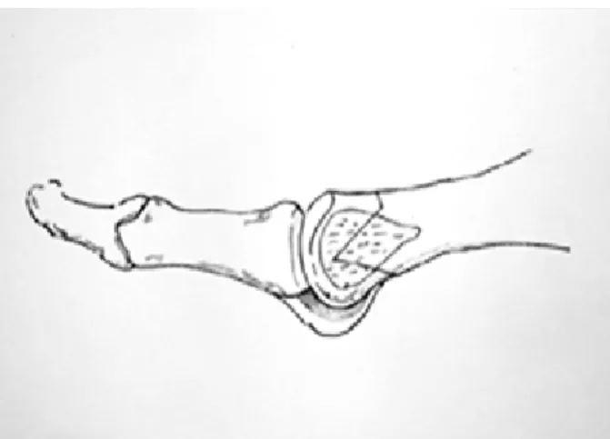
제1 중족골과 중족지관절을 약 5 cm 정도 배내측으로 절 개한 후, 이 지역의 근피 신경(천비골신경)의 감각 신경 분지 가 다치지 않게 조심해서 이 신경과 그 혈관속, 장족무지 신 건을 외측으로 분리하고 피낭 구조를 노출시킨 후 그 기저부 를 원위 지골에 둔 채 V자형의 피판을 만들었고, 중족골의 두부를 노출 시키고 oscillating saw를 이용하여 건막류를 제거 하였다. 경부 바로 가까이에서 중족골의 장축에 수직으 로 2mm 깊이의 가로로 절골술을 시행하였다(Fig. 1).
V자형의 정점은 중족골 두부 중심부의 원위부까지, 상지 정점으로부터 중족골 경부의 배부 바로 아래 2 mm까지, 하 지는 중족골 경부 아랫 부분까지 연장되었고, 상지과 하지의 적정한 각도는 60°였다.
절골술은 oscillating saw를 이용하여 행해졌으며 근위 절편은 tower clip으로 고정하고 원위 절편은 외측으로 전위
Figure 1. A transverse osteotomy just proximal to the neck made with depth of 2 mm perpendicular to long axis of the metatarsal (Lateral view).
Figure 2. The osteotomy made with an osillating saw, the distal fragment displaced laterally by digital pressure with the proximal fragment grasped with tower clip (apico-obliue view).
Figure 3. Operative photograph of the new modified chevron osteotomy for hallux valgus-distal fragment displaced laterally to the needed amount of correction.
Figure 4. Preoperative, postoperative, 12 months follow-up radiograph of hallux valgus corrected with new modified chevron osteotomy. (A) Preoperative standing radiograph shows moderate hallux valgus deformity and 1st metatarsal length is 56.6 mm. (B) Postoperative standing radiograph shows new modified chevron osteotomy and length kept the same. (C) 12 months postoperative standing radiograph shows well stabilized correction of hallux valgus and no significant shortening of 1st metatarsal length of 56.6cm from preoperative state.
preop. post op. (12 mo) Hallux valgus angle
Intermetatarsal angle metatarsal length(mm)
34.1°
11.1°
56.6
14.4°
7.9°
56.2 Table 1. Radiographic (objective) results (degree) 되었고 그 측방 전위 정도는 교정이 필요한 정도에 따라 결
정하였다(Fig. 2).
이 새로운 절골술은 원위 절편을 쉽게 그리고 각 형성 없 이 정확히 전위시킬 수 있었고, 다루기 편하다는 장점이 있 으며 이 술식을 통해 훨씬 효율적이고 정확하게 교정이 이루 어지고, 종래 chevron 술식보다 전위 후 절골 부위가 더욱 안정적이었다(Fig. 3).
골간에서부터 측면으로 튀어나온 부위를 톱을 이용하여 제거하였으며 피낭 피판을 구멍을 통해 통과 시킨 후 단순 봉 합하였다. 봉합은 일반적인 방법으로 이루어졌으며 발은 압 박 창상처치를 시행한 후 단 하지 석고부목을 착용하였다.
결 과
술 후 평균 3년(범위, 2~5년)추시한 임상적 결과로서 모 든 환자들은 새로운 절골술에 의하여 미적 및 동통의 결과에 모두 만족하였으며 특히 전예에서 술 후 동통의 소실이 있었 다. 39명의 환자중 37명이(53예) 동통이 전혀 없었으며 좋은 미적결과를 보여 수술 결과에 아주 만족하였다. 2명은 가끔 약간의 동통을 호소하였으나 참을 수 있는 정도이었다.
새로운 변형된 절골술은 절골부위의 각형성 또는 단축이 예 방되었으며, 변형의 교정후 절골부위가 기존의 절골술 보다 안정적이었으며, 보다 효과적이고 정확한 교정이 가능하였다.
Hallux-metatarso-phalangeal-interphalangeal scale8)에 따른 평균 점수는 93.2점(범위 78~100점)이었다.
방사선적 결과로 술 전 중족골간각은 평균 14.4도(범위 11~23도)에서 술 후 평균 7.9도(범위 5~14도)로 줄어들어 평균 6.5도의 교정정도를 보였으며, 술 전 무지 외반각은 평 균 34.1도에서(범위 16~48도), 술 후 평균 11.1도로 평균 23 도의 교정정도를 보였다. 또한 중족골의 단축 여부를 확인하 기 위하여 수술 전 및 수술 후 방사선 사진을 통해 종족골 길 이를 측정한 결과 술 전 56.6 mm, 술 후 12 개월 후 56.2 mm로 길이의 차이는 없었다(Table 1)(Fig. 4A, Fig 4B, Fig. 4C).
중족골 두부의 골 괴사는 3년간의 추적관찰 중 나타나지 않았다. 모든 예에서 그밖에 특별한 합병증은 없었다.
고 찰
증상이 있는 무지 외반증 치료에 건막류 절제술과 연부 조 직 재건을 동반한 많은 종류의 제1중족골 골절술이 시행되었 으나 수술 방법을 선택하는 것은 매우 어렵다12,14).
다양한 수술법에 대한 검토가 가능하지만 실제로 수술법 의 선택은 수술의가 수련 배경과 임상 경험을 토대로 하기 마 련이다.
여러 골절술의 변형술이 고안되었고 Mitchell이 행한 원위 골절술이 가장 대중적이지만, 51예중 17예에서 제2종족골두 에 족저피부경절이 발생하였다2,13). Klosok 등9)은 무지외반 증에 대한 치료로 chevron 술식과 Wilson 술식을 비교하여 Wilson 술식이 더 나은 기능적 결과를 보인다고 발표하였다.
Corless4)와 Johnson 등7)은Mitchell 절골술의 기술적인 어려움과 함께 절골부의 불안정성으로 인한 전이, 배측각 변 형, 불유합, 무혈성 괴사, 제 1족지의 단축 및 관절 강직이 발생하는 등 문제점을 지적하고, 이러한 단점을 보완하기 위 하여 Mitchell 절골술의 원리를 이용한 chevron 절골술을 시행하여 좋은 결과를 얻었다고 보고하였다. Donnelly 등7) 은 절골각의 변화 및 screw 고정을 부가한 변형된 chevron 절골술(modified chevron osteotomy)로 무지외반각 35도, 제 1-2중족골간각 15도까지의 무지외반증에까지 적용범위 를 넓혔다. 그러나 변형된 chevron 술식은 전통적인 절골술 과 유사한 해부학적인 교정을 보이면서도 보다 안정적 이었 고10), Chevron절골술이 경증과 중증의 무지외반증 치료에 믿을만한 방법18)이라고 하였으나 술 후 합병증으로 골절편의 전위, 후면 각 형성, 불유합, 무혈성 괴사, 무지외반변형의 재발 등이 동반될수 있다. Trnka 등18)은 4명의 환자가 chevron 술식을 이용한 절골 부위에서 술 중 불안정성을 보 였다고 발표하였다. 기저부를 통한 중족골 근위부 골절술이 Loison11), Balaceson1), Trethowan 등17)에 의해 기술된 적 이 있다. 많은 저자들이 중족골 골간 골절술을 선호하였고 이것은 Wilson 술식으로 대표된다13).
저자들의 새로운 chevron 절골술식은 중족골 두부를 내측 각형성 없이 외측으로 전위 시켜, 후면 각 형성이 없을 뿐 아 니라 내측 또는 외측 기울임, 절골술 시행 부위에서의 골 단 축 등이 발생하지 않아, 저자들은 이 새로운 변형 술식이 절 골 부위에서 더욱 안정적이며 종전의 방법보다 훨씬 정확한 교정이 가능하다고 생각되었다. 또한 이번 연구에서 제1 중 족골의 유의한 단축이 나타나지 않음이 입증되었고, 혈전 정 맥염이나 절단 부위의 불유합, 중족골두의 무혈성괴사 등의 합병증이 없었으며 술 후 환자들은 6~8주 내에 일상생활로 복귀할 수 있었다.
결 론
술 후 평균 3년간 추시한 39명의 환자에게 55예의 수술이 시행되었고, 모든 예에서 동통이 만족스럽게 경감되었으며 미적 결과에 대해서도 만족하였다.
술후 제1,2중족골간각은 평균 5.5도, 무지 외반각은 평균 23도 교정되었으며, 교정이 되지 않은 경우는 없었으며, 이 새로운 술식은 변형의 교정후 절골부위가 기존의 chevron 절골술 보다 더욱 안정적 이었으며, 정확하고 효과적인 변형 의 교정이 가능하여 권장할 수술기법으로 사료된다.
REFERENCES
1) Balacesce J: Un caz de hallux valgus simetric. Rev. Chir, 7: 128-135, 1903.
2) Canale PB, Aronsson DD, Lamont RL and Manoli A:
2nd The Mitchell procedure for the treatment of adolescent hallux valgus. J Bone and Joint Surg, 75-A: 1610-1617, 1993.
3) Carr CR and Body BM: Correctional osteotomy for metatarsus primus varus and hallux valgus. J Bone and Joint Surg. 59-A: 1353-1367, 1968.
4) Coreless JR: A Modification of Mitchell procedure. J Bone and Joint Surg, 58B: 136-138, 1976.
5) Donnelly RE, Saltzman CL, Kile TA and Johnson KA:
Modified chevron osteotomy for hallux valgus. Foot Ankle Int. 15(12): 642-645, 1994.
6) Harold BK and Gary LP: Arthrodesis versus resection arthroplasty for failed hallux vallgus operation. Clin orthop, 347: 208-214, 1998.
7) Johnson KA, Cofield RH and Morrey BF: Chevron osteotomy for hallux valgus. Clin Orthop, 142: 44-47, 1979.
8) Kitaoka HB, Alexander IJ, Adelaar RS, Nunlay JN, Myerson MD and Sanders M: Clinical rating systems for the Ankle-hindfoot, midfoot, hallux and lesser. Foot Ankle, 15: 349-353, 1994.
9) Klosok JK, Pring DJ and Jessop JH: Chevron or Wilson metatarsal osteomomy for hallux valgus. A prospective randomized trial. J Bone and Joint Surg, 75-B: 825-829, 1993.
10) Lewis RJ and Feffer HL: Modified chevron osteotomy of the first metatarsal. Clinc. Orthop, 157: 105-109, 1981.
11) Loison M: Note sur lee traitement chirurgicalee du hallux valgus d apres letude radio-graphique de la deformation.
Bull. Soc. Chir. paris 27: 528, 1901.
12) Mann RA, Rudicel S and Graves SC: Repair of hallux valgus with a distal soft-tissue procedure and proximal metatarsal osteotomy. J Bone and Joint Surg, 74-A:
124-129, 1992.
13) Mitchell CL, Fleming JL, Allen R, Glenney C and Sanford GA: Osteotomy-Bunionectomy for hallux valgus.
J Bone and Joint Surg, 40-A: 41-60, 1958.
14) Okuda R, Kinoshita M, Morikawa J, Jotoke T and Abe M: Distal soft tissue procedure andproximal metatarsal osteotomy in hallux valgus. Clinc. Orthop, 379: 209-217, 2000.
15) Piggott H: The nature history of hallux valgus in adolescence and early adult life. J Bone and Joint Surg, 42-A: 749-760, 1960.
16) Saltzman CL, Brandser EA, Berbaum KS, et al:
Reliability of standard foot radiographic meas urement.
Foot Ankle, 15: 661-665, 1994.
17) Trethowan J: Hallux valgus. In: Choye CC ed. Systems of Surgery. New York, PB Hoeber : 1046-1049, 1923.
180 Trnka HJ, Zembsch A, Easley ME, Salzer M, Ritschl P and Myerson MS: The Chevron osteotomy for correction of hallux valgus. J Bone and Joint Surg, 82-A: 1373-1378, 2000.
19) Wilson CL: A method of fusion of the metatarso- phalangeal joint of the great toe. J Bone and Joint Surg, 40-A: 384-385, 1958.