서 론
포상 연부 육종(Alveolar Soft Part Sarcoma) 은 1 9 5 2년 C h r i s t o p e r s o n에 의해 처음 보고되었으 며3 ), 그 희귀성으로 인해 종종 다른 양성 및 악성 종 양과 감별을 요한다. 비교적 젊은층에서 호발하며 성장 속도가 느리지만, 발견 당시 종종 전이성 병변 이 동반되어 예후가 불량하다8 ). 저자들은 골반골에 서 원발성으로 발생해 다발성 골 및 뇌에 전이된 1 예와 하퇴부에 원발성으로 발생한 1예에 대한 치료 를 경험하였기에 그 희귀성에 비추어 문헌과 함께 보고하고자 한다.
증 례 증례 1
3 5세 여자 환자로 넘어진 후 발생한 우측 고관절 동통 및 운동 범위 제한을 주소로 내원하였으며, 과 거력상 5개월 전 우측 골반 부위의 골종양 발견 후 타병원에서 항암 및 방사선 치료를 받은 적이 있었 다(Fig. 1A). 신체 검사상 우측 고관절에 압통을 동반한 운동 범위 제한과 우측 골반골 부위에 종괴 가 촉지되었다.
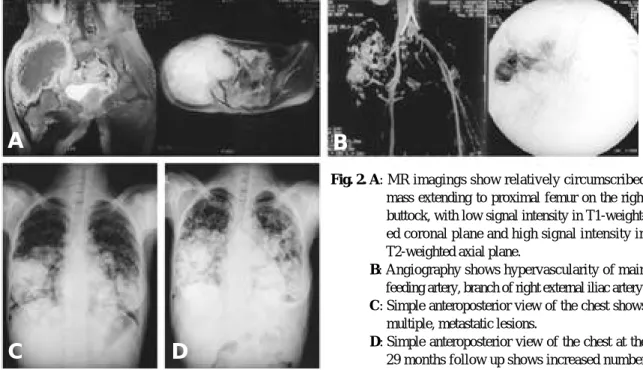
단순 방사선 사진상 우측 대퇴골 전자하부에 골 파괴와 병적 골절 및 비구 상방의 장골에 골 흡수 및 종괴 음영 소견을 보였으며(Fig. 1B), 흉부에 전이 성 다발성 폐종양 음영 소견 보였다(Fig. 2C). 자기
※통신저자: 정 성 택
광주시 동구 학동 8번지
전남대학교 의과대학 정형외과학교실
Tel: 062) 227-1640, Fax: 062) 225-7794, E-mail: [email protected]
포상 연부 육종
- 2례 보고 -
전남대학교 의과대학 정형외과학교실
정성택・서형연・신상규・박용철
포상 연부 육종(Alveolar soft part sarcoma)은 성인에서 하지 대퇴부, 소아에서 두경부 에 주로 발생한 것으로 알려져 있고, 비교적 천천히 성장함에도 불구하고 전이된 후에 발견된 경우가 많아 예후는 좋지 않은 매우 드문 종양이다. 조직학적으로 가포상( p s e u d o a l v e o l a r p a t t e r n )의 종양 세포들이 특징적으로 관찰되며 폐, 뇌, 골격 순으로 전이된다. 저자들은 비 교적 드문 부위인 골반골에서 발생해 뇌에 전이된 1예와 하퇴부에 원발성으로 발생한 1예에 대한 치료를 경험하였기에 그 희귀성에 비추어 문헌과 함께 보고하고자 한다.
색인 단어: 포상 연부 육종, 뇌 전이 Volume 9, Number 2, December, 2003
Fig. 1. A : Initial roentgenogram shows osteolytic lesion on the right ilium and huge soft mass on the right buttock. . Fig. 1.B : Preoperative roentgenogram shows pathologic fracture on the subtrochanteric region and remaining mass
and osteolytic lesion on right ilium.
A B
Fig. 2. A: MR imagings show relatively circumscribed mass extending to proximal femur on the right buttock, with low signal intensity in T1-weight- ed coronal plane and high signal intensity in T2-weighted axial plane.
Fig. 1.B: Angiography shows hypervascularity of main feeding artery, branch of right external iliac artery.
Fig. 1.C: Simple anteroposterior view of the chest shows multiple, metastatic lesions.
Fig. 1.D: Simple anteroposterior view of the chest at the 29 months follow up shows increased number
A
C D
B
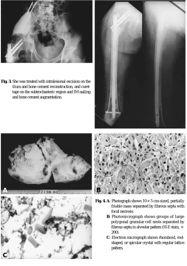
Fig. 3. She was treated with intralesional excision on the ilium and bone cement reconstruction, and curet- tage on the subtrochanteric region and IM nailing and bone cement augmentation.
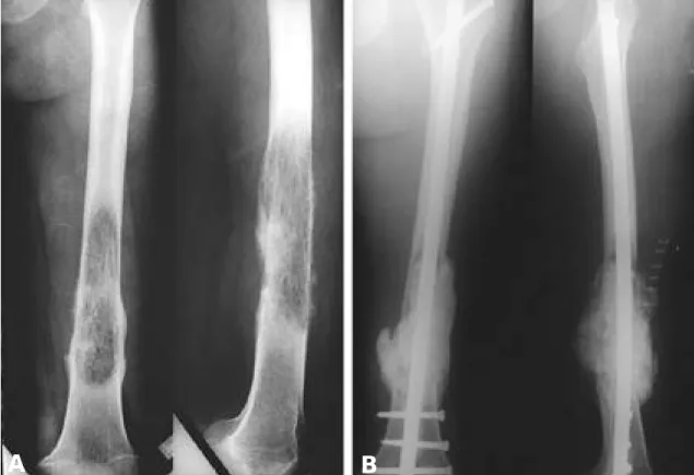
Fig. 4. A: Photograph shows 10×5 cm-sized, partially friable mass separated by fibrous septa with focal necrosis.
Fig. 1.B: Photomicrograph shows groups of large polygonal granular cell nests separated by fibrous septa in alveolar pattern (H-E stain, × 200).
Fig. 1.C: Electron micrograph shows rhomboid, rod- shaped, or spicular crystal with regular lattice pattern.
A
C
B
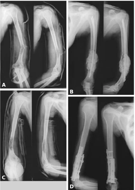
정 및 골시멘트를 이용한 보강술을 시행하였다( F i g . 6A, B). 술후 12, 13개월째 각각 좌측과 우측의 상 완골 원위부에 동일 병변 발견되어 좌측은 소파술 후 E n d e r정을 이용한 골수강내 고정술을 시행하였 으며, 우측은 소파술 후 이중 금속판을 이용한 내고 정 및 골시멘트를 이용한 보강술을 시행하였다( F i g . 7A, B, C, D).
Fig. 6. A : At postoperative 9 months, permeative destructive lesion on the left femur.
A B
Fig. 5. Brain CT at postoperative 3 weeks shows oste- olytic lesion and soft mass on left occipital region.
Fig. 7.A : At postoperative 12 months, pathologic fracture on the left distal humerus.
Fig. 1.B : Curettage, and IM nailing and bone cement augmentation was performed.
Fig. 1.C : At postoperative 13 months, pathologic fracture on the right distal humerus.
Fig. 1.D : Curettage, and dual plate fixation and bone cement augmentation was performed.
A
C
B
D
통성 종괴가 있었으며, 최근에 그 크기가 증가하는 소견을 보여 1개월 전에 타병원에서 신경초종의증으 로 진단 받고 종괴 절제술을 시행하였으며 조직 검 사상 포상 연부 육종 의심되어 전원되었다. 신체 검 사상 좌측 하퇴부에 동통이나 압통을 보이지 않는 부드러운 종괴가 촉지되었다.
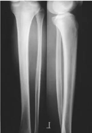
Fig. 8. Preoperative roentgenogram shows no specific finding on left calf region.
Fig. 9. T2-weighted MR image demonstrates ill-defined high attenuated lesion in posterolateral aspect of upper calf region.
Fig. 10. Postoperative roentgenogram shows wide mar- ginal excision including lateral gastrocnemius
의 경계가 불분명한 종괴는 T1 강조 영상에서 감소 및 T2 강조 영상에서 증가된 신호 강도를 보였으며 조영제 주입후 이 부위는 조영 증강을 보였다( F i g .
9). 흉부 단층 촬영 및 골주사 검사상 특이 사항은 없었다. 신경증상은 없었으며, 단순 두부 방사선 소 견상 이상소견은 발견되지 않았다. 수술전 타병원에 서 얻은 조직 검사상 포상 연부 육종으로 진단하고, AJCC 분류상 IIIA 단계로 간주하고 수술하였다.
수술은 종괴에서 5 cm의 안정 변연을 두어 좌측 후외방의 가자미근 및 외측 비복근과 그 피부, 그리 고 비골을 포함하는 광범위 절제술을 시행하였다 (Fig. 10). 육안 소견상 불분명한 경계를 가진 섬유 조직내에 혈종으로 생각되는 물질이 관찰되었으며 (Fig. 11A, B), 조직학적으로 가성 포상 형태의 큰 종양 세포소가 엷은 내피막의 혈관에 의해 분리되어 있으며 종양 세포는 원형 및 다각형의 진하고 연한 음영을 보이고 호산구성의 세포질이 관찰되었다 (Fig. 12). 술후 방사선 치료를 시행하였으며, 17개 월째 국소 재발은 보이지 않으며 계속 외래 관찰 중 이다.
Fig. 11. A: Photograph shows wide marginal resection of the mass including lateral gastrocnemius and soleus muscle, fibula and skin.
Fig. 11.B: Cut surface of the mass shows ill-defined grayish brown tissue with focal necrosis and hemorrage.
A B
Fig. 12. Photomicrograph shows groups of large polyg- onal granular cell nests separated by fibrous septa in alveolar pattern(H-E stain, ×200).
세 이하 여자에서 높은 빈도를 보이며, 발생 부위는 대개 성인에서 주로 하지 대퇴부, 소아에서는 두경 부에 호발한다. 본 연구에서는 비교적 드문 지역인 골반부와 하퇴부에 1예씩 발생하였다.
주된 전이 부위는 폐, 골격, 뇌 등이며1 , 5 , 1 5 , 2 0 ), 특 히 이 종양은 다른 악성 육종보다 더 뇌에 전이를 잘 하는 것으로 알려져 있다1 8 , 2 1 ). 따라서 폐 전이된 경 우나, 신경학적인 증상이 있는 경우에는 뇌 단층 촬 영 등 전이를 진단하기 위한 영상 검사를 시행하여 야 한다. 본 연구에서는 신경학적인 증상이 있는 전 이성 뇌병변에 대해서 절제술을 시행하였다. 포상 연부 육종의 경우 비교적 젊은 연령에 발생하고, 생 존율이 양호함을 고려시 전이성 병변에 대해서도 적 극적인 치료를 시행하는 것이 잔여 삶동안 환자의 만족감을 높일 수 있다고 생각된다.
임상 양상은 천천히 자라는 무통성 종괴가 주증상 이고 증상이 경미하여 쉽게 간과하는 경우가 많으며 폐나 뇌에 전이된 후에 병원을 찾는 경우가 많다8 ). 그러나 골을 침범시 약화에 의한 동통이 발생할 가 능성이 있다.
방사선학적으로 골에 침범한 경우 골 용해( o s t e- olytic) 및 침투상( p e r m e a t i v e )으로 나타나며 섬유 육종, 골모세포종, 거대세포종, 악성섬유성 이형성 증 등과 비슷한 소견을 보이며1 1 ), 자기 공명 영상의 T1 강조 영상에서 감소 및 T2 강조 영상에서 증가 된 신호 강도를 보이며 조영제 주입후 이 부위는 조 영 증강을 보인다1 9 ).
혈관 촬영상 혈관성이 풍부한 동정맥 문합( A V shunts) 및 소실 지연(delayed washout) 등의 소 견 보여 혈관 기형 및 혈관이 풍부한 연부 조직 종양
른다. 조직학적으로 악성 신세포암종, 부신결절종, 과립세포종, 폐포성 횡문근 육종 등과 유사하므로 환자의 임상증상, 방사선학적 검사와 조직학적 검 사, 특히 Periodic acid Schiff 염색 및 전자 현미 경 소견 등이 상기 질환과의 감별에 도움이 된다
3 , 4 , 5 , 1 5 , 1 6 ). 세포 유전학적 분석으로 1 7 q 2 5와 X p 1 1 . 2
에서 염색체의 재정열이 보고 되었으며1 4 ), 삼염색체 성(trisomy) 7, 8과 단일염색체성(monosomy) 18 등과 연관성도 보고되었다2 3 ). 추가적인 세포 유전학 적 분석 및 분자학적인 연구가 진행중이다.
치료는 원발 및 전이 병소의 근치 절제술이 주를 이루며, 불완전한 수술적 절제인 경우 방사선 치료 로써 국소적인 조절을 용이하게 할 수 있다1 , 4 , 5 , 2 2 ). 골 내 전이로 인한 골절의 경우 다른 골전이성 골종양 의 치료와 같이 소파술과 시멘트 보강술 및 내고정 술을 시행하였다. 증례 1의 경우 술후 즉각적인 안 정성은 물론 골절의 유합도 얻을 수 있었으며, 추시 상 골 병변의 국소 재발은 보이지 않았다.
화학 요법은 그 효과에 대해 연구가 더 필요한 실 정이다.
예후 인자로는 종양의 절제 가능성이 가장 중요하 며, 그 외에 진단시 전이 유무, 종양의 크기 등이 있 으며1 ), 특히 소아에서는 두경부에 호발해 조기 발견 되며 크기가 더 작고 절제가 용이해 성인보다 예후 가 좋다2 ). 5년 생존율은 Portera 등2 0 )은 발견당시 국소적인 형태는 8 7 %이나 단발성 전이가 존재하는 경우 2 0 %로 보고하였다. 그러나 다발성 전이를 보 인 경우 평균 생존 기간이 4 0개월로 비교적 길었다.
본 연구에서는 발견 당시 전이가 없었던 1례의 경우 광범위 절제술후 2년 추시상 국소 및 전신의 재발
소견은 없었다. 진단 당시 이미 다발성 폐 전이가 있 었던 1례의 경우 최초 수술후 뇌 전이까지 발생하였 으나 뇌의 종괴 및 골격의 다발성 병적 골절에 대해 수술적 치료 후 증상 호전 및 2 4개월의 추시기간 중 생존 중이다. 따라서 진단시 전이가 있어도 생존 기 간이 비교적 길기 때문에 보다 적극적인 접근으로 환자의 삶의 질을 유지할 수 있다고 생각된다.
REFEREMCES
01) Baum ES, Fickensher L, Nachman JB : Pulmonary resection and chemotherapy for metasta- tic alveolar soft part sarcoma. C a n c e r 4 7 : 1 9 4 6 , 1981.
02) Casanova M, A Ferrari A, Bisogno G et al . : Alveolar soft part sarcoma in children and adoles- cent: A case report from the soft-tissue sarcoma Italian cooperative group. Annals of Oncology, 11:1445-1449, 2000.
03) Christopherson WM, Foote FW and Stewart FW: Alveolar soft part sarcoma. Structurally char- acteristic tumors of uncertain histogenesis. C a n c e r 5:100, 1952.
04) Ekfors TO, Kalimo H, Rantakokko V: Alveolar soft-part sarcoma. A report to two cases with some histochemical and ultratructural observations.
Cancer 43:1672, 1979.
05) Enzinger FM, Weiss SW: Soft tissue tumors. 3rd edition. St Louis: CV Mosby. 1067-1074, 1995.
06) Farquharson M : Alveolar soft part sarcoma.
British Med J, 2:1068-1069, 1960.
07) Fender FA: Liposarcoma. Report of a case with intracranial metastasis. Am J Pathol 9:909, 1993.
08) Franz ME and Sharron WW: Soft tissue tumors.
The CV Mosby company 780, 1983.
09) Horn RC and Stout AP: Granular cell myoblas- toma. Surg. Gynecol Obstet, 76: 315, 1943.
10) Johnson RWP and Somerville PG: A malignant soft-tissue paraganglioma of the leg. J Pathol Bacteriol, 86:169, 1963.
11) Kim KS, Ko SH, Kim KJ et al.: Alveolar soft part
sarcoma with metastasis to bone. J Korean Orthop Assoc, 29:336-341, 1994.
12) Klemperter P: Myoblastoma of the striated mus- cle. Am J Cancer, 20:324, 1934.
13) Kolodny A: Angioendoepitheliuma of bone. A r c h Surg, 12:854, 1926.
14) Lattes R: Alveolar soft-part sarcoma; Tumors of the Soft Tissue 1:251, 1981.
15) Leeberman PH, Brennan MF and Kimmel M, et a l.: Alveolar soft part sarcoma. C a n e r, 63:1-13, 1989.
16) Ladanyi M, Lui MY, Antonescu CR et al.: The der(17)t(X;17)(p11;q25) of human alveolar soft part sarcoma fuses the TFE3 transcription factor gene to ASPL, a novel gene at 17q25. Oncogene, 20:48-57, 2001.
17) Macfarlane A, Macgregor AB: Malignant non- chromaffin paraganglioma of the thigh. Arch. Dis.
Child. 33:55, 1958.
18) Ogose A, Moritis T, Kobayashi H et al.: Brain metastases in musculoskeletal sarcomas. Jpn J Clin Oncol, 29(5):245-247, 1999.
19) Pang LM, Roebuck DJ, Griffith JF et al.: Aleolar soft-part sarcoma: a rare soft-tissue malignancy with distinctive clinical and radiological features.
Pediatr Radiol, 31:196-199, 2001.
20) Portera CA, Viet H, Patel SR et al.: Aleolar soft part sarcoma. Clinical course and patterns of metas- tasis in 70 patients treated at a single institution.
American cancer Society 585-591, 2001.
21) Salvati M, Cervoni L, Caruso R et al.: Sarcoma metastatic to the brain: A series of 15 cases. S u r g Neurol, 49:441-444, 1998.
22) Stein JR: Alveolar soft-part sarcoma. J Bone Joint Surg, 38-A:1126, 1956.
23) Tornoczky T, Kalman E, Sapi Z et al . : Cytogenetic abnormalities of alveolar soft-part sar- comas using interphase fluorescent in situ hybridization: tirsomy for chromosome 7 and monosomy for chromosomes 8 and 18 seem to be characteristic of the tumor. Virchows Arch , 438:173-180, 2001.
24) Willis RA : Pathology of tumors. 4th ed, Butterworth and Co., 890-893, 1967.
thigh in adults. It is very rare tumor that has poor prognosis due to its late detection after distant metastasis in spite of its slow growth rate. It is histologically characterized by pseudoalveolar pattern tumor cells. And metastasis usually occur in the site of lung, brain and skeleton in order lately.
We have managed two cases of the sarcoma, one which took place in relatively rare part, pelvic bone and has spread to the brain, the other which primarily occured in the calf. For its varity, we report these two cases with reviewing of the literatures.
Key Words: Alveolar soft part sarcoma, Brain metastasis
Address reprint requests to Sung-Taek Jung,M.D.
Department of Orthopedics, Chonnam National University Hospital 8 Hak-dong, Dong-gu, Gwangju, 501-757, Korea
TEL: 82-62-227-1640, FAX: 82-62-225-7794, E-mail: [email protected]