Vol. 2, No. 2, pp 59~63, 2008
Received Jul 14, 2008; 1st revised Oct 21, 2008; 2nd revised Nov 5, 2008; accepted Nov 6, 2008 Corresponding author:Myun-Whan Ahn, MD
Department of Orthopedic surgery, College of Medicine, Yeungnam University 317-1, Daemyungdong, Namgu, Daegu, 705-717, Korea
Tel: +82-53-620-3640, Fax: +82-53-628-4020, E-mail: [email protected]
Effect of Bone Marrow Cell Collection Techniques and Donor Site Locations on In-vitro Growth of Bone Forming Cells
Sung Min Chung#, Eun-Bok Kim$, So-Young Park$, Min Chul Sung#, Hyun Kook Youn#, and Myun-Whan Ahn#
Department of Orthopedic surgery, College of Medicine, Yeugnam University#, Department of Physiology, College of Medicine, Yeugnam University$ P
Puurrppoossee:: This study evaluated the influence of bone marrow cell collection techniques and donor site locations on the in- vitro growth of bone-forming cells.
M
Maatteerriiaallss && MMeetthhooddss:: Sixty six samples of bone marrow cell collections (BMCC) or bone marrow aspirates (BMA) from 15 patients were obtained. Thirty eight samples for culture were composed of 23 BMA from 7 tibial condyles and 16 ilia, with the other 15 BMCC from the contralateral ilia. The other 28 samples were used for the analysis of alkaline phosphatase activities. After counting total cell number, mesenchymal stem cells (MSC) obtained from samples were incubated for 14 days. Alkaline phosphatase staining was used to count the number of stained colonies to show osteogenic differentiation.
R
Reessuullttss:: The average MSC counts of BMA from tibial condyles and ilia were 1.42×10⁶and 7.35×10⁶respectively, with 4.80
×106from ilial BMCC (p=0.010). MSC cultures could not be produced from tibial condyles in all 7 samples. However, 9 of 15 BMCC samples and 9 of 16 ilial BMA samples were successfully cultured (p=0.018). The average of cell counts in the suc- cessful cultures was 7.92×10⁶, whereas that in the failed cultures was 2.85×10⁶(p=0.000). Multiple regression analysis showed that colony count was associated with the patient’s age and total cell numbers, but not with collection methods such as BMCC or BMA (p=0.000, R=0.648, beta; age=-0.405, cell number=0.356). The discriminating formula indicated that more than 5.25×10⁶cells were needed for successful culture.
C
Coonncclluussiioonn:: For successful cultures in vitro and for grafts, the total number of collected bone forming cells is more impor- tant than donor sites or collection methods. For young patients, grafting of bone-marrow-derived osteoprogenitor cells is promising.
Keywords: Bone marrow cells, Bone marrow collection, Bone marrow aspiration
INTRODUCTION
New minimally invasive techniques for collecting bone marrow derived cells from various donor sites1,2,3,4, such as metaphysis of long bones, allow for in vitro and in vivo bone formation. Aspiration volume from the bone marrow5, harvesting technique6,7,8, donor site location,9and the age- related decline10,11,12,13,14 can affect the in vitro growth of
osteoprogenitor cells. Although bone marrow aspirate is the most common, bone marrow cell collection from the ground bone sludge and reaming debris of the long bone can also provide vital mesenchymal stem cells15. Reaming debris may also be an alternative to bone tissue grafting.
Here, we clarified the influence of bone marrow collec- tion techniques and donor site locations on the growth of osteoprogenitor cells in vitro.
MATERIALS AND METHODS
Fifteen patients who underwent spinal fusion using auto- genous iliac bone grafts were recruited for the study. Ten patients were male and 5 were female. The mean age (±
SE) at the time of surgery was 43±20 years old (range, 13 to 70 years old). Bone marrow cell collections (BMCC) were obtained from bone marrow sludge during harvesting of bone grafts in group I. Bone marrow aspirates (BMA) were obtained percutaneously from the iliac crest on the contralateral side in group II and from the proximal metaph- ysis of the tibia in group III. Sixty six samples of BMCC or BMA were obtained. Thirty eight samples for culture were composed of 23 BMA from 7 tibial condyles and 16 ilia, with the other 15 BMCC from the contralateral ilia. The other 28 samples were used for the analysis of alkaline phosphatase activity. Five ml of each sample was obtained using a 5 cc syringe containing heparin solution and a bone marrow aspiration needle. Two ml of the heparinized bone marrow suspension was used for the study on colony forma- tion containing alkaline phosphatase staining and 2 ml for the alkaline phosphatase activity assay. For culture of bone marrow derived cells16, 2 ml of each bone-marrow suspen- sion was mixed with two volumes of saline and one volume of Ficoll and centrifuged at 1,500 rpm for 10 minutes. The buffy coat was isolated and washed with two volumes of saline. After calculating the total number of cells using a hemocytometer, each sample was plated in a 100-mm-diam- eter dish. Cells were incubated in 8 ml Dulbecco’s Modified Eagle Medium (DMEM) (GIBCO, Carlsbad, CA, USA) containing 10% fetal bovine serum (GIBCO), 10 nM dex- amethasone, and 1% antibiotic/antimycotic (GIBCO) at 37�C, 95% O2and 5% CO2. Culture media were changed on day 7 and then every 3 days for 1 week. On day 14, the total num- ber of colonies stained with alkaline phosphatase was counted and alkaline phosphatase activity was measured in each culture dish. For alkaline phosphatase staining, the medium was removed and the cell layer was rinsed with phosphate buffered saline (PBS) two times. Cells were incubated with 2% paraformaldehyde for 30 minutes and then rinsed with PBS three times at 25�C. Cells were then incubated with 1.5 ml naphthol AS-BI alkaline solution (Sigma, St. Louis, MO, USA) with fast red violet LB for 15 minutes, and the total number of red alkaline phosphatase- positive colonies in each 100-mm dish was manually count- ed. For the alkaline phosphatase activity assay, the medium
was removed and the culture dish was rinsed with PBS three times. The cell layer was disrupted with 1 ml of 0.5%
Triton X in 25 mM glycine-NaOH and then 100 μL of solu- tion was transferred to a microcentrifuge tube for the assay.
Fifty microliter p-nitrophenyl phosphate (Sigma) was added to microcentrifuge tube, which was incubated for 30 min- utes at 37�C in 95% O2 and 5% CO2. The reaction was stopped with 50 μL of 2N NaOH and absorbance was read immediately after incubation at 405 nm in a plate reader.
The chi-square test, t-test and one-way ANOVA were used to confirm group differences. Multiple regression and discriminant analysis were used to extract factors associated with successful culture.
RESULTS
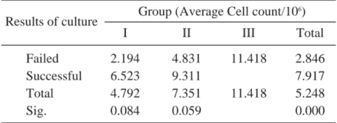
Average cell counts in group I, II and III were 4.80*106, 7.35*106, and 1.42*106, respectively (p=0.010) (Table 1), with a significant difference only between groups II and III.
MSC counts were variable within each group. BMCC cell counts from ilia in group I ranged from 150,000 to 13,120,000. BMA cell counts from ilia in group II ranged from 19,500 to 15,000,000, and those from tibial condyles in group III ranged from 13,000 to 5,920,000.
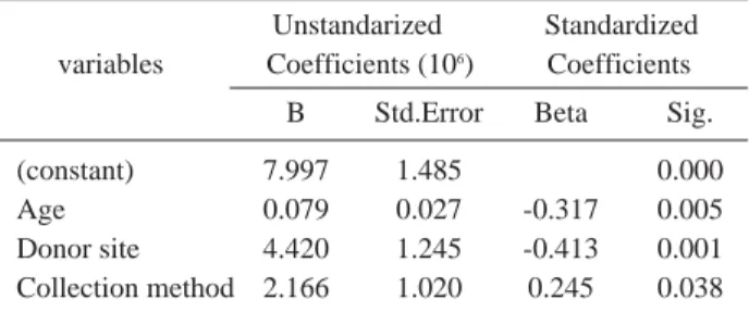
Multiple regression analysis suggested that the age, donor site location, and collection method would contribute to cell count values (Table 2). Cell counts were increased in younger patients, allowing for sufficient harvesting from the proximal metaphysis of the tibia. Percutaneous aspiration of bone marrow was more efficient than bone marrow cell col- lection from ground bone or bone sludge.
Six of 15 BMCC samples and 7 of 16 BMA samples from the ilium failed in culture, whereas all 7 samples of MSC
Table 1. Average numbers of cell count associated with the method of collection and donor site of bone marrow cells
Results of culture Group (Average Cell count/106)
I II III Total
Failed 2.194 4.831 11.418 2.846
Successful 6.523 9.311 7.917
Total 4.792 7.351 11.418 5.248
Sig. 0.084 0.059 0.000
Group I: Bone marrow collection (bone marrow sludge) from ilium
Group II: Bone marrow aspiration from contralateral ilium Group III: Bone marrow aspiaration from proximal tibia
aspirated from the tibial condyle failed in culture (p=0.018) (Table 3).
Average cell counts of successful and failed cultures were 7.92×10⁶and 2.85×10⁶, respectively (p=0.000). Average colony counts in group I and II in cases of successful cul-
ture were 210 and 184, respectively (p=0.761) (Table 4).
Multiple regression analysis indicated that colony count was associated with the patient's age and total cell numbers, but not with collection methods (p=0.000, R=0.648, beta;
age=-0.405, cell number=0.356) (Table 5). The discriminat- ing formula indicated that 5.25×10⁶or more cells were needed for successful culture (Table 6).
Average alkaline phosphatase activity in the cultured mesenchymal stem cells in group I, II, and III were 0.593, 0.862, and 0.087, respectively, with group III significantly lower than the other two groups. Multiple regression showed that alkaline phosphatase activity was related to cell count, but not age, donor site location, or collection method Table 2. Selected variables according to the result of stepwise
method of multiple regression-Dependent variable=cell count Unstandarized Standardized variables Coefficients (106) Coefficients
B Std.Error Beta Sig.
(constant) 7.997 1.485 0.000
Age 0.079 0.027 -0.317 0.005
Donor site 4.420 1.245 -0.413 0.001
Collection method 2.166 1.020 0.245 0.038 R=0.547 R2=0.300 p-value=0.000
Table 3. Results of culture associated with the method of col- lection and donor site of bone marrow cells
Culture Result of culture in relation to groups
I II III Total
Faile 16 17 7 20
Success 19 19 - 18
Total 15 16 7 38
p-value=0.018
Group I: Bone marrow collection (bone marrow sludge) from ilium
Group II: Bone marrow aspiration from contralateral ilium Group III: Bone marrow aspiaration from proximal tibia Table 4. Average numbers of alkaline-phosphatase-positive
colony count associated with the method of collection and donor site of bone marrow cells
Numbers of ALP - positive
Group colony count
I II Total
Mean 210 184 197
SE 169 147 141
N 19 119 118
p-value=0.761
Table 5. Selected variables according to the result of stepwise method of multiple regression- Dependent
variable=numbers of count of colony
Unstandarized Standardized Variable Coefficients Coefficients B (Std.Error) Beta Sig.
(constant) 199.121 76.894 0.014
Age -3.574 1.268 -0.405 0.008
Cell count 1.188E-05 4.795E-06 0.356 0.018 R=0.648 R2=0.420 p-value=0.000
Table 6. Results of discriminant analysis-Dependent variable= successful culture
Canonical Discriminant Function Coefficients Function=1
Cell count 2.57355E-07
(Constant) -1.350631485
* Successful culture : 5.25×10⁶of cell numbers or more, base on calculation of the discriminating formula
Table 7. Average numbers of alkaline phosphatase activity associated with the method of collection and donor site of bone marrow cells
Group ALP-positive Activity (absorbance)
I II III Total
Mean 0.593 0.862 0.088 0.610
SE 0.273 0.275 0.005 0.161
N 9 13 6 28
p-value=0.187
Table 8. Selected variables according to the result of stepwise method of multiple regression-Dependent
variable=Activity of alkaline phosphatase
Unstandarized Coefficients Standardized
variables Coefficients
B Std.Error Beta
(constant) -0.043 0.180
Cell count 1.541E-07 3.180E-08 0.689 R=0.688 R2=0.474 p-value=0.000
(Tables 7 and 8).
DISCUSSION
To avoid donor site morbidity, minimal invasive percuta- neous surgical techniques, bone substitutes, and induction materials are used. Bone marrow aspiration is the most common5, where bone marrow and marrow cells are collect- ed using a bone drill or biopsy trephine7to supply bone forming cells to the nonunion site of the long bone and the bone substitute. Reaming debris of the long bone is an alter- native substitute for bone grafts, but the outcomes from this process are not clearly understood. The metaphysis of long bones, such as the tibia1and the radius and coronoid processes of the ulna can be local alternative sources near the lesion. Thus, donor site location9, harvesting tech- nique6,7,8 and age-related decline of osteoprogenitor cells11,12,13,14, as well as aspiration volume, can affect in vitro and in vivo growth of bone forming cells. The relationships between these factors are not well understood.
Here we show that total colony formation in vitro was related to the patient’s age and the total number of MSC obtained, with age more important in the regression analy- sis. In contrast, donor site location was more important than age or collection method for total cell count. Thus, the metaphysis of long bones is not a good source of stem cells, especially in elderly patients. The mean cell count (±SD) of all samples of patients younger than 20 years old was 7.497×10⁶(range, 2.260×10⁶to 13.600×10⁶) and all sam- ples except one were cultured successfully, meaning that younger patients have more abundant and more active mar- row cells. Thus, bone marrow derived osteoprogenitor cell grafts derived from young patients have greater potential for survival, with more cells needed in elderly donors.
The metaphysis of long bones are an alternative source of marrow cells. However, we found that all tibia samples failed in culture. Most also yielded low cell counts (less than 1.5×10⁶) and one sample with a total cell number of 5.920×10⁶still failed in culture. Thus, the ilium is consid- ered to be a better source for bone marrow derived stem cell.
Reamed bone debris and bone sludge seemed to be advantageous for in vivo growth because of induction mate- rials contained in the bone matrices, but they did not pro- vide sufficient stem cells for in vitro growth.
CONCLUSION
For successful in vitro culture and grafting, the total num- ber of collected bone forming cells is more important than donor site or collection method. In young patients, the graft of bone-marrow-derived osteoprogenitor cells is promising.
The ilium is the best source of osteoprogenitor cells, but future work may improve yields from long bones.
REFERENCES
01. Chen YC, Chen CH, Chen PL, Huang IY, Shen YS, Chen CM: Donor site morbidity after harvesting of proxi- mal tibia bone. Head Neck 2006; 28: 496-500.
02. Zhu SS, Hu J, Li N, Zhou HX, Luo E: Autogenous coro- noid process as anew donor source for reconstruction of mandibular condyle: an experimental study on goats. Oral Surg Oral Med Oral Pathol Oral Radiol Endod 2006; 101:
572-580.
03. Brutus JP, Loftus JB: Gerdy’s tubercle as a source of cancellous bone graft for Surgery of the upper extremity:
description of technique. J Hand Surg Am 2006; 31: 147- 149.
04. McLain RF, Fleming JE, Boehm CA, Muschler GF:
Aspiration of osteoprogenitor cells for augmenting spinal fusion: comparison of progenitor cell concentrations from the vertebral body and iliac crest. J Bone Joint Surg Am 2005; 87: 2655-2661.
05. Muschler GF, Boehm C, Easley K: Aspiration to obtain osteoblast progenitor cells from human bone marrow: the influence of aspiration volume. J Bone Joint Surg Am, 1997; 79: 1699-1709.
06. Brawley SC, Simpson RB: Results of an alternative auto- genous iliac crest bone graft harvest method. Orthopedics 2006; 29: 342-346.
07. Lakhey S, Shrestha BP, Pradhan RL, Pandey B, Rijal KP: Results of autogenous trephine biopsy needle bone grafting in fractures of radius and ulna. JNMA J Nepal Med Assoc 2005; 44: 84-86.
08. Khanna G, Lewonowski K, Wood KB: Initial results of anterior interbody fusion achieved with a less invasive bone harvesting technique. Spine 2006; 31: 111-114.
09. Pradel W, Tenbieg P, Lauer G: Influence of harvesting technique and donor site location on in vitro growth of osteoblastlike cells from facial bone. Oral Surg Oral Med
Oral Pathol Oral Radiol Endod 2006; 101: 285-290.
10. Quarto R, Thomas D, Liang CT: Bone progenitor cell deficits and the age associated decline in bone repair capac- ity. Calcif Tissue Int 1995; 56: 123-129.
11. Bak B, Andreassen TT: The effect of aging on fracture healing in the rat. Calcif Tissue Int 1989; 45: 292-297.
12. Irving JT, LeBolt SA, Schneider EL: Ectopic bone for- mation and aging, Clin Orthop Relat Res 1981; 154: 249- 253.
13. Nishimoto SK, Chang CH, Gendler E, Stryker WF, Nimni ME: The effect of aging on bone formation in rats:
biochemical and histological evidence for decreased bone
formation capacity. Calcif Tissue Int 1985; 37: 617-624.
14. Howes R, Bowness JM, Grotendorst GR, Martin GR, Reddi AH: Platelet-derived growth factor enhances dem- ineralized bone matrix-induced cartilage and bone forma- tion. Calcif Tissue Int 1988; 42: 34-38.
15. Trinkaus K, Wenisch S, Siemers C, Hose D, Schnettler R: Reaming debris: a source of vital cells! First results of human specimens. Unfallchirurg 2005; 108: 650-656.
16. Behrens P, Wolf E, Bruns J: In vitro culture of human autologous osteoblast cells on natural bone mineral.
Orthopade 2000; 29: 129-134.