대한관절경학회지 제14권 제1호 2010 Journal of Korean Arthroscopy Soc.
Volume 14, Number 1, February, 2010
증 례
— 25 — 급성 전방십자인대 파열 시 내측 반월상 연골의 손상은 약 50%에서 동반되며, 만성 전방 십자인대 손상 시는 반월상 연 골 손상 빈도가 80%까지 올라간다고 보고하고 있다5). 반월상 연골의 양동이 손잡이형 파열은 반월상 연골 파열의 10% 정 도의 빈도를 나타내고 있으며4), 본 파열의 유형에서 약 10~15%에서 봉합이 가능하다고 보고 된다6). 더욱이 동일 슬 관절에 발생하는 내외측 반월상 연골의 양동이 손잡이형 파 열은 보고가 드물며2,3,9), 저자들의 검색으로는 최초의 국내 보 고로 사료된다. 이에 저자들이 경험한 내외측 반월상 연골에 동시에 발생한 양동이 손잡이형 파열의 임상 소견 및 방사선 소견을 문헌 고찰과 함께 보고하고자 한다.
증례 보고
26세 남자환자로 1개월 전부터 발생한 우측 슬관절 동통과 잦은 잠김 현상을 호소하며 내원하였다. 환자는 계단 오르다 가 회전력에 의한 손상을 내원 전 5개월 전 및 1달 전에 입었 으며, 5 개월 전 개인 방사선과병원 에서 MRI 검사(Fig. 1) 를 시행하였으나 증상이 경미하여 특별한 치료는 시행하지 않았다 본원 내원 시 이전 MRI를 재고한 결과 외측 반월상 연골의 양동이 손잡이형 파열 소견을 보였다(Fig. 1). 과거력 상 5년전 우측 슬관절 전방십자인대 대퇴 부착부 파열로 타 병원에서 봉합술 시행 받았다. 이학적 소견상 대퇴 사두근 위 축 건측에 비해 2 cm 정도였다. McMurray 검사상 내외측 반월상 연골 모두에서 양성 소견을 보였으며 Lachman 검사 및 전방 전위 검사 모두 양성 소견이었다. 단순방사선 사진상 은 특이 소견 없었으며, MRI 검사상 전방십자인대 파열 소견 과 내, 외측 반월상 연골의 양동이 손잡이형 파열 소견(Fig.
2)이 관찰되었다. 관절경 검사 소견상 내측 반월상 연골은 원 위치로 정복된 상태였으나 탐식자로 전위 유발 시 중심부로 전위되는 소견을 보였다(Fig. 3 A).
내
내측 측 및 및 외 외측 측 반 반월 월상 상 연 연골 골에 에 동 동시 시 발 발생 생한 한 양 양동 동이 이 손 손잡 잡이 이형 형 파 파열 열 -
- 증 증례 례 보 보고 고 - -
서울보훈병원 정형외과
윤정로∙김택선∙양재혁∙강규복∙김영찬
Simultaneous Bucket-handle Tears of both Medial and Lateral Meniscus - A Case Report -
Jung-Ro Yoon, M.D., Taik-Sun Kim, M.D., Jai-Hyuk Yang, M.D., Kyu-Bok Kang, M.D., Young-Chan Kim M.D.
Department of Orthopaedic Surgery, Seoul Veterans Hospital, Seoul, Korea
Most of bucket handle meniscal tears are associated with anterior cruciate ligament (ACL) deficiency. Lateral meniscus lesions are more common with acute ACL deficiency, where medial meniscus lesions are more associated with chronic ACL deficiency. We reported an ACL deficient knee with bucket handle tears of medial and lateral meniscus of the same knee. The report suggests the need for increased awareness of the possible presence of this. Additionally, we discuss injury mechanism, clinical symptoms, specific signs on Magnetic Resonance Imaging (MRI), and treatment options.
KEY WORDS: Bucket handle tear, Medial and lateral menisci, Simultaneous
�Address reprint request to Kyu-Bok Kang, M.D.
Department of Orthopaedic Surgery, Seoul Veterans Hospital, 6-2 Dunchon-dong, Kangdong-gu, Seoul 134-060, Korea Tel: 82-2-2225-1352, Fax: 82-2-487-0754
E-mail: [email protected]
외측 반월상 연골은 양동이 손잡이형 파열로 중앙 전위 반 월상 연골이 뒤집혀있는 상태였다(Fig. 3 B). 전방십자인대 는 대퇴 부착부는 관찰되지 않았으며 잔여 인대가 후방 십자 인대의 활액막에 유착되어 있었다. 내,외측 반월상 연골에 대 하여는 봉합술을 시행하였다 외측 반월상 연골은 5개월 이상 경과된 파열이었으나 관절경하에서 중앙부로 전위된 단편의 회전방향을 저자들이 보고했던 방법을 이용 관찰하였고10), 이 를 정복 후 봉합할 수 있었다. 회전 방향은 외측 반월상 연골
의 경우 하방으로 회전을 시작하여 180도 회전 후 대퇴 과간 에 감돈 되어 있던 상태로 일반적인 전내측 도달법 외에 추가 로 내측부 인대 앞쪽에 근접한 전내측에 부도달법을 이용하 여 두개의 탐식자로 힘을 가하여 원래 위치로 정복할 수 있었 다. 내측 반월상 연골은 술 전 MRI상 양동이 손잡이형 파열 소견이 관찰 되었으나, 관절경 검사 시는 정복된 상태였다. 봉 합술은 내,외측 모두 3개의 inside-out 봉합술을 시행하였 고, suture hook (Linvatec, Largo FL, USA)에 No. 0
— 26 — 대한관절경학회지 제 14 권 제 1 호 2010년
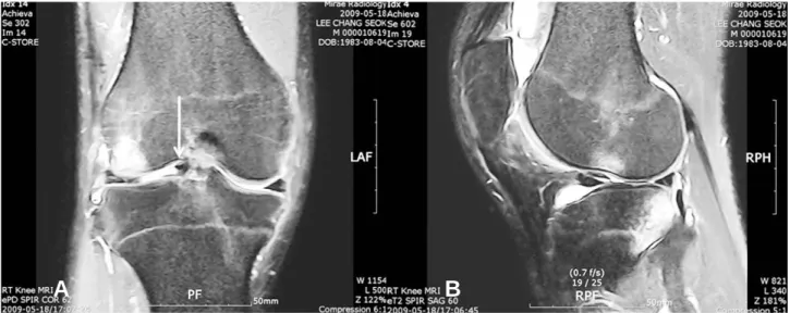
Fig. 2. Preoperative MR images. (A) Coronal T2-weighted MR image shows three structures in the intercondylar notch: PCL (whitish arrow) - normal posterior cruciate ligament; M (black arrows) - displaced bucket-handle fragment from medial and lateral meniscal tears. Arthroscopic findings showed that the anterior portion of the medial meniscus (MM) was continued to the ligament-like tissues which had covered the the anterior portion of the anterior cruciate ligament (ACL). (B) Sagittal T1- weighted MR image demonstrating a low-signal band anterior and parallel to the posterior cruciate ligament (double PCL sign). (C) Sagittal T1-weighted MR image through the lateral compartment shows flipped meniscus delta sign in the anterior horn of the lateral meniscus.
B C
A
Fig. 1. MR images, 5 months prior to a operation. (A) Coronal T2-weighted MR image revealing displaced central fragment of a lat- eral meniscus (whitish arrow). (B) Sagittal T2-weighted MR image through the lateral compartment shows flipped meniscus delta sign in the anterior horn of the lateral meniscus.
B
A
PDS (polydioxanone suture)를 넣어 2개의 all-inside 봉 합술을 병용하였다. 전방십자인대에 대해서는 이차적 수술을 계획하였으나 환자가 개인적인 문제로 미루고 있는 상태이 다. 술 후 1년 경과된 상태로 Lachman 검사 grade 2의 불안 정성이 있으나 관절운동제한 소견은 없으며 Lysholm knee score 78점, Tegner score 5 의 결과를 보였다.
고 찰
전방십자인대 파열과 반월상 연골의 파열은 흔히 동반되 나, 내외측 반월상 연골 동시에 파열이 동반되는 경우에 대해 서는 빈도에 대해서는 7%1), 20%8)등 다양하게 보고하고 있다 더우기 내외측 반월상 연골 동시에 발생한 양동이 손잡이형 파열 저자들의 검색상 3예가 보고 되고 있으며, 급성 전방 십 자인대와 동반된 경우가 2례2,9), 만성 전방 십자인대 손상과 동반되었던 경우 1례3)였다. 저자들의 경우는 만성 전방 십자 인대 부전에서 병발한 경우로 특이한 점은 내원 5개월전 외측 반월상 연골의 양동이 손잡이형 파열이 발생하였으며, 일상 생활에는 지장이 없는 상태로 지내다가, 3주전 내측 반월상 연골의 양동이 손잡이형 파열로 잠김 현상으로 내원하게 되 었다. 반월상 연골의 양동이 손잡이형 파열에서는 잠김 현상 이 흔한 임상 증상이지만 외측 반월상 연골에서는 이와 같은 증상 없이 양동이 손잡이형 파열이 발생할 수 있음7)을 반영하 는 경우였다.
반월상 연골의 양동이 손잡이형 파열의 진단에 있어서 MRI 검사가 유용하며, 진단에 대한 민감도 및 특이도는 90%
이상으로 보고하고 있다. 이 유형의 파열을 진단하기 위한 MRI 소견은 반원상 연골의 나비넥타이 모양의 소실(absent bow tie sign), 관상면상 절단증후(coronal truncation sign), 이중 후방십자인대 징후(double posterior cruciate ligament sign), 전위된 연골판 징후(flipped meniscus
sign), 대퇴 과간 내 연골 단편(fragment in the inter- condylar notch) 등이 있다4). 추가로 내,외측 반월상 연골에 동시 발생한 양동이 손잡이형 파열의 경우는 사중십자인대 징후(quadruple cruciate sign)의 소견이 도움이 될 수 있 다고 보고하였다2). 본 예에서는 나비넥타이 모양의 소실 (absent bow tie sign)의 소견을 관찰 되지 않았고, 그 외 전 술 된 4가지 소견은 관찰 되었다(Fig. 2). 4십자 징후 (quadruple cruciate sign)의 소견은 전방 십자인대의 대퇴 부착부가 떨어져 후방십자인대 활액막에 붙어 있는 관계로 관찰 되지 않았고 대신 내,외측 반월상 연골의 전위 단편 과 후방 십자인대의 3개 구조물이 대퇴 과간에서 관찰 되었다 (Fig. 2 A).
내외측 반월상 연골 동시에 발생한 양동이 손잡이형 파열 의 치료는 보고된 3예 중 전방십자인대의 급성 파열과 동반된 1예에서만 inside-out 술기와 FasT-FixTM를 이용한 all- inside 술기를 병용하여 반월상 연골 봉합술을 시행하였으며9), 나머지 2예(급성 손상 1예, 만성손상 1예)는 반월상 연골 절 제술의 시행하였다. 저자들의 경우는 중앙부로 전위된 단편 의 회전방향을 고려하였고10), 이를 이용전위 단편을 정복 후 봉합할 수 있었다. 특히 외측 반월상 연골의 경우와 같이 5개 월 경과된 만성 파열이고, 중앙으로 전위된 반월상 연골 단편 이 180도 회전하여 있었던 경우로 변형에 대한 정확한 이해가 정복에 도움이 될 것으로 생각된다.
저자들은 내,외측 반월상 연골에 동시 발생한 양동이 손잡 이형 파열을 경험하였으며 발생 빈도가 드물기 때문에 진단 을 위해서는 본 구조물의 발생 가능성에 대한 인식이 중요하 며 추가적으로 방사선, 관절경 소견이 진단에 도움이 될 것으 로 사료된다.
— 27 —
내측 및 외측 반월상 연골에 동시 발생한 양동이 손잡이형 파열 - 증례 보고 -∙윤정로 외
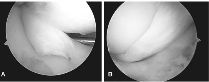
Fig. 3. (A) Probing induced displaced medial meniscus bucket handle tear during diagnostic arthroscopy. (B) Diagnostic arthroscopic appearance of the displaced lateral meniscus bucket handle tear of the same knee.
B
A
REFERENCES
01) Binfield PM, Maffulli N and King JB: Patterns of menis- cal tears associated with anterior cruciate ligament lesions in athletes. Injury, 24: 557-61, 1993.
02) Bugnone AN, Ramnath RR, Davis SB and Sedaros R:
The quadruple cruciate sign of simultaneous bicompart- mental medial and lateral bucket-handle meniscal tears.
Skeletal Radiol, 34: 740-4, 2005.
03) Cetik O, Cirpar M, Eksioglu F and Uslu M : Simultaneous bucket handle tear of both medial and lateral menisci of a knee with chronic anterior cruciate ligament deficiency. Knee Surg Sports Traumatol Arthrosc, 14:
356-9, 2006.
04) Dorsay TA and Helms CA: Bucket-handle meniscal tears of the knee: sensitivity and specificity of MRI signs.
Skeletal Radiol, 32: 266-72, 2003.
05) Keene GC, Bickerstaff D, Rae PJ and Paterson RS:
The natural history of meniscal tears in anterior cruciate ligament insufficiency. Am J Sports Med, 21: 672-9, 1993.
06) Phillips BB: Arthroscopy of lower extremity., St Louis, Mosby: pp 2811-2922,2007.
07) Shakespeare DT and Rigby HS: The bucket-handle tear of the meniscus. A clinical and arthrographic study. J Bone Joint Surg Br, 65: 383-7, 1983.
08) Tandogan RN, Taser O, Kayaalp A, et al.: Analysis of meniscal and chondral lesions accompanying anterior cru- ciate ligament tears: relationship with age, time from injury, and level of sport. Knee Surg Sports Traumatol Arthrosc, 12: 262-70, 2004.
09) Tecklenburg K, Schoepf D, Hoser C and Fink C:
Anterior cruciate ligament injury with simultaneous locked bucket-handle tears of both medial and lateral meniscus in a 19-year-old female professional ski racer: a case report. Knee Surg Sports Traumatol Arthrosc, 15:
1125-9, 2007.
10) Yoon JR, Muzaffar N, Kang JW, Lim HC, Bae JH and Nha KW: A novel technique for arthroscopic reduction and repair of a bucket-handle meniscal tear. Knee Surg Sports Traumatol Arthrosc, 17: 1332-5, 2009.
— 28 — 대한관절경학회지 제 14 권 제 1 호 2010년
대부분의 반월상 연골의 양동이 손잡이형 파열은 전방 십자인대와 동반하여 발생하며, 전방십자 인대의 급성 손상 시는 외측 반월상 연골의 양동이 손잡이형 파열이 흔한 반면 만성 전방 십자인대 손상 시는 내측에서 흔히 발생한다고 보고된 다. 저자들은 전방십자 인대의 불안정성과 동반 된 내,외측 반월상 연골의 동시적 양동이 손잡이형 파열에 대해 보고하고 자 한다.저자들은 전방십자 인대의 불안정성과 동반 된 내,외측 반월상 연골의 동시적 양동이 손잡이형 파열의 발생 가능 성에 대한 인식을 높이고자 하였으며, 추가적으로 본 손상의 발생기전, 임상증상, 자기공명영상 검사상 특이 소견 및 치료 에 대해 문헌 고찰과 함께 보고하고자 한다.
색인 단어: 양동이 손잡이형 파열, 내외측 반월상 연골, 동시 발생 초 록