Urological Oncology
Prostate-Specific Antigen Density as a Powerful Predictor of
Extracapsular Extension and Positive Surgical Margin in Radical Prostatectomy Patients with Prostate-Specific Antigen Levels of Less than 10 ng/ml
Jin-Seok Chang, Hoon Choi, Young-Seop Chang, Jin-Bum Kim, Mi Mi Oh1, Du Geon Moon1, Jae Hyun Bae1, Jun Cheon1
Department of Urology, Konyang Universtiy College of Medicine, Daejeon, 1Department of Urology, Korea University School of Medicine, Seoul, Korea
Purpose: To assess the ability of preoperative variables to predict extracapsular ex- tension (ECE) and positive surgical margin (PSM) in radical prostatectomy patients with prostate-specific antigen (PSA) levels of less than 10 ng/ml.
Materials and Methods: From January 2008 to December 2009, 121 patients with pros- tate cancer with PSA levels lower than 10 ng/ml who underwent radical prostatectomy were enrolled in the study. The differences in clinical factors (age, PSA, PSA density [PSAD], digital rectal examination [DRE] positivity, positive magnetic resonance imaging [MRI], Gleason sum, positive core number, and positive biopsy core percent- age) with ECE and the presence of positive margins were determined and their in- dependent predictive significances were analyzed.
Results: The ECE-positive patients had higher PSA, PSAD, and MRI-positive percen- tages, and PSM patients had higher PSA, PSAD, MRI-positive percentages, Gleason sum, and positive biopsy core percentages for prostate cancer. In the multivariate anal- ysis, PSAD and MRI positivity were the best independent predictors for ECE, and PSA and PSAD were the best independent predictors of PSM. By receiver operating charac- teristic curve analysis, PSAD had better discriminative area under the curve value than did PSA for ECE (0.765 vs 0.661) and PSM (0.780 vs 0.624). The best predictive PSAD value was 0.29 ng/ml/cc for ECE and 0.27 ng/ml/cc for PSM.
Conclusions: PSAD has relevance to ECE (plus MRI findings) and PSM (plus PSA).
PSAD might be a powerful predictor of ECE and PSM preoperatively in patients under- going a radical prostatectomy with PSA levels of less than 10 ng/ml.
Key Words: Extracapsular extension; Positive surgical margin; Prostatectomy; PSAD
This is an Open Access article distributed under the terms of the Creative Commons Attribution Non-Commercial License (http://creativecommons.org/licenses/by-nc/3.0) which permits unrestricted non-commercial use, distribution, and reproduction in any medium, provided the original work is properly cited.
Article History:
received 20 March, 2011 accepted 20 September, 2011
Corresponding Author:
Hoon Choi
Department of Urology, Konyang Universtiy College of Medicine, 685, Gasuwon-dong, Seo-gu, Daejeon 302-718, Korea
TEL: +82-42-600-9226 FAX: +82-42-542-3790 E-mail: [email protected]
INTRODUCTION
Prostate cancer is best cured by radical prostatectomy when the disease is organ-confined and the pathologic stage after surgery is known to be related with cancer control. Predicting the pathological stage of a prostate can- cer is important for deciding on a treatment strategy, but current clinical methods, including imaging or examina-
tion tools, are still of limited value for given patients, espe- cially those with intermediate risk [1,2].
Prostate-specific antigen (PSA) has been used as a crite- rion for consideration for prostate biopsy, and 30% to 35%
of men with PSA less than 10 ng/ml will be found to have prostate cancer [3]. Usually, 80% of men with PSA less than 4.0 ng/ml have pathologically proven organ-confined dis- ease, and 66% of men with PSA levels between 4.0 and 10.0
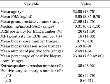
TABLE 1. Perioperative characteristics of 121 radical prosta- tectomy patients with PSA<10 ng/ml
Variable Value
Mean age (yr) Mean PSA (ng/ml)
Mean gram:prostate volume (range) Median ng/ml/cc:PSAD (range) DRE positivity for ECE number (%) MRI positivity for ECE number (%) Mean biopsy core number (range) Mean biopsy Gleason score (range) Mean number of positive core (range) Mean percentage of positive biopsy
core (range)
Extracapsular extension number (%) Positive surgical margin number (%) pT2
pT3
62.60 (46-75) 6.65 (3.35-9.79)
27.89 (12-75) 0.31 (0.07-1.10)
26 (21.49) 18 (14.88) 8.7 (5-13) 6.68 (6-9) 2.48 (1-6) 26.03 (7.69-62.50)
35 (28.92) 30 (24.79) 8 (6.61)
PSA: prostate-specific antigen, PSAD: prostate-specific antigen density, DRE: digital rectal examination, ECE: extracapsular ex- tension, MRI: magnetic resonance imaging
ng/ml have organ-confined disease [4]. In other words, 20%
to 34% of prostate cancer patients have extraprostatic dis- ease regardless of low to intermediate PSA levels (less than 10 ng/ml). Shinohara et al reported the ability of the serum PSA level to predict the pathologic stage [5]. On the other hand, Partin et al failed to utilize serum PSA to determine the pathologic stage of clinically localized prostate cancer.
They could not find a correlation between serum PSA and pathologic stage [6].
Because of variable PSA production by individual pros- tate cancer cells and wide variation in the contribution of benign prostatic hyperplasia to serum PSA levels, risk analysis based on only the PSA level seems incomplete if the PSA level is low to intermediate.
Extracapsular extension (ECE) and positive surgical margin (PSM) are important findings because of their im- plications for tumor biology, disease recurrence, and can- cer survival [7,8]. In this study, we tried to determine the correlation between preoperative predictive factors such as digital rectal examination (DRE) findings, magnetic res- onance imaging (MRI) findings, PSA with prostate-specific antigen density (PSAD), and biopsy characteristics and ECE and PSM as pathological outcomes in 121 radical pros- tatectomy patients with PSA less than 10 ng/ml.
MATERIALS AND METHODS
From January 2008 to December 2009, we evaluated 121 prostate cancer patients with PSA lower than 10 ng/ml who underwent radical prostatectomy performed by the stand- ard transperitoneal approach with the interfacial techni- que as previously reported [9]. Patients who had under- gone previous surgery for benign prostatic hypertrophy or who had received hormonal or radiation therapy previous to robot-assisted laparoscopic radical prostatectomy (RALP) were excluded. These 121 men underwent trans- rectal prostate biopsies owing to elevated PSA levels of ≥4 ng/ml or abnormal DRE findings or abnormal hypoechoic lesions on transrectal ultrasonography (TRUS). For PSA testing, patients’ serum was obtained before prostatic manipulations. Prostate volume was defined by measuring the height (H), width (W), and length (L) of the prostate from two selected orthogonal views and calculating the vol- ume (V) as that of the corresponding ellipsoid formula:
V=0.52xWxHxL [10]. PSAD was obtained by dividing se- rum PSA levels by the individual prostate volumes meas- ured by TRUS. All patients underwent a DRE and endor- ectal MRI to determine preoperative prostate status.
Tumors that MRI showed to be attached to the prostate cap- sule or tumors with a localized bulge were classified as sus- pected extracapsular extension. Systematic biopsies were obtained for 6 to 13 cores from separate regions, with at least 3 cores from each of both lobes. The total core number and positive core number of biopsies per patient were verified. Biopsy specimens were taken from different areas of the prostate assigned to proper Gleason scores.
Pathological staging of the radical prostatectomy speci-
men was performed and organ-confined cancer was des- ignated as long as the capsule was not penetrated.
Histopathologic data of radical prostatectomy specimens including capsule penetration and surgical margin were recorded. ECE was defined as a tumor extending outside of the prostate into the periprostatic soft tissues; accord- ingly, tumor invasion of the prostatic capsule without pen- etration was not ECE.
Once the patients were identified, we performed uni- variate analysis to determine the differences by pathologic outcome (ECE positivity and PSM) in age, PSA, PSAD, DRE positivity, and MRI positivity. Biopsy characteristics (Gleason sum, number of positive cores, and positive biopsy core percentage for prostate cancer) were also compared.
Next, the significant predictors were entered into a logistic regression test to determine their independent predictive significance. All statistical analysis was performed by us- ing the Mann Whitney U-test, Pearson's chi-square test for univariate analysis, and logistic regression for multi- variate analysis. Receiver operating characteristic (ROC) curves were used to compare the diagnostic power and de- termine the cutoff value associated with ECE and PSM positivity. SPSS version 12.0 (SPSS Inc., Chicago, IL, USA) was used and a p-value below 0.05 was considered statisti- cally significant.
RESULTS
Table 1 lists the patients’ characteristics. The patients’
mean age was 62.6 years (range, 46 to 75 years). The mean pre-biopsy PSA level was 6.65 mg/l, mean prostate volume was 27.89 g, and mean PSAD was 0.31 (range, 0.07 to 1.10).
Twenty-six patients had palpable tumors and 18 patients had ECE on MRI. The median number of total cores was
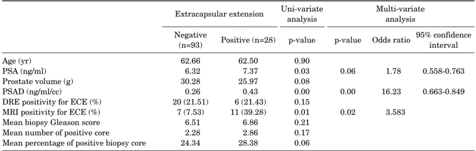
TABLE 2. Uni-variate and multi-variate analysis of clinical and biopsy features for predicting ECE of 121 radical prostatectomy patients
Extracapsular extension Uni-variate
analysis Multi-variate
analysis Negative
(n=93) Positive (n=28) p-value p-value Odds ratio 95% confidence interval Age (yr)
PSA (ng/ml) Prostate volume (g) PSAD (ng/ml/cc)
DRE positivity for ECE (%) MRI positivity for ECE (%) Mean biopsy Gleason score Mean number of positive core
Mean percentage of positive biopsy core
62.66 6.32 30.28 0.26 20 (21.51)
7 (7.53) 6.51 2.28 24.34
62.50 7.37 25.97 0.43 6 (21.43) 11 (39.28)
6.86 2.86 28.38
0.90 0.03 0.08 0.00 0.15 0.01 0.21 0.17 0.06
0.06 0.00 0.02
1.78 16.23 3.583
0.558-0.763 0.663-0.849
PSA: prostate-specific antigen, PSAD: prostate-specific antigen density, DRE: digital rectal examination, ECE: extracapsular ex- tension, MRI: magnetic resonance imaging
FIG. 1. (A) Receiver operating characteristic (ROC) curves comparing prostate-specific (PSAD, dotted line) and PSA (line) for extracapsular extension. (B) ROC curves comparing PSAD (dotted line) and prostate-specific antigen (PSA, line) for positive surgical margin.
8.7. The percentage of positive cores was derived from the number of positive biopsy cores divided by the total number of biopsy cores per patient multiplied by 100. The median positive core number and the percentage of positive cores were 2.48 (range, 1 to 6) and 26.03% (range, 7.69% to 62.50%), respectively.
Table 2 shows the association of clinical and biopsy fea- tures with ECE in radical prostatectomy patients. By uni- variate analysis, ECE-positive patients had higher PSA (7.37 vs. 6.32 ng/ml), PSAD (0.43 vs. 0.26 ng/ml/cc) level, and MRI positivity (39.28% vs. 7.53%). In the multivariate analysis, PSAD and MRI positivity were the best in- dependent predictors of ECE. In the ROC curve analysis of PSA and PSAD, PSAD showed better discriminative AUC value (0.765) than did PSA (0.661) for ECE (Fig. 1A).
In Table 3, the analysis of associated factors for PSM showed that PSM patients had higher PSA (7.42 vs. 6.28
ng/ml), PSAD (0.44 vs. 0.25 ng/ml/cc) level, and MRI pos- itivity for ECE (26.01% vs. 12.24%). As for biopsy charac- teristics, Gleason sum (6.97 vs. 6.25) and percentage of bi- opsy cores positive for prostate cancer (29.92% vs. 24.14%) were higher in PSM patients. In the multivariate analysis, however, only PSA and PSAD were independent predictors of PSM statistically. The ROC curve showed diagnostic val- ue of PSA and PSAD for PSM, and PSAD had better dis- criminative AUC value (0.780) than did PSA (0.624) (Fig.
1B). The best predictive PSAD value for ECE was 0.29 ng/ml/cc (sensitivity 0.72, specificity 0.68) and 0.27 ng/ml/cc (sensitivity 0.82, specificity 0.67) for PSM.
DISCUSSION
Pathological stage is usually predicted preoperatively to evaluate the possibility of cure by a particular therapy.
TABLE 3. Uni-variate and multi-variate analysis of clinical and biopsy features for predicting positive surgical margin (PSM) of 121 radical prostatectomy patients
Surgical margin Uni-variate
analysis Multi-variate
analysis Negative
(n=98) Positive (n=23) p-value p-value Odds ratio 95% confidence interval Age (yr)
PSA (ng/ml) Prostate volume (g) PSAD (ng/ml/cc)
DRE positivity for ECE (%) MRI positivity for ECE (%) Mean biopsy Gleason score Mean number of positive core
Mean percentage of positive biopsy core
62.67 6.28 29.69 0.25 21 (21.43) 12 (12.24)
6.25 2.34 24.14
62.47 7.42 26.97 0.44 5 (21.74)
6 (26.0) 6.97 2.73 29.91
0.87 0.02 0.11 0.00 0.39 0.02 0.14 0.37 0.03
0.04 0.00 0.07 0.10
2.38 20.29 1.83 1.81
0.515-0.734 0.689-0.871
PSA: prostate-specific antigen, PSAD: prostate-specific antigen density, ECE: extracapsular extension, DRE: digital rectal examina- tion, MRI: magnetic resonance imaging
Reliable assessment of organ-confined prostatic cancer would be the best available method for complete removal without increasing the incidence of avoidable PSMs.
Several established reports have tried to show the proba- bility of PSM or ECE positivity by use of preoperative varia- bles, such as clinical stage, PSA, and biopsy features [7,10-12].
Despite preoperative staging with the use of these pa- rameters, pathologic evaluation shows that a significant number of patients undergoing radical prostatectomy have extraprostatic disease and positive resection margins.
Patients with organ-confined prostatic cancer have sig- nificantly greater 5 year disease-free survival than do those with disease that is not organ-confined, and several studies have shown that PSM is an independent adverse predictor of pathologic outcome and significantly corre- lates with the progression-free survival rate [11,13-15].
Former studies have shown that combining Gleason score, clinical stage, and PSA allows the estimation of or- gan-confined prostate cancer. However, for the inter- mediate-risk patient group, little additional information may be expected from those characteristics for predicting organ-confined disease [16,17]. In our study of 121 prostate cancer patients with a low to intermediate PSA level, biop- sy Gleason score, DRE positivity, and mean number of pos- itive cores showed no significant relationship. It is well known that biopsy features, such as tumor length, the pro- portion of cancer involvement, number of positive cores, and the proportion of positive cores are predictive parame- ters of ECE and cancer-related outcomes [18-20]. The his- topathological features of prostate cancer grade and extent in needle biopsies have been used to predict cancer grade, tumor volume, and pathological stage in the prostate. The Gleason grade on needle biopsies has been demonstrated to be predictive of stage, and the number of positive cores in the biopsy was the most useful predictor of tumor size [21-23]. Equally, maximum tumor length was the best in-
dependent predictor of ECE and the number of positive bi- opsies was the most useful single parameter with a positive predictive value of 83% in 274 lobes and a negative pre- dictive value of 55% [7,13].
Pretreatment PSA, highest biopsy Gleason sum, and percentage of positive biopsy cores for cancer were the three most significant independent predictors of pathologic stage after radical prostatectomy in a recent study [24]. But cancer volume in the prostatectomy specimens was weakly or not correlated with the number of positive biopsies, total length of cancer in biopsies, and the percent of Gleason grade 4/5 on biopsies. Therefore, with these critical param- eters from the biopsy specimens, Noguchi et al were unable to estimate cancer volume in the radical prostatectomy specimen on an individual basis [25]. In our data, PSM pa- tients had a higher percentage of positive biopsy cores with no statistical significance.
Generally, PSA is a strong predictor of tumor aggre- ssiveness. Noldus and Stamey reported a strong correla- tion (r=0.70) between preoperative PSA and the pathologi- cally measured cancer volume. This correlation was not shown when they investigated larger numbers of patients [26]. PSA alone cannot predict the cancer volume reliably.
PSA elevation produced by benign prostatic hyperplasia and advancing age decreases the reliability of serum PSA as a predictive parameter for cancer volume [5,6]. In an at- tempt to improve the ability to predict organ-confined dis- ease, Seaman et al tested the efficacy of PSAD. These inves- tigators found greater accuracy predicting organ-confined disease with PSAD. Patients who have low PSAD values are ideal candidates for radical prostatectomy, and PSAD was a better predictor of final pathologic stage than Gleason score [27]. Taneja et al reported that the AUC for predicting the ECE of prostate tumors was 0.62 and 0.69 for complexed serum PSA and complexed PSAD, re- spectively [28]. These findings were supported by those of Naya et al who reported an AUC of 0.54 and 0.70 for com-
plexed PSA and complexed PSAD, respectively [29]. Our study showed a better discriminative AUC value of PSAD for ECE (0.765) and for PSM (0.780) than previous reports.
These results could be significant because of the narrow spectrum of enrolled patients with PSA less than 10 ng/ml.
In general, PSAD is a better predictor of prostate cancer in men with PSA levels of 4-10 ng/ml, especially when ultra- sound-determined measurements of prostate volume are available. This suggests that PSAD is a powerful parame- ter for estimating tumor volume in the gray zone of prostate cancer regardless of patient race or age. Our data showed more accuracy of PSAD in predicting stage, which could be related to the small volume of the Asian race; more interna- tional study will be needed [27]. As we know, this is the first study about the relationship between PSM and PSAD.
Furthermore, like our result for ECE, Ishida et al reported that the pathological stage was found to be significantly correlated with the MRI findings but not the DRE findings [11].
Accurate cancer staging is critical for selecting the most appropriate treatment option in prostate cancer.
Currently available predictive efforts for stage are compli- cated, so we tried to simplify the related factors. In our study, we were able to show a significant predictive power and cutoff value of PSAD in terms of ECE and PSM com- pared with PSA. This study had some limitations, includ- ing its retrospective manner and lack of data about actual pathologically measured cancer volume. Although no sin- gle parameter should be used in clinical practice to increase the ability to predict the presence of unfavorable pathologic outcomes, PSAD can be a useful and very effective predictor of ECE and PSM as a tool to reflect the tumor burden, and we can counsel patients with discretion regarding the like- lihood of complete cancer control.
CONCLUSION
PSAD has relevance to ECE (plus MRI findings) and PSM (plus PSA). Although various clinical factors could sig- nificantly increase adequate cancer control, PSAD could be a useful and powerful predictor for ECE and PSM as a pre- operative characteristic in radical prostatectomy patients with PSA less than 10 ng/ml. However, our study was con- ducted in two centers with 94 RALP cases and 27 RRP cases under three surgeons. Thus, larger and single-center stud- ies will be needed.
Conflicts of Interest
The authors have nothing to disclose.
REFERENCES
1. Hara I, Miyake H, Hara S, Yamanaka N, Ono Y, Eto H, et al. Value of the serum prostate-specific antigen-alpha 1-antichymotrypsin complex and its density as a predictor for the extent of prostate cancer. BJU Int 2001;88:53-7.
2. Smith JA Jr, Scardino PT, Resnick MI, Hernandez AD, Rose SC,
Egger MJ. Transrectal ultrasound versus digital rectal examina- tion for the staging of carcinoma of the prostate: results of a pro- spective, multi-institutional trial. J Urol 1997;157:902-6.
3. Zheng XY, Xie LP, Wang YY, Ding W, Yang K, Shen HF, et al. The use of prostate specific antigen (PSA) density in detecting prostate cancer in Chinese men with PSA levels of 4-10 ng/mL. J Cancer Res Clin Oncol 2008;134:1207-10.
4. Rietbergen JB, Hoedemaeker RF, Kruger AE, Kirkel WJ, Schröder FH. The changing pattern of prostate cancer at the time of diagnosis: characteristics of screen detected prostate cancer in a population based screening study. J Urol 1999;161:1192-8.
5. Shinohara K, Wolf JS Jr, Narayan P, Carroll PR. Comparison of prostate specific antigen with prostate specific antigen density for 3 clinical applications. J Urol 1994;152:120-3.
6. Partin AW, Carter HB, Chan DW, Epstein JI, Oesterling JE, Rock RC, et al. Prostate specific antigen in the staging of localized pros- tate cancer: influence of tumor differentiation, tumor volume and benign hyperplasia. J UroI 1990;143:747-52.
7. Graefen M, Haese A, Pichlmeier U, Hammerer PG, Noldus J, Butz K, et al. A validated strategy for side specific prediction of organ confined prostate cancer: a tool to select for nerve sparing radical prostatectomy. J Urol 2001;165:857-63.
8. Ohori M, Kattan MW, Koh H, Maru N, Slawin KM, Shariat S, et al. Predicting the presence and side of extracapsular extension:
a nomogram for staging prostate cancer. J Urol 2004;171:1844-9.
9. Choi H, Ko YH, Kang SG, Kang SH, Park HS, Cheon J, et al. Biopsy related prostate status does not affect on the clinicopathological outcome of robotic assisted laparoscopic radical prostatectomy.
Cancer Res Treat 2009;41:205-10.
10. Gancarczyk KJ, Wu H, McLeod DG, Kane C, Kusuda L, Lance R, et al. Using the percentage of biopsy cores positive for cancer, pre- treatment PSA, and highest biopsy Gleason sum to predict patho- logic stage after radical prostatectomy: the Center for Prostate Disease Research nomograms. Urology 2003;61:589-95.
11. Ishida M, Nakashima J, Hashiguchi A, Mizuno R, Shinoda K, Kikuchi E, et al. Are predictive models for cancer volume clinically useful in localized prostate cancer? Int J Urol 2009;16:936-40.
12. Lee JT, Lee S, Yun CJ, Jeon BJ, Kim JM, Ha HK, et al. Prediction of perineural invasion and its prognostic value in patients with prostate cancer. Korean J Urol 2010;51:745-51.
13. Nakanishi H, Troncoso P, Babaian RJ. Prediction of ex- traprostatic extension in men with biopsy Gleason score of 8 or greater. J Urol 2008;180:2441-5.
14. D’Amico AV, Whittington R, Malkowicz SB, Schultz D, Schnall M, Tomaszewski JE, et al. A multivariate analysis of clinical and pathological factors that predict for prostate specific antigen fail- ure after radical prostatectomy for prostate cancer. J Urol 1995;154:131-8.
15. Mian BM, Troncoso P, Okihara K, Bhadkamkar V, Johnston D, Reyes AO, et al. Outcome of patients with Gleason score 8 or high- er prostate cancer following radical prostatectomy alone. J Urol 2002;167:1675-80.
16. Kattan MW, Stapleton AM, Wheeler TM, Scardino PT.
Evaluation of a nomogram used to predict the pathologic stage of clinically localized prostate carcinoma. Cancer 1997;79:528-37.
17. Partin AW, Kattan MW, Subong EN, Walsh PC, Wojno KJ, Oesterling JE. Combination of prostate specific antigen, clinical stage, and Gleason score to predict pathological stage of localized prostate cancer. A multi-institutional update. JAMA 1997;277:
1445-51.
18. Graefen M, Haese A, Pichlmeier U, Hammerer PG, Noldus J, Butz K, et al. A validated strategy for side specific prediction of organ
confined prostate cancer: a tool to select for nerve sparing radical prostatectomy. J Urol 2001;165:857-63.
19. Naya Y, Slaton JW, Troncoso P, Okihara K, Babaian RJ. Tumor length and location of cancer on biopsy predict for side specific ex- traprostatic cancer extension. J Urol 2004;171:1093-7.
20. Tsuzuki T, Hernandez DJ, Aydin H, Trock B, Walsh PC, Epstein JI. Prediction of extraprostatic extension in the neurovascular bundle based on prostate needle biopsy pathology, serum prostate specific antigen and digital rectal examination. J Urol 2005;173:
450-3.
21. Cookson MS, Fleshner NE, Soloway SM, Fair WR. Correlation be- tween Gleason score of needle biopsy and radical prostatectomy specimen: accuracy and clinical implications. J Urol 1997;157:
559-62.
22. Wang X, Brannigank RE, Rademaker AW, McVary KT, Oyasu R.
One core positive prostate biopsy is a poor predictor of cancer vol- ume in the radical prostatectomy specimen. J Urol 1997;158:
1431-5.
23. Cheng L, Slezak J, Bergstralh EJ, Myers RP, Zincke H, Bostwick DG. Preoperative prediction of surgical margin status in patients with prostate cancer treated by radical prostatectomy. J Clin Oncol 2000;18:2862-8.
24. Gancarczyk KJ, Wu H, McLeod DG, Kane C, Kusuda L, Lance R, et al. Using the percentage of biopsy cores positive for cancer, pre-
treatment PSA, and highest biopsy Gleason sum to predict patho- logic stage after radical prostatectomy: the Center for Prostate Disease Research nomograms. Urology 2003;61:589-95.
25. Noguchi M, Stamey TA, McNeal JE, Yemoto CM. Relationship be- tween systematic biopsies and histological features of 222 radical prostatectomy specimens: lack of prediction of tumor significance for men with nonpalpable prostate cancer. J Urol 2001;166:104-9.
26. Noldus J, Stamey TA. Limitations of serum prostate specific anti- gen in predicting peripheral and transition zone cancer volumes as measured by correlation coefficients. J Urol 1996;155:232-7.
27. Seaman EK, Whang IS, Cooner W, Olsson CA, Benson MC.
Predictive value of prostate-specific antigen density for the pres- ence of micrometastatic carcinoma of the prostate. Urology 1994;43:645-8.
28. Taneja SS, Hsu EI, Cheli CD, Walden P, Bartsch G, Horninger W, et al. Complexed prostate-specific antigen as a staging tool:
results based on a multicenter prospective evaluation of com- plexed prostate-specific antigen in cancer diagnosis. Urology 2002;60(4 Suppl 1):10-7.
29. Naya Y, Fritsche HA, Cheli CD, Stamey TA, Bartsch G, Brawer MK, et al. Volume indexes of total, free, and complexed pros- tate-specific antigen enhance prediction of extraprostatic disease extension in men with nonpalpable prostate cancer. Urology 2003;62:1058-62.