Results of Revision Surgery and Causes of Unstable Total Knee Arthroplasty
In-Soo Song, MD, Doo-Hoon Sun, MD, Jae-Gyun Chon, MD, Sung-Won Jang, MD, Dong-Hyuk Sun, MD
Department of Orthropaedic Surgery, Sun General Hospital, Daejeon, Korea
Received October 29, 2012; Accepted June 23, 2013 Correspondence to: In-Soo Song, MD
Department of Orthropaedic Surgery, Sun General Hospital, 29 Mokjung- ro, Jung-gu, Daejeon 301-725, Korea
Tel: +82-42-220-8220, Fax: +82-42-220-8220 E-mail: [email protected]
Over the past two decades primary total knee arthroplasty (TKA) has proven to be a highly successful operation, with survivorship rates approaching 95% after a 15-year follow-up period.1) However, the results of revision TKA over the same period have been less encouraging. Failure rates in a range of 11% to 60% have been reported after a generally shorter follow-up interval.2,3) The poor results of TKA have been attributed to multiple factors, including size mismatching,3-5) ligamentous instability or soft tissue incompetence,3-6) pain of an unclear etiology,7) loosening8) and infection,5,9) and extensor mechanism problems.6,8)
Background: The aim of this study was to evaluate causes of unstable total knee arthroplasty and results of revision surgery.
Methods: We retrospectively reviewed 24 knees that underwent a revision arthroplasty for unstable total knee arthroplasty. The average follow-up period was 33.8 months. We classified the instability and analyzed the treatment results according to its cause.
Stress radiographs, postoperative component position, and joint level were measured. Clinical outcomes were assessed using the Hospital for Special Surgery (HSS) score and range of motion.
Results: Causes of instability included coronal instability with posteromedial polyethylene wear and lateral laxity in 13 knees, coronal instability with posteromedial polyethylene wear in 6 knees and coronal and sagittal instability in 3 knees including post breakage in 1 knee, global instability in 1 knee and flexion instability in 1 knee. Mean preoperative/postoperative varus and valgus angles were 5.8°/3.2° (p = 0.713) and 22.5°/5.6° (p = 0.032). Mean postoperative α, β, γ, δ angle were 5.34°, 89.65°, 2.74°, 6.77°.
Mean changes of joint levels were from 14.1 mm to 13.6 mm from fibular head (p = 0.82). The mean HSS score improved from 53.4 to 89.2 (p = 0.04). The average range of motion was changed from 123° to 122° (p = 0.82).
Conclusions: Revision total knee arthroplasty with or without a more constrained prosthesis will be a definite solution for an unstable total knee arthroplasty. The solution according to cause is very important and seems to be helpful to avoid unnecessary over-constrained implant selection in revision surgery for total knee instability.
Keywords: Unstable total knee, Total knee instability, Revision arthroplasty
Instability is the third most common cause of failure of a total knee arhtroplasty among many causes of revision arthroplasty as listed above.10) These instability can lead to dislocation after TKA, although it is uncommon. Disloca- tion after TKA usually is a difficult problem to address.
The most frequently reported causes of instability and dis- location after TKA are malpositioning of the implant, flex- ion-extension gap mismatch, excessive soft tissue release, extensor mechanism incompetence and inappropriate se- lection of the primary implant. Delayed rupture of poste- rior cruciate ligament (PCL), breakage of the polyethylene insert,11) breakage of the polyethylene post and neurologic diseases are less common causes.7,12,13) Modes of instabil- ity according to its direction can be classified as coronal (varus-valgus), sagittal (anteroposterior [AP]), flexion, recurvatum, and global instability. We analyzed the mode of instability and assumed the cause of the instability.10,14,15)
This study was designed to classify factors leading to
Copyright © 2014 by The Korean Orthopaedic Association
This is an Open Access article distributed under the terms of the Creative Commons Attribution Non-Commercial License (http://creativecommons.org/licenses/by-nc/3.0) which permits unrestricted non-commercial use, distribution, and reproduction in any medium, provided the original work is properly cited.
Clinics in Orthopedic Surgery • pISSN 2005-291X eISSN 2005-4408
the TKA instability and to evaluate the clinical and radio- logic outcome of revision arthroplasty for unstable TKA.
We hypothesized that unstable TKA could be expressed by various types and an evaluation of the causes of instability would be helpful to choice the implants or surgical tech- niques in the revision TKA.
METHODS
Patient Enrollment
We studied a consecutive series of 63 knees in 61 patients that underwent revision TKA for various problems after TKA at our institution (Sun General Hospital, Daejeon, Korea) between December 2004 and December 2010. The procedures were performed by a single surgeon (ISS) and all cases were performed at the same institution. There was no external funding source for this study. An Institutional Review Board approval was obtained for this study. Revi- sion arthroplasty for other causes unrelated to instability were 39 knees of 39 patients as follows: infection of 20 knees, periprosthetic fracture of 4 knees, implant loosen- ing of 13 knees, and implant malposition of 2 knees. So, the subjects of this study included the remaining 24 knees of 22 patients that underwent revision TKA for TKA instability. Those 24 knees were eligible for review in a follow-up period with an average of 33.8 months (range, 6 to 70 months). The mean interval between the primary TKA and revision TKA was 82.5 months (range, 14 to 228 months). Of the 24 knees, there were 7 knees that un- derwent primary TKA in our hospital and 17 knees that underwent arthroplasty in another institution. The study subjects consisted of 4 men and 18 women and the mean age of the patients was 71.0 years (range, 54 to 85 years) at the time of revision surgery. Thirteen knees were on the left side and 11 knees were on the right side. The original diagnosis for primary TKAs was osteoarthritis in 23 knees and rheumatoid arthritis in 1 knee. Laxity in coronal plane and/or sagittal plane was present in all patients. This in- stability was confirmed by preoperative stress radiographs (AP drawer stress view, valgus-varus stress view) and physical examinations including AP drawer stress test, valgus-varus stress test and posterolateral drawer test.
All patients expressed various symptoms, which may be associated with instability, including sense of giv- ing way, start-up pain, recurrent effusion, anterior knee pain, pes, and hamstring tendinitis. No infection was clini- cally suspected in any patient. We decided to conduct a re- vision surgery in cases that demonstrated significant laxity in AP direction and/or valgus-varus direction and/or pos- terolateral rotational instability. And we certified the rela-
tionship between the instability in radiographs and clinical expression. No loose component was demonstrated by radiographs in any patient and we made confirmed the implant stability during the operation. Twenty-four knees of unstable TKA were assigned to 10 knees (42%) of pos- terior stabilized design and 14 knees (58%) of a cruciate- retaining (CR) type.
Surgical Technique and Component Evaluation
A longitudinal midline skin incision was used in all cases and care was taken to incorporate and not cross the pre- vious skin incision. In case of simultaneously existing incisions we chose the most lateral previous incision to ensure minimal breakage of the blood supply. Exposure was obtained by a medial parapatellar arthrotomy in 22 knees (92%) and additional tibial tubercle osteotomy in 2 knees (8%). We checked the component stability and confirmed the causes of preoperative instability. We per- formed polyethylene exchange to thicker plastic when only posteromedial wear of the polyethylene existed and made certain the regained stability. Prosthesis and cement were removed using various tools, such as a small power saw and thin osteotome while preserving as much normal bone as possible. A standard cemented prosthesis was used if both collateral ligaments were felt to be competent, and a constrained prosthesis was used if one or both collateral ligaments were incompetent. Hinged prosthesis was not used in any case. All revision prostheses were of cemented design. Of the 24 knees, 7 knees received a cemented stan- dard posterior cruciate ligament substituting (PS) pros- thesis (3 Vanguard CR, Biomet Inc., Warsaw, IN, USA; 1
Table 1. Prostheses Used in Revision Surgery of Unstable Total Knee Arthroplasty
Prostheses Cases (no.)
Polyethylene insert change Total (7), up-sized (5), same- sized (2)
Cemented standard PS prosthesis (6 knees) Vanguard CR, Biomet (3) Scorpio NRG, Stryker (1) Vanguard RP, Biomet (1) LCS, Depuy (1) Semiconstrained PS prosthesis (11 knees) Vanguard PS, Biomet (7)
Nexgen, Zimmer (2) Scorpio, Stryker (2) PS: posterior cruciate ligament substituting.
Scorpio NRG, Stryker Inc., Mahwah, NJ, USA; 1 Vanguard RP, Biomet Inc.; and 1 LCS, Depuy Inc., Warsaw, IN, USA) and 11 knees received a constrained revision prosthesis (7 Vanguard PS, Biomet Inc.; 2 Nexgen, Zimmer, Warsaw, IN, USA; and 2 Scorpio NRG).
We used extended stems that could be helpful in regaining an attenuated collateral ligament and preventing a toggling effect of the implant. Exceptions were 7 cases of exchange of polyethylene insert only and 2 cases with confirmed stability of collateral ligament in the opera- tion field. An extended femoral stem was used in 2 cases only, an extended tibial stem in 2 cases only and extended femoral and tibial stem together in 11 cases during the op- eration (Table 1).
Surgeries entailing revision of both femoral and tibial components were conducted in 13 knees (54%), only femoral component in 2 knees (8%), only tibial compo- nent in 2 knees (8%), and polyethylene exchange alone was performed in 7 knees (30%) (Table 2). Collateral liga- ment repair in medial femoral epicondyle was used in one knee. Soft tissue balancing of the knee was assessed and the wound was closed.
Range of motion was started on the first postopera- tive day and weight bearing as tolerated was allowed on
the second postoperative day. Mean operation time for re- vision surgery was 95 minutes (range, 45 to 185 minutes).
Classification and Evaluation
We classified the unstable 24 knees into 5 types: isolated coronal instability, sagittal instability with the knee flexed to 90°, combined coronal with sagittal instability, flexion instability and global instability with regard to its reasons.
All 24 knees had complete radiographic follow-ups. Ra- diographs were obtained before and after surgery, includ- ing AP radiographs obtained with the patient standing and supine.
A lateral radiograph and a skyline patellar radio- graph were obtained to assess the alignment of the limb and component position using α, β, Υ, δ angle and status of the joint line. Joint lines were determined on AP radio- graphs obtained before and after surgery with the patient supine by measuring the distance between the tip of the fibular head and the distal margin of the lateral femoral condyle preoperatively and between the tip of the fibular head and the distal margin of the lateral femoral com- ponent postoperatively. The skyline patellar radiographs were examined for patellar tilt, subluxation or dislocation according to the classification of Bindelglass and Vince.16)
We checked the valgus and varus stress radiographs before surgery and at the last follow-ups. Clinical out- comes were assessed according to the knee rating score of the Hospital for Special Surgery (HSS).
Statistical analyses for the evaluation of radiographs and clinical results were performed with paired t-test. All statistical analyses were performed with the SPSS ver. 14.0 (SPSS Inc., Chicago, IL, USA), and p < 0.05 was consid- ered statistical significant.
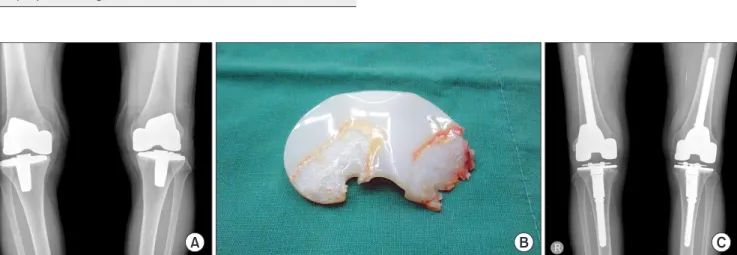
Fig. 1. (A) The radiograph shows coronal plane instability and varus deformity on both postoperative knees. (B) We found posteromedial bearing wear in the operative field. (C) We performed a revision total knee arthroplasty with femoral and tibial component change.
Table 2. Exchange of the Components in Revision Total Knee Arthroplasty
Exchange of the components No. of knees
Revision of both femoral and tibial components 13
Revision of femoral component 2
Revision of tibial component 2
Polyethylene exchange alone 7
RESULTS
Instability associated with frank dislocation in preopera- tive period was demonstrated in four cases (one case of flexion instability, two cases of coronal with sagittal insta- bility, and one case of global instability), while the other 20 cases did not have frank dislocation. Of the 24 cases, coronal plane (mediolateral) instability with concomitant posteromedial polyethylene wear and lateral ligament at- tenuation showing 3° more difference than the opposite site in preoperative varus stress view was shown in 13 cases (in both sides for one patient) (Fig. 1). Coronal insta- bility with polyethylene wear alone was shown in 6 cases.
Coronal with sagittal plane (AP) instability was shown in 3 cases (Fig. 2). Among these 3 cases, 2 cases had a medial collateral ligament (MCL) and a PCL rupture (1 case of PS design and 1 case of CR design) and another case present- ed with MCL rupture and post fracture of the polyethylene insert (Fig. 3).
Flexion instability with spin-out of the polyethylene insert during squatting was shown in one case. Globally, one case showed rotational instability and varus thrust gait on walking. Only in 24 cases we didn’t find any sagit-
tal instability (Table 3). Of 13 cases of coronal instability with posteromedial polyethylene wear and lateral ligament attenuation presented 10 cases with revision arthroplasty with long stems in femoral and/or tibial implants (Fig. 4).
Of 6 cases with posterolateral polyethylene wear under- went 4 cases a bearing exchange to upsize and the remain- ing 2 cases underwent a bearing exchange to the same size.
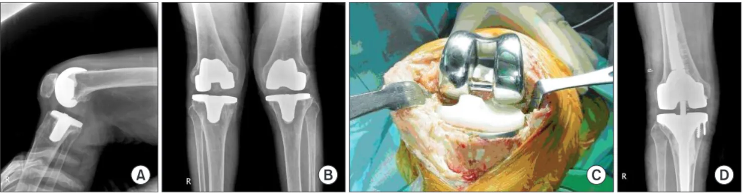
One case of flexion instability with spinout of polyethylene insert underwent bearing exchange to upsize and attained Fig. 2. Radiographs demonstrating sagittal plane instability (A) and coronal plane instability (B). We presumed posterior cruciate ligament and medial collateral ligament attenuation. (C) We performed a revision arthroplasty with femoral component change.
Fig. 3. The right knee shows posterior instability (A) and valgus instability (B) suggesting coronal and sagittal instability. We found a post fracture of the polyethylene insert (C) and performed a revision surgery with femoral and tibial component change (D).
Table 3. Classification of Unstable Total Knee Arthroplasties According to Causes
Classification of unstable total knee arthroplasties No. of knees Coronal instability with posteromedial polyethylene wear
and lateral ligament attenuation 13
Coronal instability with posteromedial wear of polyethylene
insert only 6
Coronal and sagittal instability 3
Flexion instability 1
Global instability 1
stability (Table 4).
The mechanical axis deviation at standing radio- graphs was changed from preoperative mean 3.4 mm (range, 14.0 to 0.0 mm) to postoperative mean 1.4 mm (range, 3.8 to 0.0 mm) and didn’t have statistical signifi- cance between preoperation and postoperation (p = 0.63).
The preoperative and postoperative varus angle was mean 5.8° (range, 13.0° to 1.0°) and mean 3.2° (tange, 7.2° to 1.0°;
p = 0.713) on varus stress radiographs. The preoperative and postoperative valgus angle was mean 22.5° (range, 32.0° to 11.0°) and mean 5.6° (tange, 8.0° to 2.0°; p = 0.032) on valgus stress radiography. The postoperative α angle was mean 5.34°, β angle was mean 89.65°, γ angle was mean 2.74°, and δ angle was mean 6.77° in the implant position analyses. The outlier over three degree was 1 case (4.17%) in α angle and 2 cases (8.33%) in γ angle. Levels of joint line in preoperation and at last follow-up were mean 14.1 mm and mean 13.6 mm (range, 4.8 to 21.0 mm) from tip of the fibular head (p = 0.82). Patellar tilts by Bindel- glass and Vince16) classification were 10 cases (41.6%) in
central and 14 cases (58.3%) in lateral tilt. None of the cases showed a medial tilt (0.0%). The range of motion at preoperation and last follow-ups were preoperatively mean 123° (range, 110° to 130°) and at last follow-up mean 122°
(range, 95° to 130°) postoperatively (p = 0.82). The mean HSS score improved from preoperative 53.4 (range, 38.0 to 62.0) to postoperative 89.2 (range, 72.0 to 95.0) with statis- tical significance (p = 0.04).
DISCUSSION
Causes of revision arthroplasty after total knee replace- ment are diverse and unclear. Hossain et al.17) cited com- mon causes for revision as infection (2.9%), instability (1.7%), and aseptic loosening (1.4%). Clinical instability has been estimated to be present in 1%–2% of patients following a TKA procedure and in 10%–20% after a TKA revision.14) Instability after total knee replacement is being increasingly reported in the literature.18-22) Knee disloca- tion after total knee replacement was first reported in 4 patients in a series of 220 patients by Insall et al.23) in 1979.
This TKA instability may or may not be accompanied by dislocation and can be classified according to causes.20,22)
First, mediolateral instability or coronal instability may be as frequent a reason for revision of TKA and it can be due to incorrect ligament balancing or lack of identifi- cation of an incompetent collateral ligament. Inadequate medial structural releases that can evoke the delayed MCL rupture or attenuation frequently lead to delayed coronal instability. We didn’t experience such circumstances be- cause we always attended to check the adequate medial release and mediolateral balance, but there were several cases of coronal instability showing posterolateral poly- ethylene insert wear. Treatments of unstable TKA with coronal instability don’t need revision surgery using con- strained type with long stem. And only the exchange of
Table 4. Operative Procedures in Revision Total Knee Arthroplasty for Unstable Total Knee Arthroplasty
Classification of unstable total knee arthroplasty Operative procedure in revision total knee arthroplasty Coronal instability with posteromedial polyethylene wear and lateral ligament attenuation (13) Revision with long stem (10)
Revision without long stem (3) Coronal instability with posteromedial wear of polyethylene insert (6) Upsized bearing exchange (4) Same sized bearing exchange (2)
Coronal and sagittal instability (3) Revision with long stem (3)
Flexion instability (1) Upsized bearing exchange (1)
Global instability (1) Revision with long stem (1)
Fig. 4. The left knee showed global instability on radiography and clinical examination (A). We performed a revision arthroplasty on the left knee with femoral and tibial component change (B).
inserted polyethylene demonstrated sufficient stability in the treatment of 6 cases of coronal instability with postero- medial polyethylene wear in this study. But, lateral liga- ment attenuation is very important in choice of treatments in such cases.
In this study, 13 cases with lateral ligament attenua- tion needed a revision of the implants. Of those 13 cases, a constrained implant type with long stem was used in 10 cases. So, an analysis of the cause of instability is most important in the revision of an unstable TKA and this analysis procedure is very important to prevent a re-revi- sion of the recurrence of instability. Extensor mechanism incompetence, inadequate balancing of the PCL, exces- sive release of posterolateral structures, polyethylene post fracture, hyperextension or a broken polyethylene insert may all contribute to an AP TKA dislocation. Flexion instability means that the flexion gap is too loose. Factors contributing to early flexion instability included poor pos- terior offset restitution, PCL incompetence or component malpositioning. Late forms of flexion instability may be associated with a delayed rupture or degeneration of the PCL and rotational instability. In our study, 1 case showed locked posterior dislocation and coronal with sagittal in- stability 7 years after original procedure.
The knee demonstrated a broken polyethylene post by fatigue fracture that was assumed as a late form of flex- ion instability. So we performed revision surgery using constrained implant with femoral and tibial long stem and obtained the stability of the knee with excellent clinical results. In particular, CR prostheses may have a PCL prob- lem, which can cause a loose flexion gap, sagittal instability and polyethylene dislocation, regardless of whether there is a delayed PCL rupture or PCL attenuation. Accordingly, its conditions require a total knee revision to the PS pros- thesis system. Many elderly patients will be expected to have an incompetent PCL function. Considering possible PCL problems after CR prosthesis, we prefer PS prosthesis that has an effective joint motion with post-cam mecha- nism and are more stable to dislocation than CR prosthe- sis.
We can divide such TKA instability by type into early instability and late instability for consideration. An early instability can result from a component malalign- ment, incorrect mechanical axis, gap imbalance, ligament rupture (PCL or MCL) and extensor mechanism abnor- mality, while late instability may result from polyethylene wear, polyethylene post wear or fracture, ligament attenu- ation, extensor mechanism dysfunction and others.23,24) In the research of the author, two cases indicated sagittal and coronal instability with PCL and MCL rupture due to
trauma after 3 years and 5 years, respectively. As well, one case indicated a sagittal instability with polyethylene post fracture without a trauma history.
According to the reports of several authors, most ligament reconstructions cannot solve the problem of instability due to collateral ligament attenuation, which ultimately may progress into knee dislocation or polyeth- ylene dislocation.11,24,25) The principle of treatment for TKA instability is to exchange unstable knee to stable knee, but the exchange to thicker polyethylene must carefully consider the variation in the flexion and extension gap. It is considered that there will be few cases in which stabil- ity can be ensured with upsized polyethylene alone. Ac- cording to recent reports, patients undergoing revision of femoral and tibial components had better outcomes than those undergoing isolated polyethylene exchange.15) But, our study contained of 7 cases with posteromedial poly- ethylene bearing wear leading to coronal instability and to an exchange of isolated polyethylene bearing, and its final results demonstrated excellent results without recurrent instability.
The problem of PCL in the CR type of implant can be solved by exchanging to PS type. But an overall evalua- tion and solutions for coronal and global instability must be carefully considered as this only solves the problem of sagittal instability. In any case, the use of a more con- strained type of implant must be considered for TKA in- stability and the semi-constrained prosthesis or the hinged type of implant can be used. Efforts must be made to care- fully raise by stage the level of constraint to obtain stability.
The most fundamental point of such revision sur- gery is to obtain equal flexion and extension gap. For this, an accurate evaluation of the integrity of each ligament must be performed. Although the current diversified pos- terior stabilized knee arthroplasty as an advanced post- cam mechanism can compensate for a certain amount of loose flexion gap, it applies considerable stress to polyeth- ylene post and ultimately may cause fatigue fracture on such post.26,27)
Some authors asserted that coronal instability can be divided into reconstructable MCL and non-reconstructa- ble MCL according to the stability of MCL. And the semi- constrained type of implants are used for reconstructable MCL, whereas linked or hinged implants are necessary for the case of absent or non-reconstructable MCL.22,28,29) A hinged revision implant can be used in cases of absence of MCL or non-reconstructable MCL, unstable flexion gap, poorly functioning extensor mechanism and revision of previous hinge, but it has not been used in our study series. An increasing component constraint might reduce
the instability.
Revision TKA usually requires a more constrained prosthesis than primary TKA. However, doing so may increase the forces transmitted to the fixation and implant interfaces, which might lead to premature aseptic loosen- ing. A more constrained type of prosthesis was not always required in the cases of simple polyethylene wear or post fracture with TKA instability, but a more constrained type of prosthesis was always required when instability was ac- companied by two planes or more.24,25)
The research of our series has its shortcoming as the volume of cases is not enough to classify the types of un- stable TKA. An additional limitation was the simple coro- nal instability due to posteromedial wear of polyethylene.
To sum up, the present study shows that those cases
of knee instability after primary TKA have various causes and an analysis of the causes of instability could be helpful to choice the implant and the surgical techniques in the re- vision TKA. A revision TKA with or without a more con- strained prosthesis regardless of the implant types would be a definite solution to TKA instability, but the solution according to the causes is very effective and seems to have a chance of avoidance of unnecessary over-constrained implant selection in a revision surgery for an unstable TKA.
CONFLICT OF INTEREST
No potential conflict of interest relevant to this article was reported.
REFERENCES
1. Ranawat CS, Flynn WF Jr, Deshmukh RG. Impact of mod- ern technique on long-term results of total condylar knee arthroplasty. Clin Orthop Relat Res. 1994;(309):131-5.
2. Ahlberg A, Lunden A. Secondary operations after knee joint replacement. Clin Orthop Relat Res. 1981;(156):170-4.
3. Thornhill TS, Dalziel RW, Sledge CB. Alternatives to ar- throdesis for the failed total knee arthroplasty. Clin Orthop Relat Res. 1982;(170):131-40.
4. Samuelson KM. Bone grafting and noncemented revi- sion arthroplasty of the knee. Clin Orthop Relat Res.
1988;(226):93-101.
5. Goldberg VM, Figgie MP, Figgie HE 3rd, Sobel M. The re- sults of revision total knee arthroplasty. Clin Orthop Relat Res. 1988;(226):86-92.
6. Rand JA, Bryan RS. Revision after total knee arthroplasty.
Orthop Clin North Am. 1982;13(1):201-12.
7. Ochsner JL Jr, Kostman WC, Dodson M. Posterior disloca- tion of a posterior-stabilized total knee arthroplasty: a report of two cases. Am J Orthop (Belle Mead NJ). 1996;25(4):310- 2.
8. Insall JN, Dethmers DA. Revision of total knee arthroplasty.
Clin Orthop Relat Res. 1982;(170):123-30.
9. Cameron HU, Hunter GA. Failure in total knee arthroplas- ty: mechanisms, revisions, and results. Clin Orthop Relat Res. 1982;(170):141-6.
10. McAuley JP, Engh GA, Ammeen DJ. Treatment of the unsta- ble total knee arthroplasty. Instr Course Lect. 2004;53:237- 41.
11. Ng TP, Chiu KY. Recurrent dislocation of total knee arthro- plasty: an unusual cause. J Arthroplasty. 2003;18(8):1067-70.
12. Erceg M, Maricevic A. Recurrent posterior dislocation fol- lowing primary posterior-stabilized total knee arthroplasty.
Croat Med J. 2000;41(2):207-9.
13. Dawson-Bowling S, Tavakkolizadeh A, Cottam HL, Butler- Manuel PA. Multiple sclerosis and bilateral dislocations of total knee replacements: a case report. Knee Surg Sports Traumatol Arthrosc. 2008;16(2):148-51.
14. Villanueva M, Rios-Luna A, Pereiro J, Fahandez-Saddi H, Perez-Caballer A. Dislocation following total knee arthro- plasty: a report of six cases. Indian J Orthop. 2010;44(4):438- 43.
15. Azzam K, Parvizi J, Kaufman D, Purtill JJ, Sharkey PF, Aus- tin MS. Revision of the unstable total knee arthroplasty:
outcome predictors. J Arthroplasty. 2011;26(8):1139-44.
16. Bindelglass DF, Vince KG. Patellar tilt and subluxation fol- lowing subvastus and parapatellar approach in total knee arthroplasty: implication for surgical technique. J Arthro- plasty. 1996;11(5):507-11.
17. Hossain F, Patel S, Haddad FS. Midterm assessment of causes and results of revision total knee arthroplasty. Clin Orthop Relat Res. 2010;468(5):1221-8.
18. Ochsner JL Jr, Kostman WC, Dodson M. Posterior disloca- tion of a posterior-stabilized total knee arthroplasty: a report of two cases. Am J Orthop (Belle Mead NJ). 1996;25(4):310- 2.
19. Lombardi AV Jr, Mallory TH, Vaughn BK, et al. Dislocation following primary posterior-stabilized total knee arthro-
plasty. J Arthroplasty. 1993;8(6):633-9.
20. Mills HJ, McKee MD, Horne G, Waddell JP. Dislocation of posteriorly stabilized total knee arthroplasties. Can J Surg.
1994;37(3):225-8.
21. Gebhard JS, Kilgus DJ. Dislocation of a posterior stabilized total knee prosthesis: a report of two cases. Clin Orthop Relat Res. 1990;(254):225-9.
22. Sharkey PF, Hozack WJ, Booth RE Jr, Balderston RA, Roth- man RH. Posterior dislocation of total knee arthroplasty.
Clin Orthop Relat Res. 1992;(278):128-33.
23. Insall J, Scott WN, Ranawat CS. The total condylar knee prosthesis: a report of two hundred and twenty cases. J Bone Joint Surg Am. 1979;61(2):173-80.
24. Pritsch M, Fitzgerald RH Jr, Bryan RS. Surgical treatment of ligamentous instability after total knee arthroplasty. Arch Orthop Trauma Surg. 1984;102(3):154-8.
25. Bartel DL, Marshall JL, Schieck RA, Wang JB. Surgical repositioning of the medial collateral ligament: an ana- tomical and mechanical analysis. J Bone Joint Surg Am.
1977;59(1):107-16.
26. Harwin SF. Revision total knee arthroplasty using the Kine- max Plus Superstabilizer prosthesis: minimum 10-year follow-up. Knee. 2006;13(2):93-7.
27. Delp SL, Kocmond JH, Stern SH. Tradeoffs between motion and stability in posterior substituting knee arthroplasty de- sign. J Biomech. 1995;28(10):1155-66.
28. Barrack RL. Evolution of the rotating hinge for complex to- tal knee arthroplasty. Clin Orthop Relat Res. 2001;(392):292- 9.
29. Kim YH, Kim JS. Revision total knee arthroplasty with use of a constrained condylar knee prosthesis. J Bone Joint Surg Am. 2009;91(6):1440-7.