서 론
1)방사성요오드 스캔은 갑상선 분화암의 위치 및 전이 여부 를 확인하는데 민감도와 특이도가 높은 방법이다[1,2]. 방사 성요오드가 갑상선에 특이적으로 섭취되는 것은 NIS (sodium-iodide symporter)가 갑상선 세포에 발현되어 있기 때문이며[1,2] 이는 갑상선 이외의 장기에서도 발현된다[3].
접수일자: 2007년 9월 21일 통과일자: 2007년 10월 24일
책임저자: 이은경, 서울대학교 의과대학 내과학교실
흉선은 생애 초기 T 세포의 면역기능의 발달에 관여하다 가 나이가 들수록 지방으로 대체되는 기관으로, 이 역시 NIS를 발현하기 때문에 방사성 요오드가 섭취되며, 정상 흉 선의 섭취 빈도는 1~8.2%로 보고되었다[4]. 그러나 갑상선 분화암 환자에서 종격동에 방사성요오드 섭취가 발견되면 생리적인 섭취와 갑상선 분화암의 전이여부를 감별하는 것 이 중요하다[5].
저자들은 갑상선 유두암으로 수술한 후 방사성요오드 스 캔에서 지속적으로 종격동에 섭취 증가를 보여 수술적으로 종격동 종괴를 제거한 환자를 경험하였기에 종격동 종괴의 면역염색 및 문헌 고찰과 함께 보고하는 바이다.
갑상선 전이암으로 오인된 흉선의 방사성요오드 섭취
서울대학교 의과대학 내과학교실, 병리학교실1, 흉부외과학교실2, 핵의학교실3
이은경․김경원․박소연
1․박영주․김영태
2․정준기
3․조화영․구윤희․민희석․박성회
1․박도준․조보연 Mediastinal Uptake Misinterpreted as Metastasis in Papillary Thyroid Cancer
Eun Kyung Lee, Kyung Won Kim, So Yeon Park1, Young Joo Park, Young Tae Kim2, June Key Chung3, Hwa Young Cho, Yun Hyi Ku, Hee Suk Min,
Seong Hoe Park1, Do Joon Park, Bo Youn Cho
Department of Internal Medicine, Department of Pathology1, Department of Thoracic Surgery2, Department of Nuclear Medicine3, Seoul National University College of Medicine
ABSTRACT
Radioactive iodine (RAI) therapy is used for the removal of remnant thyroid tissue or metastatic thyroid cancer cells in differentiated thyroid cancer. The main mechanism of the therapy is destruction of cells by radioactive iodine that penetrates the cells though the action of the sodium-iodide symporter (NIS). We experienced a case of a 26-year-old woman with mediastinal uptake as detected on a radioiodine scan, who was previously diagnosed with papillary thyroid cancer. For diagnostic tests including chest computed tomography (CT) and a radioiodine scan, the stimulated thyroglobulin level did not show a definite cause of the mediastinal uptake. During regular follow-up, the thymus became triangular with clear margins. The patient had neither specific symptoms nor physical findings related to the presence of a thymic mass. A subsequent CT scan showed an irregular margin of the thymus, suggestive of thymic metastasis. The patient underwent a mediastinectomy. The removed specimen was composed of normal thymic tissue. Moreover, we demonstrated the presence of human NIS by immunohistochemical analysis. After thymectomy, the mediastinal uptake was markedly decreased as compared to the previous scan. This case suggests that a clinician should be suspicious for the functional uptake of thymus when metastasis is unlikely in a clinical situation. (J Kor Endocr Soc 22:460~464, 2007)
ꠏꠏꠏꠏꠏꠏꠏꠏꠏꠏꠏꠏꠏꠏꠏꠏꠏꠏꠏꠏꠏꠏꠏꠏꠏꠏꠏꠏꠏꠏꠏꠏꠏꠏꠏꠏꠏꠏꠏꠏꠏꠏꠏꠏꠏꠏꠏꠏꠏꠏꠏꠏꠏꠏꠏꠏꠏꠏꠏꠏꠏꠏꠏꠏꠏꠏꠏꠏꠏꠏꠏꠏꠏꠏꠏꠏꠏꠏꠏꠏꠏꠏꠏꠏꠏꠏꠏꠏꠏꠏꠏꠏꠏꠏꠏꠏꠏꠏꠏ Key Words: iodine-131, sodium-iodide symporter (NIS), thymus, thyroid papillary carcinoma
증 례
환 자: 여자, 26세 주 소: 종격동 종괴
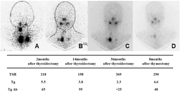
현병력: 환자는 3년 9개월 전 갑상선암이 의심되어 갑상 선전절제술 및 우측 보존적근치목수술(modified radial neck dissection)을 받아 갑상선 유두암(T3N0M0)으로 진단받았 다. 갑상선전절제술 2개월 후 방사성요오드 치료(30 mCi)를 시행하였고, 치료 4일째 시행한 방사성요오드 스캔에서 종 격동의 섭취가 증가되었다(Fig. 1A). 당시 갑상선글로불린은 5.5 ng/mL이었다. 종격동의 섭취증가 원인을 감별하기 위하 여 흉부 전산화 단층촬영을 시행하였고, 종격동에서 큰 이상
소견은 발견되지 않았다(Fig. 2A). 이후 2차례 정기적으로 시행한 방사성요오드 치료 후 스캔에서도 지속적으로 종격 동의 섭취 증가가 관찰되었다(Fig. 1B, C). 흉부 전산화 단 층촬영에서 방사성 요오드가 섭취된 부분과 동일한 위치에 흉선 증식(최대직경 2 cm)이 관찰되었으나(Fig. 2B) 변연이 깨끗하고 림프절 전이 소견은 보이지 않아 경과를 관찰하였 다.
갑상선전절제술 후 14개월 후 갑상선호르몬 보충을 중단 하였을 때 갑상선글로불린이 2.0 ng/mL로 측정되었고 이후 갑상선호르몬 보충 시 1 ng/mL 미만으로 유지되었다. 갑상 선전절제술 후 3년이 경과된 시점에서 방사성요오드 치료를 위해 갑상선호르몬 복용을 중단하자 갑상선글로불린이 2.3
Fig. 1. Radioiodine scans and stimulated thyroglobulin levels. (A) for 2 months after thyroidectomy (post-radioiodine treatment scan). (B) for 14 months after thyroidectomy (post-radioiodine treatment scan). (C) for 36 months after thyroidectomy (post-radioiodine treatment scan). (D) for 8 months after thymectomy (follow-up diagnostic scan): The mediastinal uptake was decreased after thymectomy. TSH, thyroid stimulating hormone; Tg, thyroglobulin; TgAb, antithyroglobulin antibody.
Fig. 2. Chest computed tomography (CT). (A) for postoperative 3 months (B) for postoperative 14 months: The thymus looks like an arrow head and it enlarged than previous CT (A). (C) for postoperative 3 years: The size and shape of thymic lesion was changed. The irregular nodular shape and enhancement in the lesion seems to be a metastatic lesion.
ng/mL로 이전보다 증가하였다. 또한 방사성요오드 치료 4 일째 치료 후 스캔에서 종격동의 섭취가 전보다 증가되고, 흉부 전산화 단층촬영에서 종괴의 크기가 커지면서(2.7 × 0.9 cm) 변연이 불규칙해져서 갑상선 유두암의 종격동 전이 를 배제할 수 없어 이를 수술적으로 확인하기 위해 입원하 였다. 환자는 입원 당시 갑상선호르몬 복용 중이었고, 흉부 증상이나 종괴에 의한 압박증상이 전혀 없었으며, 근무력감 이나 어지러움을 호소하지 않았다.
신체 검사: 혈압은 115/85 mmHg, 맥박수 분당 68회, 호 흡수 분당 20회, 체온은 36℃이었다. 환자는 의식은 명료하 였고, 경부에 이전 수술 반흔이 있었으며 경부 림프절은 촉 지되지 않았다. 그 외 진찰소견에서 이상은 없었다.
검사 소견: 말초혈액검사에서 혈색소 11.1 g/dL, 백혈구 4,550/mm3, 혈소판 263,000/mm3이었고, C-반응단백은 정상 범위였다. 갑상선호르몬 복용 중 시행한 갑상선기능검사에
서 유리 T4 1.29 ng/dL (0.93~2.13 ng/dL), 갑상선자극호르 몬 < 0.05 uIU/mL (0.4~4.1 uIU/mL), 갑상선글로불린 < 1 ng/mL, 항갑상선글로불린 항체 68 ng/dL(0~100 ng/dL)이 었다.
임상경과: 환자는 종격동 종괴를 수술적으로 제거하였다.
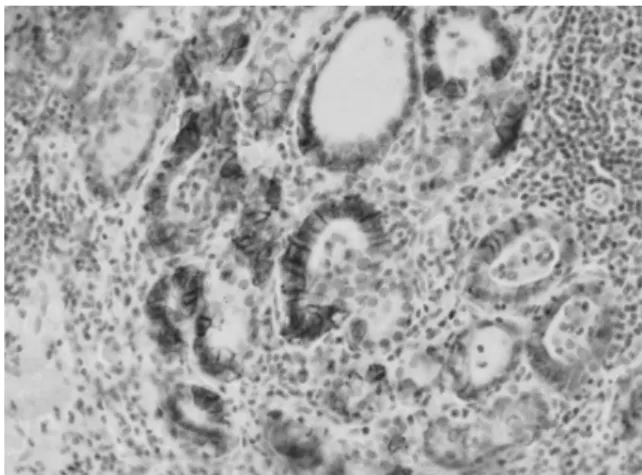
병리소견에서 정상 흉선 조직이었고 갑상선조직은 관찰되지 않았다. 면역염색소견에서 제거된 흉선의 thyroid transcription factor-1(TTF-1)과 갑상선글로불린은 음성이었 고, human NIS(hNIS)는 양성이었다(Fig. 3). 흉선에서 hNIS가 발현된 곳은 대식세포였다. 이전 수술 시 제거된 조 직에서 원발 종양인 갑상선암 조직의 일부 종양세포(Fig. 4) 와 갑상선암 주위의 만성 갑상선염 소견을 보이는 부위의 기저막(Fig. 5)에서 hNIS가 발현되었다. 흉선절제술 8개월 후 진단적 목적으로 시행한 방사성요오드 스캔(10 mCi)에 서 종격동의 섭취가 이전 스캔 결과보다 감소되었다(Fig.
1D). 환자는 현재까지 재발의 증거 없이 외래에서 경과 관 찰 중이다.
고 찰
본 증례는 갑상선 유두암으로 갑상선전절제술 후 방사성 요오드의 종격동 섭취가 발견되어 종격동 절제술을 받았고 이후 방사성요오드의 흉선 섭취가 소실되었다.
131I은 NIS라는 단백 통로를 통해 갑상선에 특이적으로 섭취되는데, hNIS는 갑상선뿐 아니라 침샘, 젖샘, 췌장, 위 점막, 난소, 폐, 신장, 결장, 흉선 등에서 발현된다[3,6,7]. 본 증례에서와 같이 갑상선전절제술 후 방사성요오드의 종격동 섭취가 발견된 경우 생리적인 섭취를 고려해 보아야 한다[8].
이는 스트레스, 갑상선 기능항진증 같은 질병이나 항암요법 등으로 인한 반동현상 때문에 흉선 증식이 나타날 수 있기 Fig. 3. Immunohistochemistry for hNIS protein in thymic
tissue. Only a few thymic macrophages were stained by anti-hNIS (H&E, ×40).
Fig. 4. Immunohistochemical detection for hNIS protein in thyroid tissue. hNIS protein was detected in the basolateral membrane of the tissues of chronic thyroiditis around the papillary cancer (H&E, ×40).
Fig. 5. Immunohistochemistry for hNIS protein in tissue section of papillary thyroid carcinoma. Most of tumor cells reveal loss of hNIS expression except for focal membranous and cytoplasmic staining in a few tumor cells (H&E, ×100).
때문이다[9,10]. 아울러 종격동에는 흉선 외에도 방사성요오 드를 섭취하는 다른 조직들이 있고, 원발성 폐암, 결핵, 기관지 확장증 등에서도 섭취되므로 함께 감별해야 한다[6]. 여러 연 구자들이 생리적인 흉선 섭취를 전이암과 구분하는 방법으 로 아령(dumb-bell)모양이나 화살촉 모양, 반복적인 방사성 요오드 치료 후 흉선의 섭취율 증가, 지연(동위원소 섭취 후 7일째) 스캔에서 흉선의 섭취율 증가, 갑상선글로불린 수치가 낮으며 영상검사에서 흉선의 비대 등을 생리적인 흉선 섭취의 근거로 제시한 바 있으나 진단적이지는 않다[5,6,11].
이처럼 방사성요오드 스캔만으로는 감별이 어렵기 때문에 주로 전산화 단층촬영을 통해 추가적인 정보를 얻는다[12,13].
흉선 증식은 갑상선 기능항진증이나 부신피질 기능 저하, 그 리고 항암요법 등의 심각한 기저질환의 병력이 있는 경우 의심할 수 있고, 전산화 단층촬영 소견으로 미만성으로 대칭 적인 크기 증가가 있을 경우 가능성이 높아진다. 대부분 일 시적이며 원래의 흉선 모양을 유지하는 것이 특징이다. 흉선 종은 10~15%에서 근무력증을 호소하나 절반 정도는 증상을 동반하지 않는다. 전산화 단층촬영 소견은 원형이나 타원형, 혹은 소엽형 종괴로 나타난다. 반면, 1) 사춘기 이후에, 2) 흉선의 편평하거나 오목한 외연이 국소적 또는 전체적으로 볼록해지거나, 3) 변연이 불규칙해지거나, 4) 내부에 불규 칙적인 조영 증강을 보이는 경우, 5) 석회화나 내부 출혈을 동반하는 경우, 6) 주변 지방조직으로 파급되는 형상을 보이 거나, 7) 국소 임파절 종대를 동반하거나, 8) 혈관이나 기도 의 위치를 변경시키는 등의 전산화 단층촬영 소견은 전이암 을 강력히 시사하는 소견이다. 또한 크기와 모양만으로는 악 성과 양성을 감별하기 힘들기 때문에 금기증이 없는 한 수 술적 제거만이 확진 방법이다. 수술 후에는 방사성요오드 스 캔에서 방사성요오드 섭취가 감소함을 확인할 수 있다. 본 증례는 갑상선호르몬 중단 시 갑상선글로불린이 증가하고, 심각한 기저 질환의 병력 없이 종격동 종괴의 크기가 커지 고 변연이 불규칙해지며 모양이 볼록해져서 갑상선 유두암 의 전이를 배제하기 어려웠던 경우이다.
Spitzweg 등[14]은 정상 흉선에서 NIS, 갑상선자극호르 몬수용체, thyroid peroxidase, 그리고 갑상선글로불린과 같 은 갑상선의 주요 단백 유전자를 발견하였다. 이 중 NIS의 경우 흉선 수질세포 내 Hassall's corpuscle, 상피세포, thymocyte에서는 발현되지만[15], 그 외 혈관이나 림프조직 및 결합조직 등에는 발현되지 않는다고 알려져 있다. 이는 정상 흉선을 관찰한 결과로, 이와는 달리 본 증례 환자의 흉 선조직은 갑상선글로불린에 대한 면역염색결과는 음성이었 고, hNIS 면역 표지자에 대해서도 음성인 가운데 hNIS 양 성인 대식세포가 흉선 내에서 발견되었으며 이는 CD68을 통해서도 확인되었다. hNIS 양성 대식세포의 기원과 hNIS 양성 대식세포와 흉선의 요오드 섭취의 인과 관계에 대해서 는 설명하기 어렵다. 그간 보고되었던 흉선에 요오드가 섭취
된 갑상선암 환자 증례들에서는 hNIS에 대한 면역염색반응 에 대해 보고된 바가 없어 추가적인 연구가 필요하다.
본 환자는 흉선 제거술 후 시행한 방사성요오드 스캔에서 종격동의 요오드 섭취가 감소하여 이전 요오드 섭취된 부위 가 흉선임이 확인되었고, 재발의 증거 없이 외래에서 경과 관찰하고 있다.
요 약
갑상선 분화암 치료에 있어서 잔여 갑상선조직의 제거 및 갑상선 전이암을 치료하는데 사용되는 방사성요오드 치료는 NIS(sodium-iodide symporter) 통로를 통해 방사성요오드가 세포 내로 들어가 세포를 파괴하는 것이 치료 기전이다. 본 증례는 26세 갑상선 유두암 환자로 동위원소 치료 중 종격 동의 요오드 섭취가 있어 흉부 전산화 단층촬영과 갑상선글 로불린으로 전이 여부를 확인하였으나 이상소견이 없었다.
경과 관찰 중 흉부 전산화 단층촬영에서 깨끗한 변연을 지 닌 무증상 흉선 종괴가 발견되었으나 증상이 없어 관찰하였 다. 그러나 이후 갑상선호르몬 중단 시 갑상선글로불린이 증 가하고, 흉부 전산화 단층촬영에서 흉선의 변연이 불규칙해 져서 전이암을 배제하기 위해 환자는 종격동 절제술을 시행 받았다. 제거된 조직의 병리 소견은 정상 흉선으로 갑상선조 직은 관찰되지 않았으며, human NIS에 양성이었다. 흉선을 제거한 후 시행한 방사성요오드 스캔에서 이전과 비교하였 을 때 종격동 섭취가 감소함을 관찰할 수 있었다. 본 증례와 같이 갑상선암 환자에서 갑상선전절제술 후 종격동에 방사 성 요오드가 섭취될 경우 모양과 크기가 변화하더라도 생리 적인 흉선에 의한 섭취일 수 있음을 문헌고찰과 함께 보고 하는 바이다.
참 고 문 헌
1. Chung JK: Sodium iodide symporter: its role in nuclear medicine. J Nucl Med 43:1188-1200, 2002 2. Mazzaferri E, Kloos R: Carcinoma of follicular
epithelium : radioiodine and other treatments. In:
Braverman LE, Utiger RD ed. Werner and Ingbar's The Thyroid : A fundamental and clinical text, 9th ed.
pp 934-966, Philadelphia, Lippincott Williams &
Wilkins, 2005
3. Vayre L, Sabourin JC, Caillou B, Ducreux M, Schlumberger M, Bidart JM: Immunohistochemical analysis of Na+/I- symporter distribution in human extra-thyroidal tissues. Eur J Endocrinol 141:382-386, 1999
4. Meller J, Becker W: The human sodium-iodine
symporter (NIS) as a key for specific thymic iodine-131 uptake. Eur J Nucl Med 27:473-474, 2000 5. Wilson LM, Barrington SF, Morrison ID, Kettle AG,
O'Doherty MJ, Coakley AJ: Therapeutic implications of thymic uptake of radioiodine in thyroid carcinoma.
Eur J Nucl Med 25:622-628, 1998
6. Davidson J, McDougall IR: How frequently is the thymus seen on whole-body iodine-131 diagnostic and post-treatment scans? Eur J Nucl Med 27:425-430, 2000
7. Wapnir IL, van de Rijn M, Nowels K, Amenta PS, Walton K, Montgomery K, Greco RS, Dohan O, Carrasco N: Immunohistochemical profile of the sodium/iodide symporter in thyroid, breast, and other carcinomas using high density tissue microarrays and conventional sections. J Clin Endocrinol Metab 88:1880-1888, 2003
8. Park C, Lee M: Thymic radioiodine uptake mimicking metastatic papillary carcinoma in the anterior mediastinum. Korean J Nucl Med 36:87-89, 2002 9. Im JG: Diseases of mediastinum. In : Im JG, Lee KS
ed. Thoracic radiology 1st ed. pp 433-459, Seoul,
Ilchokak, 2000
10. Nakamura T, Murakami M, Horiguchi H, Nagasaka S, Ishibashi S, Mori M, Ishikawa SE: A case of thymic enlargement in hyperthyroidism in a young woman.
Thyroid 14:307-310, 2004
11. Salvatori M, Saletnich I, Rufini V, Troncone L:
Unusual false-positive radioiodine whole-body scans in patients with differentiated thyroid carcinoma. Clin Nucl Med 22:380-384, 1997
12. Tecce PM, Fishman EK, Kuhlman JE: CT evaluation of the anterior mediastinum: spectrum of disease.
Radiographics 14:973-990, 1994
13. Connolly LP, Connolly SA : Thym ic uptake of radiopharmaceuticals. Clin Nucl Med 28:648-651, 2003
14. Spitzweg C, Joba W, Heufelder AE: Expression of thyroid-related genes in human thymus. Thyroid 9:133 -141, 1999
15. Vermiglio F, Baudin E, Travagli JP, Caillou B, Fragu P, Ricard M, Schlumberger M: Iodine concentration by the thymus in thyroid carcinoma. J Nucl Med 37:1830-1831, 1996