• 검색 결과가 없습니다.

An Unusual Case of Left Ventricular Free Wall Rupture Caused by a Silent Myocardial Infarction

N/A
N/A
Protected

Academic year: 2022

Share "An Unusual Case of Left Ventricular Free Wall Rupture Caused by a Silent Myocardial Infarction"

Copied!
3
0
0

로드 중.... (전체 텍스트 보기)

전체 글

(1)

702 Copyright © 2012 The Korean Society of Cardiology Korean Circulation Journal

Introduction

Left ventricular free wall rupture (LVFWR) is reported to occur in 2-6% of acute myocardial infarction (MI) cases. LVFWR is presumed responsible for as much as 20-80% of infarct-related deaths.

1)

There is a history of previous MI in 25% of cases, LVFWR can often be the first presentation of ischemic heart disease.

2)

We describe the case of a 60-year-old man without history of ischemic heart disease who was admitted with chest pain; LVFWR and bacterial pericarditis were detected by urgent echocardiography and managed successfully.
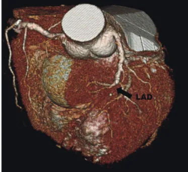
Case

A 60-year-old man was a smoker and drinker with no history of co- ronary artery disease, hypertension and hyperlipidemia except dia-

Case Report

http://dx.doi.org/10.4070/kcj.2012.42.10.702 Print ISSN 1738-5520 • On-line ISSN 1738-5555

An Unusual Case of Left Ventricular Free Wall Rupture Caused by a Silent Myocardial Infarction

Xin Jin, MD 1 , Sang-Hoon Seol, MD 2 , Seung-Hyeon Park, MD 2 , Joo-Won Lee, MD 2 , Bo-Min Park, MD 2 , Dong-Kie Kim, MD 2 , Ki-Hun Kim, MD 2 , Doo-Il Kim, MD 2 , Ho-Ki Min, MD 3 , and Yeon-Mee Kim, MD 4

1

Division of Cardiology, Department of Internal Medicine, YanBian Second People’s Hospital, YanBian, China

2

Division of Cardiology, Department of Internal Medicine ,

3

Division of Chest Surgery and

4

Pathology, Haeundae Paik Hospital, Inje University College of Medicine, Busan, Korea

Left ventricular free wall rupture (LVFWR) is a serious complication of myocardial infarction. It presents with a very high mortality rate and can be rescued by accurate diagnosis and emergency surgery. LVFWR can occur with sudden overt clinical symptoms or present insidi- ously. This report highlights the case of a man with no prior history of coronary artery disease, who presented with LVFWR and pericardial effusion that evolved to severe bacterial pericarditis. (Korean Circ J 2012;42:702-704)

KEY WORDS: Myocardial infarction; Rupture; Pericarditis.

Received: December 16, 2011 Revision Received: February 8, 2012 Accepted: March 13, 2012

Correspondence: Sang-Hoon Seol, MD, Division of Cardiology, Depart- ment of Internal Medicine, Inje University College of Medicine, Haeundae Paik Hospital, 875 Haeun-daero, Haeundae-gu, Busan 612-862, Korea Tel: 82-51-797-3009, Fax: 82-51-797-3070

E-mail: [email protected]

• The authors have no financial conflicts of interest.

This is an Open Access article distributed under the terms of the Creative Commons Attribution Non-Commercial License (http://creativecommons.

org/licenses/by-nc/3.0) which permits unrestricted non-commercial use, distribution, and reproduction in any medium, provided the original work is properly cited.

betes, which was diagnosed several years ago. The man presented in the emergency room with complaints of chest pain for 2 days. The patient was conscious and physical examination showed heart rate 128 beats/min, blood pressure 95/70 mm Hg, temperature 36.5˚C.

Heart sound was faint without murmur and bruits. Initial electrocar-

diogram (ECG) showed atrial fibrillation and pathological Q waves

in V 1-6. Chest X-ray revealed marked cardiomegaly. Blood tests

were negative for Troponin I and creatine kinase-MB (CK-MB). In-

flammatory markers were raised (white cell count of 23.12×10⁹/L,

neutrophil percentage 88.4%, C reactive protein 16.84 mg/dL), and

hemoglobin was mildly reduced (11.7 g/L). Kidney and liver enzy-

mes were severely elevated (aspartate aminotransferase 2325 IU/L,

alanine aminotransferase 1005 IU/L, γ-guanosine triphosphate 130

IU/L, lactate dehydrogenase 2989 IU/L, blood urea nitrogen 124.2

mg/dL, Creatinine 7.29 mg/dL). These lab results suggested that the

patient was almost experiencing multiple organ dysfunction syn-

drome. Echocardiography showed reduced left ventricular (LV) sys-

tolic function and LV dilatation, especially at the LV apex. A large

amount of pericardial effusion was noted. There was a small ab-

normal shunt from the LV apex to the pericardial space (Fig. 1). Thus,

LVFWR was strongly suspected. In order to establish a diagnosis of

LVFWR, chest computerized tomography (CT) was conducted and

showed LV pseudoaneurysm with rupture in the anteroinferior wall

with associated hemopericardium (Fig. 2). The patient underwent

emergency LV reconstructive surgery (Dor procedure). The pericardial

space was full of dirty fibrotic material. A small tear was found on

the LV apex. The postoperative pathology revealed focal coagula-

(2)

703 Xin Jin, et al.

http://dx.doi.org/10.4070/kcj.2012.42.10.702 www.e-kcj.org

tion necrosis and abscess presented with blood clots and fibrin clots.

Blood culture showed Streptococcus pneumonia. Postoperatively, intravenous antibiotic treatment was continued and the patient was discharged after 2 weeks with a good recovery. Three months later, coronary CT showed total occlusion of the mid left anterior descending coronary artery. Right coronary artery and left circumflex coronary artery was normal without stenosis (Fig. 3). At present, the aspirin therapy has been continued after discharge.

Discussion

Left ventricular free wall rupture is a dramatic complication of MI in which there is a rupture of infarcted LV free wall tissue. The rup- ture is commonly insidious with bleeding into the pericardial sac and subsequent cardiac tamponade.

3)

It is third to cardiogenic shock and arrhythmias as the leading cause of death following an MI.

4)

The most common location of LVFWR is in the anterior or lateral walls.

5)

It often appears within the first week following an acute MI,

usually on the 4th or 5th day post-MI, although the time to mani- festation may range from a few minutes to more than one month after the acute MI.

6-9)

Clinically, ruptures can be divided into 3 types:

acute rupture, which results in death within a few minutes due to massive hemorrhage into the pericardial cavity; subacute rupture, which is characterized by a smaller tear that may temporarily be sealed by a clot or fibrinous pericardial adhesion and may be compa- tible with life for several hours or even longer; and chronic rupture with false aneurysm formation, which occurs when the leakage of blood is slow and when surrounding pressure on the epicardium temporarily controls the hemorrhage.

10)

The recent significant in- crease in the number of LVFWR diagnosed before death and the number of surgical repairs attempted is largely due to the wide- spread availability and use of cardiac imaging, particularly echocar-

A   B  

Fig. 1. Echocardiography reveals (A) an pseudoaneurysm was formed on the left ventricular apex with shunt flow from the left ventricular cavity to the pericardial space, (B) left ventricular apex was obviously dilated to become an aneurysm and a large amount of effusion can be seen.

Fig. 2. Chest computed tomography shows the rupture on the apex of left ventricular wall.

Fig. 3. Coronary computed tomography shows that the mid left anterior

descending coronary artery (arrow) was completely obstructed. LAD: left

anterior descending coronary artery.

(3)

704 Ventricular Free Wall Rupture Caused by Silent MI

http://dx.doi.org/10.4070/kcj.2012.42.10.702 www.e-kcj.org

diography.

11)

Echocardiography provides invaluable diagnostic infor- mation and indicates the possible extent and location of a rupture prior to surgery. The most frequent echocardiographic finding in the case of LVFWR is a localized pericardial effusion overlying the infarcted akinetic area. Other signs include echogenic ‘specks’ within the effusion and visible wall defects.

12)

In the current case, we found that pericardial effusion by echocardiography established the di- agnosis of LVFWR by chest CT. On the basis of Troponin I, CK-MB and ECG, we thought that the anterior MI had occurred previously and led to LVFWR. At first, we had suspected coronary atheroscle- rotic heart disease was the reason of MI. In addition, we also could not rule out bacterial endocarditis, other sources of infection and embolization. The patient had diabetes (immunocompromised host) and atrial fibrillation. Atrial fibrillation could cause embolization (such as septic embolism) into coronary artery, then MI due to just embolism or abscess (caused by septic embolism) could occur without atherosclerotic coronary artery disease. The spread of in- fection affected the pericardial cavity and caused severe bacterial pericarditis. For this reason, CT angiography of the patient showed no evidence of coronary artery disease in other vessels except in the left anterior descending coronary artery occlusion. A patho- logic report could not differentiate whether MI was atherosclerotic, or embolic, or secondary change according to abscess. The etiology of MI remains unclear.

In conclusion, rupture of the LV free wall is an often fatal com- plication of acute MI. Sometimes it presents insidiously. Physicians need to be aware of the risk factors, clinical signs, ECG and so on.

This diagnosis can be made more accurately by using echocardiog- raphy and chest CT. Most of all, survival depends on early assess- ment of the patient, prompt investigation and diagnosis, and urgent surgical treatment.

References

1. Figueras J, Cortadellas J, Soler-Soler J. Left ventricular free wall rup-

ture: clinical presentation and management. Heart 2000;83:499-504.

2. Shirani J, Berezowski K, Roberts WC. Out-of-hospital sudden death from left ventricular free wall rupture during acute myocardial infarc- tion as the first and only manifestation of atherosclerotic coronary ar- tery disease. Am J Cardiol 1994;73:88-92.

3. Vanezis AP, Quadery R, Wasil M, Azher M. An unusual presentation of left ventricular free wall rupture following a silent myocardial infarc- tion. BJMP 2009;2:41-3.

4. Pohjola-Sintonen S, Muller JE, Stone PH, et al. Ventricular septal and free wall rupture complicating acute myocardial infarction: experi- ence in the Multicenter Investigation of Limitation of Infarct Size. Am Heart J 1989;117:809-18.

5. David TE. Surgery for post infarction rupture of the free wall of the ventricle. In: David TE editor. Mechanical Complications of Myocardial Infarction. Austin: RG Landes Company;1993. p.142.

6. Pollak H, Diez W, Spiel R, Enenkel W, Mlczoch J. Early diagnosis of sub- acute free wall rupture complicating acute myocardial infarction. Eur Heart J 1993;14:640-8.

7. Mann JM, Roberts WC. Rupture of the left ventricular free wall during acute myocardial infarction: analysis of 138 necropsy patients and com- parison with 50 necropsy patients with acute myocardial infarction without rupture. Am J Cardiol 1988;62:847-59.

8. Batts KP, Ackermann DM, Edwards WD. Postinfarction rupture of the left ventricular free wall: clinicopathologic correlates in 100 consecu- tive autopsy cases. Hum Pathol 1990;21:530-5.

9. Park SC, Kim JH, Kim YM, et al. A case of early developed left ventricu- lar free wall rupture followed by acute inferior myocardial infarction.

Korean Circ J 1999;29:73-8.

10. Exadaktylos NI, Kranidis AI, Argyriou MO, Charitos CG, Andrikopoulos GK. Left ventricular free wall rupture during acute myocardial infarction:

early diagnosis and treatment. Hellenic J Cardiol 2002;43:246-52.

11. Reardon MJ, Carr CL, Diamond A, et al. Ischemic left ventricular free wall rupture: prediction, diagnosis, and treatment. Ann Thorac Surg 1997;64:1509-13.

12. Wehrens XH, Doevendans PA. Cardiac rupture complicating myocardi-

al infarction. Int J Cardiol 2004;95:285-92.

수치

Fig. 2. Chest computed tomography shows the rupture on the apex of left  ventricular wall.

참조

관련 문서

Chest computed tomography revealed large left ventricular aneurysm with rupture in the anteroinferior wall and associated hemopericardium (Fig.2).. The patient

Among the study subjects who had a baseline LVEF of less than 45% soon after acute MI, left ventricular systolic dysfunction was recovered (follow-up LVEF ≥45%) in almost 50%

Fatal rupture of both left ventricular free wall and ventricular septum (double rupture) during acute myocardial infarction: analysis of seven patients stu- died

Background: Matrix Metalloproteinase (MMP) inhibition has emerged as a potential therapeutic strategy for the left ventricular dilatation that occurs after myocardial infarction.

After observing a 1 cm rupture on the right ventricular wall and a necrotic hemorrhagic scar with a rupture on the left ventricular apical wall, repair of the ruptured areas with

Relationship between Serial Measurement of Plasma Brain Natriuretic Peptide and Left Ventricular Remodeling or Long-term Prognosis after Acute Myocardial Infarction..

Here we describe a case of a patient with right ventricular (RV) dysfunction caused by acute RV infarction and with cardiogenic shock, whose condition improved after

eNOS, endothelial nitric oxide synthase; iNOS, inducible nitric oxide syn- thase; MI, myocardial infarction; MI + Losar, myocardial infarction with losartan treatment.. Numbers