DOI: 10.4196/kjpp.2009.13.6.511
511
ABBREVIATIONS: 3D-QSAR, three dimensional quantitative structure activity relationship; PBS, phosphate-buffered saline; MTT, 3-[4,5- dimethylthiazol-2-yl]-2,5-diphenyltetrazolium bromide; CoMFA, com- parative molecular field analysis; CoMSIA, comparative molecular similarity indices analysis.
Received November 25, 2009, Revised December 3, 2009, Accepted December 17, 2009
Corresponding to: Chaeuk Im, College of Pharmacy, Chung-ang University, Heuksuk-dong, Dongjak-gu, Seoul 156-756, Korea. (Tel) 82-2-820-5603, (Fax) 82-2-816-7338, (E-mail) [email protected]
Cytotoxic Activity and Three-Dimensional Quantitative Structure Activity Relationship of 2-Aryl-1,8-naphthyridin-4-ones
Yong Jin Kim, Eun Ae Kim, Mi Lyang Chung, and Chaeuk Im College of Pharmacy, Chung-ang University, Seoul 156-756, Korea
A series of substituted 2-arylnaphthyridin-4-one analogues, which were previously synthesized in our laboratory, were evaluated for their in vitro cytotoxic activity against human lung cancer A549 and human renal cancer Caki-2 cells using MTT assay. Some compounds (11, 12, and 13) showed stronger cytotoxicity than colchicine against both tumor cell lines, and compound 13 exhibited the most potent activity with IC50 values of 2.3 and 13.4 μM, respectively. Three-dimensional quantitative structure activity relationship (3D-QSAR) studies of comparative molecular field analysis (CoMFA) and comparative molecular similarity indices analysis (CoMSIA) were performed. Predictive 3D-QSAR models were obtained with q2 values of 0.869 and 0.872 and r2ncv values of 0.983 and 0.993 for CoMFA and CoMSIA, respectively. These results demonstrate that CoMFA and CoMSIA models could be reliably used in the design of novel cytotoxic agents.
Key Words: CoMFA, CoMSIA, Cytotoxicity, MTT, QSAR
INTRODUCTION
The microtubules play an important role in mitosis.
Compounds that attack microtubules can inhibit or pro- mote microtubule assembly, resulting in cell arrest in mi- tosis (Jordan and Wilson, 2004). Therefore, these anti- mitotic compounds have been used as anticancer drugs. For example, vinca alkaloids inhibit tubulin polymerization in- to microtubules and taxoids promote microtubule assembly (Rowinsky and Donehower, 1992; Verwij et al., 1994).
Colchicine is another antimitotic agent, but it is not used clinically due to its high toxicity (Hastie, 1991; Chen et al., 1997).
Some flavonoid derivatives interfere with tubulin poly- merization to produce potent cytotoxic activity against sev- eral cancer cell lines (Shi et al., 1995; Manthey et al., 2001).
To find more potent antimitotic compounds, some re- searchers have synthesized new flavonoid analogues, such as 2-arylquinolin-4-ones (Xia et al., 1998; Lai et al., 2005;
Nakamura et al., 2005), 2-arylnaphthyridin-4-ones (Chen et al., 1997; Zhang et al., 1999), and 2-arylquinazolin-4-ones (Hamel et al., 1996; Hour et al., 2000; Xia et al., 2001).
Some of these compounds, especially 2-arylnaphthyridin- 4-ones, exhibit potent cytotoxicity; thus, we synthesized a series of substituted 2-arylnaphthyridin-4-one analogues and conducted cytotoxicity assays in human tumor cell lines (Chung, 2004).
The 3D-QSAR model yields some information about the structural requirements for more potent activity of ligands
when the structure of the receptor is not known (Rieger et al., 2001). In this paper, we report the cytotoxic activity and 3D-QSAR analysis of sixteen synthesized compounds to further the development of more potent cytotoxic agents.
METHODS Materials
Phosphate-buffered saline (PBS) was purchased from Boehringer Mannheim (Indianapolis, IN). 3-[4,5-dimethyl- thiazol-2-yl]-2,5-diphenyltetrazolium bromide (MTT), col- chicine, trypsin, dimethyl sulfoxide (DMSO), and other re- agents were purchased from Sigma (St. Louis, MO). Human lung cancer A549 and renal cancer Caki-2 tumor cell lines were obtained from Chong Kun Dang Pharmaceutical Corp., and 2-aryl-1,8-naphthyridin-4-one analogues were synthesized in our laboratory.
In vitro cytotoxic assay
The prepared compounds were tested for their cytotox- icity against the human lung cancer A549 and human renal cancer Caki-2 cell lines. The cytotoxic activity was de- termined using a MTT-based colorimetric assay. Briefly, cell lines were seeded at a density of approximately 5×10³ cells/well in 96-well plates. Each well contained 180 μl of medium to which 20 μl of 10×concentration of the prepared compounds or PBS were added. After 4 days of culture,
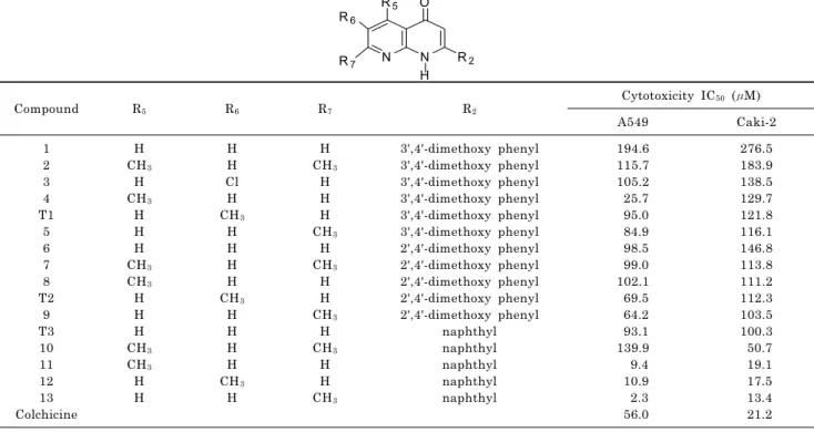
Table 1. In vitro cytotoxic activity of 2-aryl-1,8-naphthyridin-4-ones compared with colchicines
N N
O
R2 H R5 R6
R7
Compound R5 R6 R7 R2
Cytotoxicity IC50 (μM)
A549 Caki-2
1 2 3 4 T1
5 6 7 8 T2
9 T3 10 11 12 13 Colchicine
H CH3
H CH3
H H H CH3
CH3
H H H CH3
CH3
H H
H H Cl H CH3
H H H H CH3
H H H H CH3
H
H CH3
H H H CH3
H CH3
H H CH3
H CH3
H H CH3
3',4'-dimethoxy phenyl 3',4'-dimethoxy phenyl 3',4'-dimethoxy phenyl 3',4'-dimethoxy phenyl 3',4'-dimethoxy phenyl 3',4'-dimethoxy phenyl 2',4'-dimethoxy phenyl 2',4'-dimethoxy phenyl 2',4'-dimethoxy phenyl 2',4'-dimethoxy phenyl 2',4'-dimethoxy phenyl
naphthyl naphthyl naphthyl naphthyl naphthyl
194.6 115.7 105.2 25.7 95.0 84.9 98.5 99.0 102.1 69.5 64.2 93.1 139.9 9.4 10.9 2.3 56.0
276.5 183.9 138.5 129.7 121.8 116.1 146.8 113.8 111.2 112.3 103.5 100.3 50.7 19.1 17.5 13.4 21.2 0.1 mg of MTT was added to each well and incubated at
37oC for 4 h. Plates were centrifuged at 450×g to precipitate the formazan crystals. The medium was removed and 150 μl of DMSO was added to each well to dissolve formazan. In this assay, MTT was converted to blue formazan by mi- tochondrial enzymes. The intensity of the blue color formed was measured with an ELISA reader at 540 nm. The IC50
value was defined as the concentration of the compound that reduced the absorbance at 540 nm by 50% (Park et al., 1987; Manthey and Guthrie, 2002).
Molecular modeling and alignment
A total of sixteen compounds exhibiting cytotoxic activity with IC50 values ranging from 2.3∼276.5 μM were used to perform the 3D-QSAR analysis. The IC50 values were trans- formed into pIC50 (−log IC50) values and used as the de- pendent variable in CoMFA and CoMSIA studies. The structures of the training set and the test set are shown in Table 1. The data set was divided into two groups.
Thirteen compounds (1~13) were used for the training set.
Three compounds (T1~T3) were selected randomly as a test set and were used for external validation of the 3D-QSAR models.
Molecular modeling was performed using the Sybyl 8.1.1 software of Tripos (SYBYL, 2009). All molecules were sketched by the sketch module in SYBYL. Each structure was optimized using a conjugate gradient method based on the TRIPOS force field with Gasteiger Huckel charges and gradient convergence criteria of 0.05 kcal/mol. Simulated annealing on the energy optimized structures was carried out with 50 cycles. They were heated at 1000 K for 1000 fs to reach equilibrium and then annealed at 200 K for 1000 fs.
The 50 conformations were then minimized to obtain low
energy conformations for each compound. The structures were aligned by using the align database. Compound (13), which showed the most potent activity, was selected as a template molecule. The 1,8-naphthyridin fragment of com- pound (13) in its optimized conformation was used as a com- mon substructure in the alignment.
CoMFA and CoMSIA
CoMFA and CoMSIA analysis are based on the relation- ships between biological activity and structural properties of the compounds when the receptor structure is not known.
In the CoMFA study, the aligned molecules were put in a three-dimensional grid of 2Ao. The steric and electrostatic properties of the compounds were calculated at each grid point using Lennard-Jones potential and Coulombic poten- tial, respectively. The sp3 carbon probe atom with +1 charge and Van der Waals radius of 1.52Ao were used to calculate the CoMFA steric and electrostatic fields. The de- fault value of 30 kcal/mol was used as the maximum steric and electrostatic energy cut-off. In CoMSIA analysis, five fields are calculated: steric, electrostatic, hydrophobic, hy- drogen bond acceptor, and hydrogen bond donor fields. The probe atoms with radius 1Ao, charge +1, hydrophobicity
+1, hydrogen bond donating +1, and hydrogen bond ac- cepting +1 were used to calculate the five fields. The at- tenuation factor of 0.3 was used as the default value.
The partial least squares (PLS) were carried out using the cross-validated leave one out (LOO) method to evaluate q2 values. The model of highest q2 value and lowest stand- ard error of prediction suggested the optimum number of components. Leave one out (LOO) cross-validation was used to estimate the predictive ability of the models (Jackson et al., 2009).
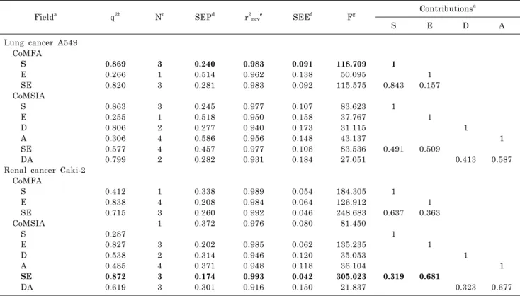
Table 2. Statistical data of CoMFA and CoMSIA for two tumor cell lines
Fielda q2b Nc SEPd r2ncve SEEf Fg Contributionsa
S E D A
Lung cancer A549 CoMFA
S E SE CoMSIA S E D A SE DA
Renal cancer Caki-2 CoMFA
S E SE CoMSIA S E D A SE DA
0.869 0.266 0.820 0.863 0.255 0.806 0.306 0.577 0.799
0.412 0.838 0.715 0.287 0.827 0.538 0.485 0.872 0.619
3 1 3 3 1 2 4 4 2
1 4 3 1 3 2 4 3 3
0.240 0.514 0.281 0.245 0.518 0.277 0.586 0.457 0.282
0.338 0.208 0.260 0.372 0.202 0.314 0.371 0.174 0.301
0.983 0.962 0.983 0.977 0.950 0.940 0.956 0.977 0.931
0.989 0.984 0.992 0.976 0.985 0.946 0.948 0.993 0.916
0.091 0.138 0.092 0.107 0.158 0.173 0.148 0.108 0.184
0.054 0.064 0.046 0.080 0.062 0.120 0.118 0.042 0.150
118.709 50.095 115.575 83.623 37.767 31.115 43.137 83.536 27.051
184.305 126.912 248.683 81.450 135.235 35.053 36.104 305.023 21.837
1 0.843
1
0.491
1 0.637
1
0.319 1 0.157
1
0.509
1 0.363
1
0.681 1
0.413
1
0.323 1 0.587
1 0.677
aFields used, S, steric; E, electrostatic; D, H-bond donor; A, H-bond acceptor; bq2, cross-validated correlation coefficient from leave-one-out(LOO); cN, optimum number of components; dSEP, standard error of prediction; er2ncv, non-crossvalidated correlation coefficient; fSEE, standard error of estimate; gF, F-test value.
RESULTS In vitro cytotoxic activity
The sixteen synthesized compounds and colchicine were evaluated for their cytotoxicity in vitro against lung cancer A549 and renal cancer Caki-2 cells. The cytotoxic activities of each compound were obtained as IC50 values, and the data are presented in Table 1. Colchicine showed moderate cytotoxicity with IC50 values of 56 and 21.2μM against A549 and Caki-2 tumor cell lines in our assay conditions, respectively. Among the synthesized compounds, Compounds 11, 12, and 13 showed stronger cytotoxic activity than col- chicine against both tumor cell lines. They have a naphthyl group at the C2-position in 1,8-naphthyridine and showed IC50 values of 9.4, 10.9, and 2.3 μM against lung cancer A549 cells and 19.1, 17.5, and 13.4 μM against renal cancer Caki-2 cells, respectively. The compound 13 gave the most potent activity and its IC50 values were 2.3 and 13.4 μM, respectively. The IC50 values of the other compounds ranged from 25.7 to 194.6 μM in A549 cells and 50.7 to 276.5 μM in Caki-2 cells.
CoMFA and CoMSIA analysis
The statistical data from CoMFA and CoMSIA models are shown in Table 2.
Lung cancer A549: By use of CoMFA default settings with steric and electrostatic fields, a cross-validated co-
efficient (q2)=0.820 with 3 optimum number of components (N) was observed. This model had a non-cross-validated co- efficient (r2ncv)=0.983, standard error of estimate (SEE)=
0.092, and F value=115.575. CoMFA with only steric field showed q2=0.869, N=3, r2ncv=0.983, SEE=0.091, and F value=
118.709. Six CoMSIA models were calculated from the same training and test sets. CoMSIA model with only steric field gave q2 value=0.863, N=3, r2ncv=0.977, SEE=0.107, and F value=83.623. These statistical parameters indicate the ac- curacy of model.
CoMFA model with steric field showed higher q2 value (0.869) than any other CoMFA and CoMSIA models.
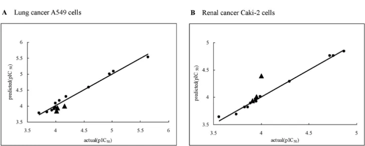
Therefore, using this model, the predicted pIC50 values for each training set and the residual were calculated (Table 3).
The predicted pIC50 values of the test set molecules and their residual values are shown in Table 4. These test set compounds were used to validate CoMFA model with steric field. A graph of the predicted pIC50 values versus actual pIC50 values for the training set and test set is shown in Fig. 1A.
Renal cancer Caki-2 cells: Six CoMSIA models with ster- ic, electrostatic, hydrogen bond acceptor, and hydrogen bond donor fields were considered. In the CoMSIA model with steric and electrostatic fields, a cross-validated co- efficient (q2)=0.872, optimum number of components (N)=3, non-cross-validated coefficient (r2ncv)=0.993, standard error of estimate (SEE)=0.042, and F value=305.023 were observed.
CoMFA model with electrostatic field gave q2=0.838, N=4, r2ncv=0.984, SEE=0.064, and F value=126.912.
Table 3. Actual and predicted activity (pIC50) of training set
Compound A549 Caki-2
Actual Predicted Residual Actual Predicted Residual
1 2 3 4 5 6 7 8 9 10 11 12 13
3.71 3.94 3.98 4.59 4.07 4.01 4.00 3.99 4.19 3.85 5.03 4.96 5.64
3.78 3.86 3.90 4.59 4.16 3.94 4.08 3.94 4.29 3.81 5.09 5.00 5.53
−0.07 0.08 0.08 0.00
−0.09 0.07
−0.08 0.05
−0.10 0.04
−0.06
−0.04 0.11
3.56 3.74 3.86 3.89 3.94 3.83 3.94 3.95 3.99 4.30 4.72 4.76 4.87
3.64 3.69 3.82 3.89 3.94 3.82 3.96 3.92 4.01 4.29 4.76 4.76 4.84
−0.08 0.05 0.04 0.00 0.00 0.01
−0.02 0.03
−0.02 0.01
−0.04 0.00 0.03
Average 0.07 0.03
Table 4. Actual and predicted activity (pIC50) of test set
Compound A549 Caki-2
Actual Predicted Residual Actual Predicted Residual
T1 T2 T3
4.02 4.16 4.03
3.85 4.00 3.95
0.17 0.16 0.08
3.91 3.95 4.00
3.94 4.01 4.39
−0.03
−0.06
−0.39
Average 0.14 0.16
Fig. 1. Scatter plot of actual activities versus predicted activities against A549 (A) and Caki-2 (B) tumor cell lines (●: training set molecules,
▲: test set molecules).
CoMSIA model with steric and electrostatic fields showed better q2 value (0.872) than any other CoMFA and CoMSIA models. With this model, the predicted pIC50 values for the training set and the residuals were calculated (Table 3).
The predicted pIC50 values of the test set and their residual values are shown in Table 4. The graph of predicted pIC50
values versus actual pIC50 values for the training set and test set is shown in Fig. 1B.
DISCUSSION
2-Phenyl-4-quinolones reportedly act as antimitotic agents that interact with tubulin at the colchicine site (Kuo et al., 1993; Li et al., 1994). The 2-phenyl-1,8-naphthyridin- 4-ones can be considered isosteres of 2-phenyl-4-quinolones.
Since the CH moiety in 4-quinolone is isosterically sub- stituted to nitrogen atom at the C8-position in 1,8-naph-
Fig. 2. Contour maps of steric and electrostatic fields in lung cancer A549 (A) and renal cancer Caki-2 (B) cells. Compound 13 is shown inside the field. Color-coding is as follows: Green indicates regions where bulky groups increase activity, whereas yellow indicates regions where bulky groups decrease activity. Blue indicates that positive charge favors high activity, whereas red indicates that negative charge favors high activity.
thyridin-4-one, the 2-phenyl-1,8-naphthyridin-4-one can be assumed to have similar biological activity to 2-phe- nyl-4-quinolones.
In terms of average cytotoxicity in the A549 tumor cell line, compounds with methyl substituted at the C6-position were less active than those substituted at the C5 or C7-position in the 1,8-naphthyridin ring, while compounds with two methyl groups at both the C5 and C7-positions or no substituted group at the C5, C6 and C7-positions in 1,8-naphthyridin were substantially less active. In renal cancer Caki-2 cells, methyl-substituted compounds at the C7-position were more potent than those substituted at the C5 or C6-position in 1,8-naphthyridin, while methyl sub- stituted compounds at both C5 and C7-positions or no sub- stituted group at C5, C6 and C7-positions in 1,8-naphthyr- idin were much less active.
The effects of substitutions at the C5, C6 and C7-positions depend on the substitution at the C2-position in 1,8-naph- thyridin, which is consistent with the previously reported
trend (Chen et al., 1997). The introduction of a naphthyl group at C2-position increased cytotoxicity and methoxy substituted compounds at both the C2`and C4`-positions were more active than those substituted at the C3`and C4`-positions in 1,8-naphthyridin.
For lung cancer A549 cells, the statistical results from the CoMFA model with steric field demonstrated high cross-validated value q2 (0.869) and non-cross-validated co- efficient value r2ncv (0.983), and these values suggest that this model was predictive. In general, a q2 value higher than 0.5 is considered an indication that the model is in- ternally predictive. This model showed optimum component number=3 and suggested that steric interaction strongly in- fluenced the cytotoxic activity.
For renal cancer Caki-2 cells, the CoMSIA model with steric and electrostatic fields showed higher q2 value than CoMFA models. Good predictivity of the model was in- dicated by high q2 (0.872) and r2ncv (0.993) coefficient values.
The relative contributions of steric field (31.9%) and electro- static field (68.1%) indicated that strong electrostatic inter- action was involved in the activity.
The small average residual values (0.07 and 0.03) of actual and predicted activities in Table 3 indicated that the pre- dicted activities from CoMFA and CoMSIA models corre- lated well with actual activities. The test set was used to validate the predictivity of these CoMFA and CoMSIA models. The residual values of the test set were calculated and their small average residual values (0.14 and 0.16) demonstrated that the predicted values of the test set also correlated well with the actual values.
Contour maps indicate the regions around molecules where increased or decreased activity was predicted by physicochemical property changes in the molecules. The CoMFA contour map of steric field in lung cancer A549 cells and the CoMSIA contour maps of steric and electrostatic fields in renal cancer Caki-2 cells are shown in Fig. 2. In the steric contour map, the green contours indicate steri- cally favored regions, whereas the yellow contours desig- nate sterically unfavorable regions. In the electrostatic con- tour map, blue and red regions are favorable for positive charge and negative charge, respectively. The molecule in the contour maps is compound (13), which showed the most potent activity.
The CoMFA steric contour map in lung cancer A549 cells (Fig. 2A) illustrated two small yellow contours above the C2-naphthyl ring and one large yellow contour below the C2-naphthyl ring of 1,8-naphthyridin. These steric contours indicate that bulky substituents at these sites are unfa- vorable for cytotoxic activity. This yellow contour map sug- gests that a flat phenyl or naphthyl ring at the C2-position increases cytotoxic activity, but non-flat and bulky sub- stituents at the C2-position decrease cytotoxicity. The pres- ence of green contours at the C4` and C8`-positions of the naphthyl ring and near the methyl group at the C7-position in 1,8-naphthyridin indicates that bulky groups around these regions increase the cytotoxic activity. This green con- tour map proposes that atoms bigger than hydrogen at the C4` and C8`-position of the naphthyl ring and that groups bigger than methyl at the C7-position of 1,8-naphthyridin might increase the activity.
The CoMSIA steric contour map in renal cancer Caki-2 cells (Fig. 2B) displayed sterically favorable green regions at the C3`, C4`, C7`, and C8`-positions of the C2-naphthyl ring and the methyl group at the C7-position in 1,8- naphthyridin. This green contour map suggests that the in-
troduction of bulky groups in these areas might increase the cytotoxic activity against renal cancer Caki-2 cells. The presence of a sterically unfavorable yellow region above the C2-naphthyl ring indicates that bulky groups around these regions decrease the cytotoxic activity, but a flat naphthyl ring at the C2-position is acceptable for the activity. The CoMSIA electrostatic contour map in renal cancer Caki-2 cells (Fig. 2B) showed the favorable regions for positive charge (blue) near the C3 and C4-position of 1,8-naphthyr- idin and the C4`-position of the C2-naphthyl ring. These blue contours indicate that electron-deficient groups at these sites enhance the cytotoxic activity, and the carbonyl group in the C4-position of 1,8-naphthyridin might be important for activity. The presence of a red contour in the C2-naphthyl ring suggests that negative electrostatic potential around this region increases the cytotoxic activity, and the elec- tron-rich C2- naphthyl ring increases the activity.
In summary, compounds 11, 12, and 13 produced stronger cytotoxicity than colchicine against both tumor cell lines.
The good predictivity of the CoMFA and CoMSIA models was shown by high q2 (0.869 and 0.872) and r2ncv (0.983 and 0.993) coefficient values. The small average residual values of the training (0.07 and 0.03) and test (0.14 and 0.16) sets indicated that the predicted activities from the CoMFA and CoMSIA models correlated well with the actual activity. The CoMFA contour map in lung cancer A549 cells suggested that atoms bigger than hydrogen at the C4` and C8`-positions of the naphthyl ring and groups bigger than methyl at the C7-position of 1,8-naphthyridin might in- crease the cytotoxicity. The CoMSIA contour map in renal cancer Caki-2 cells suggested that the introduction of bulky groups at the C3`, C4`, C7`, and C8`-positions of the C2-naphthyl ring and the C7-position in 1,8-naphthyridin and that pos- itively charged groups near the C3 and C4-positions of 1,8- naphthyridin and the C4`-position of the C2-naphthyl ring might enhance the cytotoxic activity. In addition, the car- bonyl group in the C4-position and the C2-naphthyl ring of 1,8-naphthyridin might be important for biological activity.
ACKNOWLEDGEMENTS
This Research was supported by the Chung-Ang University Research Grants in 2009.
REFERENCES
Chen K, Kuo SC, Hsieh MC, Mauger A, Lin CM, Hamel E, Lee KH. Antitumor agents. 174. 2′,3′,4′,5,6,7-Substituted 2-phenyl- 1,8-naphthyridin-4-ones: their synthesis, cytotoxicity, and inhi- bition of tubulin polymerization. J Med Chem 40: 2266−2275, 1997.
Chung ML. Synthesis and Anti-Cancer Activity of 2-Phenyl-1,8- naphthyridine-4-one Derivatives. Chung Ang Graduate School Master thesis, 2004.
Hamel E, Lin CM, Plowman J, Wang HK, Lee KH, Paull KD.
Antitumor 2,3-Dihydro-2-(aryl)-4(1H)-quinazolinone Derivatives.
Interactions with Tubulin. Biochem Pharmacol 51: 53−59, 1996.
Hastie SB. Interaction of colchicine with tubulin. Pharmacol Ther 51: 377−401, 1991.
Hour MJ, Huang LJ, Kuo SC, Xia Y, Bastow K, Nakanishi Y, Hamel E, Lee KH. 6-Alkylamino- and 2,3-dihydro-3′-methoxy-2-phenyl-
4-quinazolinones and related compounds: their synthesis, cytotoxicity, and inhibition of tubulin polymerization. J Med Chem 43: 4479−4487, 2000.
Jackson PL, Scott KR, Southerland WM, Fang YY. Enaminones 8:
CoMFA and CoMSIA studies on some anticonvulsant enami- nones. Bioor Med Chem 17: 133−140, 2009.
Jordan MA, Wilson L. Microtubules as a target for anticancer drugs. Nat Rev 4: 253−264, 2004.
Kuo SC, Lee HZ, Juang JP, Lin YT, Wu TS, Chang JJ, Lednicer D, Paull KD, Lin CM, Hamel E, Lee KH. Synthesis and cytotoxicity of 1,6,7,8-substituted 2-(4‘-substituted-phenyl)-4- quinolones and related compounds: Identification as antimitotic agents interacting with tubulin. J Med Chem 36: 1146−1156, 1993.
Lai YY, Huang LJ, Lee KH, Xiao Z, Bastow KF, Yamori T, Kuo SC. Synthesis and biological relationships of 3′,6-substituted 2-phenyl-4-quinolone-3-carboxylic acid derivatives as antimitotic agents. Bioorg Med Chem 13: 265−275, 2005.
Li L, Wang HK, Kuo SC, Wu TS, Lednicer D, Lin CM, Hamel E, Lee KH. Antitumor agents. 150. 2′,3′,4′,5′,5,6,7-Substituted 2- phenyl-4-quinolones and related compounds: Their synthesis, cytotoxicity, and inhibition of tubulin polymerization. J Med Chem 37, 1126−1135, 1994.
Manthey JA, Guthrie N. Antiproliferative Activities of Citrus Flavonoids against Six Human Cancer Cell Lines. J Agric Food Chem 50: 5837−5843, 2002.
Manthey JA, Guthrie N, Grohmann K. Biological properties of citrus flavonoids pertaining to cancer and inflammation. Curr Med Chem 8: 135−153, 2001.
Nakamura S, Kozuka M, Bastow KF, Tokuda H, Nishino H, Suzuki M, Tatsuzaki J, Morris Natschke SL, Kuo SC, Lee KH. Cancer preventive agents, part 2: synthesis and evaluation of 2-phenyl- 4-quinolone and 9-oxo-9,10-dihydroacridine derivatives as novel antitumor promoters. Bioorg Med Chem 13: 4396−4401, 2005.
Park JG, Kramer BS, Steinberg SM, Carmichael J, Collins JM, Minna JD, Gazdar AF. Chemosensitivity testing of human colorectal carcinoma cell lines using a tetrazolium-based color- imetric assay. Cancer research 47: 5875−5879, 1987.
Rieger JM, Brown ML, Sullivan GW, Linden J, and Macdonald TL.
Design, Synthesis, and Evaluation of Novel A2A Adenosine Receptor Agonists. J Med Chem 44: 531−539, 2001.
Rowinsky EK, Donehower RC. The clinical pharmacology and use of antimicrotubule agents in cancer chemotherapeutics. Pharmacol Ther 52: 35−84, 1992.
Shi Q, Chen K, Li L. Antitumor Agents. 154. Cytotoxic and antimi- totic flavonols from Polanisia dodecandra. J Nat Prod 58: 475−
482, 1995.
SYBYL Molecular Modeling Software. Tripos Inc., St. Louis, USA, 2009.
Verwij J, Clavel M, Chevalier B. Paclitaxel (Taxol) and docetaxel (Taxotere): Not simply two of a kind. Ann Oncol 5: 495−505, 1994.
Xia Y, Yang ZY, Hour MJ, Kuo SC, Xia P, Bastow KF, Nakanishi Y, Nampoothiri P, Hackl T, Hamel E, Lee KH. Antitumor agents.
Part 204: Synthesis and biological evaluation of substituted 2-arylquinazolinones. Bioorg Med Chem Lett 11: 1193−1196, 2001.
Xia Y, Yang ZY, Xia P, Bastow KF, Tachibana Y, Kuo SC, Hamel E, Hackl T, Lee KH. Antitumor Agents. 181. Synthesis and Biological Evaluation of 6,7,2′,3′,4′-Substituted-1,2,3,4-tetrahydro- 2-phenyl-4-quinolones as a New Class of Antimitotic Antitumor Agents. J Med Chem 41: 1155−1162, 1998.
Zhang SX, Bastow KF, Tachibana Y, Kuo SC, Hamel E, Mauger A, Narayanan VL, Lee KH. Antitumor agents. 196. Substituted 2-thienyl-1,8-naphthyridin-4-ones: their synthesis, cytotoxicity, and inhibition of tubulin polymerization. J Med Chem 42: 4081−
4087, 1999.