Efficacy of Veno-Venous Extracorporeal Membrane Oxygenation in Severe Acute Respiratory Failure
Jae Jun Lee,
1* Sung Mi Hwang,
1* Jae Houn Ko,
1Hyoung Soo Kim,
2Kyung Soon Hong,
3Hyun Hee Choi,
3Myung Goo Lee,
4Chang Youl Lee,
4Won Ki Lee,
5Eun Jin Soun,
6Tae Hun Lee,
6and Jeong Yeol Seo
6Departments of 1Anesthesiology and 2Thoracic and Cardiovascular Surgery, 3Division of Cardiology, Department of Internal Medicine,
4Division of Pulmonology, Department of Internal Medicine, Departments of 5Urology and 6Emergency Medicine, School of Medicine, Hallym University, Chuncheon, Korea.
Received: March 6, 2014 Revised: June 12, 2014 Accepted: June 13, 2014
Corresponding author: Dr. Hyoung Soo Kim, Department of Thoracic and
Cardiovascular Surgery, Chuncheon Sacred Heart Hospital, 77 Sakju-ro, Chuncheon 200-704, Korea.
Tel: 82-33-240-5347, Fax: 82-33-255-6244 E-mail: [email protected]
*Jae Jun Lee and Sung Mi Hwang contributed equally to this work.
∙ The authors have no financial conflicts of interest.
© Copyright:
Yonsei University College of Medicine 2015 This is an Open Access article distributed under the terms of the Creative Commons Attribution Non- Commercial License (http://creativecommons.org/
licenses/by-nc/3.0) which permits unrestricted non- commercial use, distribution, and reproduction in any medium, provided the original work is properly cited.
Purpose: The objective of this study was to evaluate our institutional experience with veno-venous (VV) extracorporeal membrane oxygenation (ECMO) in pa- tients with severe acute respiratory failure (ARF). Materials and Methods: From January 2007 to August 2013, 31 patients with severe ARF that was due to various causes and refractory to mechanical ventilation with conventional therapy were supported with VV ECMO. A partial pressure of arterial oxygen (PaO2)/inspired fraction of oxygen (FiO2) <100 mm Hg at an FiO2 of 1.0 or a pH <7.25 due to CO2 retention were set as criteria for VV ECMO. Results: Overall, 68% of patients sur- vived among those who had received VV ECMO with a mean PaO2/FiO2 of 56.8 mm Hg. Furthermore, in trauma patients, early use of ECMO had the best out- come with a 94% survival rate. Conclusion: VV ECMO is an excellent, life-sav- ing treatment option in patients suffering from acute and life-threatening respirato- ry failure due to various causes, especially trauma, and early use of VV ECMO therapy improved outcomes in these patients.
Key Words: Acute respiratory failure, extracorporeal membrane oxygenation, mechanical ventilation, survival rate
INTRODUCTION
Acute respiratory failure (ARF) is a severe and life-threatening reaction to injuries or acute infections of the lung. The most severe form of the disease causes hypox- emia, characterized by partial pressure of arterial oxygen (PaO2)/inspired fraction of oxygen (FiO2) <100 mm Hg. The prognosis of this most severe form of ARF is dismal; the mortality rate exceeds 60%.1,2
During the management of ARF, mechanical ventilation with conventional ther- apies is usually the first step; however, severe forms of ARF may be refractory to this type of management. Moreover, high inspiratory pressure may deteriorate pul- monary function and irreversibly damage the lung.3
Extracorporeal membrane oxygenation (ECMO) has the unique potential to sup-
controllable bleeding were excluded. Three of the patients initially underwent veno-arterial ECMO due to cardiogenic shock or cardiac arrest, followed by VV ECMO after re- covery of cardiac function yet without recovery of lung function. One patient who had both cardiogenic shock and ARF underwent veno-venoarterial (VVA) ECMO.
Three types of centrifugal pumps were used for ECMO.
From 2007‒May 2010, a Capiox Emergency Bypass System (Terumo, Inc., Tokyo, Japan) and a Bio-Pump (Medtronic Inc., Minneapolis, MN, USA) were used; after June 2010, a Centrifugal Rotaflow pump (Maquet Inc., Hirrlingen, Ger- many) was used. VA ECMO was performed using a 17-Fr arterial cannula (BioMedicus Medtronic Inc., Minneapolis, MN, USA) and a 21-Fr venous cannula (Biomedicus multi- stage femoral venous cannula, Medtronics Inc., Minneapolis, MN, USA), while a 21-Fr venous cannula (DLP: Medtronic Inc., Minneapolis, MN, USA) was used when switching to VV ECMO. VV ECMO was performed using 17‒28-Fr ve- nous cannulae (DLP: Medtronic Inc., Minneapolis, MN, USA or RMI: Edward’s Lifescience LLC, Irvine, CA, USA) and a 21-Fr venous cannula (Biomedus multistage femoral venous cannula, Medtronic Inc., Minneapolis, MN, USA) for drainage. Two patients underwent catheter insertion in the intensive care unit, while catheter insertion in the remaining patients was completed in the cardiac catheterization labora- tory using a 50‒80 U/kg injection of heparin, followed by catheter insertion at the femoral artery and vein for VA ECMO and at both femoral veins for VV ECMO via the Seldinger method. For anticoagulation, patients without continuous renal replacement therapy and bleeding compli- cations were managed with an activated clotting time set at port gas exchange and improve patient survival without
causing further lung damage from invasive positive pres- sure ventilation in adult patients with fulminant respiratory failure.4
The aim of this study was to present our institutional ex- perience in evaluating the outcomes of veno-venous (VV) ECMO in patients with severe ARF due to various causes, in whom mechanical ventilation with conventional respira- tory treatment could not provide adequate gas exchange.
MATERIALS AND METHODS
This study received approval from our Institutional Review Board (IRB No. 2012-92). Informed consent was not re- quired due to the retrospective study design. The study re- viewed the records of 31 patients who had received lung support by VV ECMO due to acute respiratory failure, from a total of 154 patients who had undergone ECMO due to acute cardiac failure or acute respiratory failure from Au- gust 2007 to August 2013 (Fig. 1).
The mechanical ventilator was set to have a tidal volume of 5‒6 mL/kg and a low peak inspiratory pressure (40 cm- H2O or less). The criteria for VV ECMO included the treat- ment of mechanical ventilation and optimal conventional therapy with PaO2/FiO2 <100 at an FiO2 of 1.0, with >6 cm H2O of positive end-expiratory pressure (PEEP), or a pH
<7.25 due to CO2 retention. The time interval before per- forming ECMO was the duration from the set time of an FiO2 of 1.0 to the time of the ECMO implant. Patients >75 years old and those with a terminal stage malignancy or un-
Fig. 1. Causes of acute respiratory failure. ECMO, extracorporeal membrane oxygenation; VA, venoarterial; ICH, Intracerebral hemor- rhage.
ECMO (n=154)
Acute respiratory failure (ARF) (n=31)
Traumatic ARF (n=16)
Pulmonary contusion d/t accidents (5) Near-drowning (9)
Gun-shot (1)
Paraquate intoxication (1)
Pneumonia (6) Post-op pneumonia (4) Sepsis (2)
Neurogenic pulmonary edema d/t ICH (1) Asthma (1)
Hg vapor intoxication (1) Non-traumatic ARF (n=15)
Survival to discharge (n=15) Survival to discharge (n=6)
VA ECMO (n=123)
performing ECMO, the number of pre-ECMO mechanical ventilation days, and the ECMO duration were shorter, with a low-peak blood urea nitrogen (BUN) level (p<0.05).
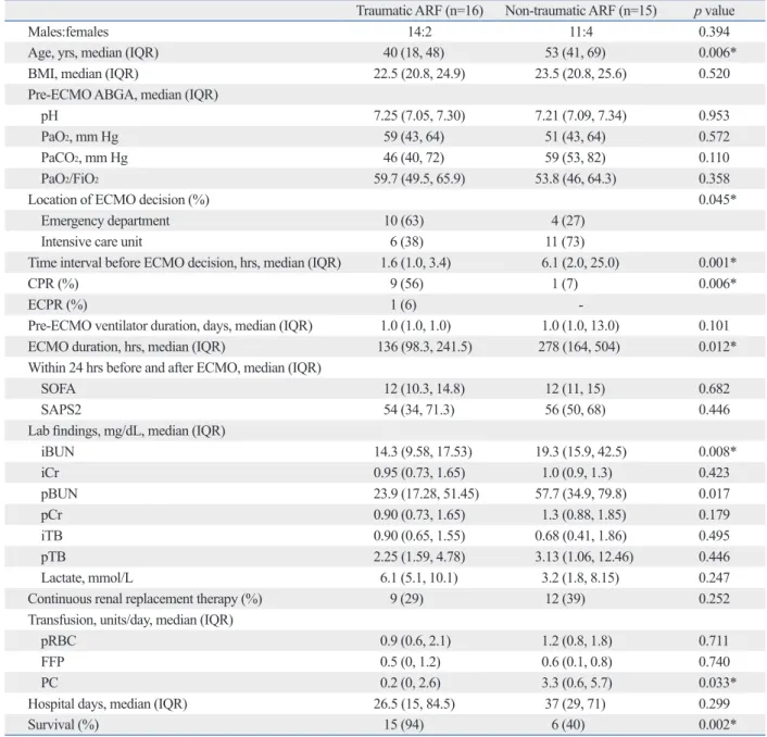
The traumatic ARF patient group had a higher survival rate than the non-traumatic patient group (p<0.002), and we also analyzed the factors related to this outcome (Table 3).
The patients with traumatic ARF were young, and cardio- pulmonary resuscitation and ECMO were performed pri- marily in the emergency room within a short period of time.
In addition, the ECMO duration was short and the initial and peak BUN levels were low (p<0.05).
The complications that occurred during ECMO included multi-organ failure in 6 patients, acute renal failure in 5 pa- tients, bed sores in 3 patients, ulcer bleeding in 3 patients, and cholecystitis in 2 patients. We also recorded one inci- dence each of extra-cannula site bleeding, leg ischemia, bronchopleural fistula, cerebral infarction, and encephalitis.
The causes of death were multi-organ failure for 6 patients (60%), encephalitis for 1 patient, and bronchopleural fistula for 1 patient; additionally the guardians of 2 patients re- fused further treatment (Table 4).
Nineteen of the 21 patients who survived were followed up for a median of 5 months (range, 1‒33 months) after discharge from hospital. Among these patients, one, who was diagnosed as brain dead, died 1 month after discharge, and another who was diagnosed with stage IV lung cancer died 14 months after discharge. Seventeen patients returned to their normal lives without complications.
DISCUSSION
Despite recent advances in critical care management, the mortality of ARF remains high. Patients were considered for VV ECMO in cases of potentially reversible acute and life- threatening respiratory failure. Recent studies suggest that VV ECMO may improve the outcomes of patients with se- vere ARF; however, indications for ECMO use remain un- certain,5-7 leading to questions of which patients are the best candidates for ECMO and whether ECMO should be initiat- ed early in the course of ARF or only in later stages of fail- ure.8 To answer these questions, the extracorporeal life sup- port organization recommended guidelines for ECMO,9 and numerous studies have reported their experiences and indi- cations of ECMO for patients with severe ARF, in addition to evaluating the factors influencing patient survival.10-13
In this cohort, 31 patients with severe ARF did not re- 140‒180 sec by 800‒1000 U/h of heparin. In the remaining
patients, partial thromboplastin time was set at 60‒80 sec with 0.4‒1.5 mg/kg/h of nafamostat mesilate (SK Chemi- cals Life Science Biz., Seoul, Korea licensed by Torii Phar- maceutical Co. Ltd., Tokyo, Japan).
ECMO flow was maintained at a mean blood pressure of
>60 mm Hg with 3.0‒4.0 L/min blood flow for VA ECMO patients with norepinephrine, as needed. For VV ECMO patients, SaO2 was maintained at >90% with a flow of 3.5‒
4.5 L/min. During ECMO, ventilators were set to a tidal volume of 5 mL/kg, a respiration rate of 10/min, a PEEP of 4‒8 cmH2O, and an FiO2 of 0.21‒0.6. Hematocrit >35%
and platelets >50000‒100000/mL were obtained with effort and transfusions performed when necessary. ECMO was removed when arterial blood gas analysis revealed pH 7.35‒7.45, PaO2 >80 mm Hg, and PCO2 <45 mm Hg under the following conditions: a gas blender FiO2 of 0.21, sweep gas of 0 L/min at an ECMO flow of 2 L/min, and the venti- lator mode set to an FiO2 of 0.6, a tidal volume of 6 mL/kg, a PEEP of 8 cmH2O, and an RR of 12‒16/min for VV ECMO or 3 L/min of O2 via nasal prong with awakening ECMO patients. VA ECMO was exchanged for VV ECMO by removing the arterial cannula and inserting the venous cannula in the femoral vein on the opposite leg for patients who did not need VA ECMO support due to severe ARF.
Statistics were calculated using IBM SPSS 21.0 (IBM Corp., Armonk, NY, USA). Categorical variables are shown as percentages and analyzed by Pearson’s χ2 test or Fisher’s exact test. Continuous variables are shown as median (inter- quartile range) and analyzed by the Mann-Whitney U test.
All p values were two sided, and p<0.05 was considered to indicate statistical significance.
RESULTS
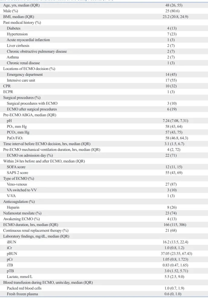
The clinical features of the 31 patients who underwent ECMO due to ARF are shown in Table 1. Of these patients, 25 were males with a median age of 48 years. Dividing pa- tients by the type of ECMO, 27 patients (87%) received VV, 3 (10%) initially received VA followed by VV, and 1 (3%) received V-AV (Table 1).
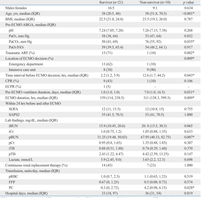
To determine risk factors for mortality, we compared the characteristics of the patients who survived to those that died (Table 2). The patients who survived were younger, had traumatic ARF, and 13 (62%) had ECMO performed in the emergency room. In addition, the time interval before
Table 1. Baseline Clinical Characteristics of the Study Patients (n=31)
Age, yrs, median (IQR) 48 (26, 55)
Male (%) 25 (80.6)
BMI, median (IQR) 23.2 (20.8, 24.9)
Past medical history (%)
Diabetes 4 (13)
Hypertension 7 (23)
Acute myocardial infarction 1 (3)
Liver cirrhosis 2 (7)
Chronic obstructive pulmonary disease 2 (7)
Asthma 2 (7)
Chronic renal disease 1 (3)
Locations of ECMO decision (%)
Emergency department 14 (45)
Intensive care unit 17 (55)
CPR 10 (32)
ECPR 1 (3)
Surgical procedures (%)
Surgical procedures with ECMO 3 (10)
ECMO after surgical procedures 6 (19)
Pre-ECMO ABGA, median (IQR)
pH 7.24 (7.08, 7.31)
PO2, mm Hg 58 (43, 64)
PCO2, mm Hg 57 (43, 75)
PaO2/FiO2 58 (46.8, 64.3)
Time interval before ECMO decision, hrs, median (IQR) 3.1 (1.5, 6.7)
Pre-ECMO mechanical ventilation duration, hrs, median (IQR) 4 (2, 72)
ECMO on admission day (%) 22 (71)
Within 24 hrs before and after ECMO, median (IQR)
SOFA score 12 (11, 15)
SAPS 2 score 55 (43, 69)
Type of ECMO (%)
Veno-venous 27 (87)
VA switched to VV 3 (10)
V-VA 1 (3)
Anticoagulation (%)
Heparin 8 (26)
Nafamostat mesilate (%) 23 (74)
Awakening ECMO (%) 4 (13)
ECMO duration, hrs, median (IQR) 166 (115, 306)
Continuous renal replacement therapy (%) 21 (68)
Laboratory findings, mg/dL, median (IQR)
iBUN 16.2 (13.5, 22.4)
iCr 1.0 (0.8, 1.2)
pBUN 37.05 (23.55, 67.43)
pCr 1.05 (0.8, 1.723)
iTB 0.83 (0.47, 1.65)
pTB 3.0 (1.52, 5.71)
Lactate, mmol/L 5.5 (2.5, 9.0)
Blood transfusion during ECMO, units/day, median (IQR)
Packed red blood cells 1.0 (0.7, 1.9)
Fresh frozen plasma 0.6 (0, 1.0)
Table 1. Continued
Platelet concentrate 1.3 (0, 4.0)
Hospital days, median (IQR) 35 (21, 74)
Outcomes (%)
Death, including patients who refused further treatment 5 (16)
Weaned followed by death 5 (16)
Survival 21 (68)
ABGA, arterial gas blood analysis; BMI, body mass index; BUN, blood urea nitrogen; CPR, cardiopulmonary resuscitation; Cr, creatinine; ECMO, extracor- poreal membrane oxygenation; ECPR, extracorporeal cardiopulmonary resuscitation; i, initial; IQR, interquartile range; p, peak; SOFA, Sepsis-related Organ Failure Assessment; SAPS, Simplified Acute Physiologic Score; TB, total bilirubin; VV, venovenous; VA, venoarterial.
Table 2. Clinical Characteristics of the Survival and Non-Survival Groups
Survivor (n=21) Non-survivor (n=10) p value
Males:females 16:5 9:1 0.634
Age, yrs, median (IQR) 38 (20.5, 48) 58 (51.8, 70.5) <0.001*
BMI, median (IQR) 22.5 (21.0, 24.8) 23.5 (19.3, 26.0) 0.787
Pre-ECMO ABGA, median (IQR)
pH 7.24 (7.05, 7.28) 7.26 (7.15, 7.38) 0.268
PaO2, mm Hg 58 (38, 64) 53 (47, 64) 0.852
PaCO2, mm Hg 50 (41, 69) 76 (55, 92) 0.035*
PaO2/FiO2 59 (39.5, 65.4) 54 (48.2, 64.1) 0.917
Traumatic ARF (%) 15 (71) 1 (10) 0.002*
Location of ECMO decision (%) 0.009*
Emergency department 13 (62) 1 (10)
Intensive care unit 8 (38) 9 (90)
Time interval before ECMO decision, hrs, median (IQR) 2.2 (1.2, 5.9) 12.6 (1.7, 44.2) 0.043*
CPR (%) 9 (43) 1 (10) 0.106
ECPR (%) 1 (5) - -
Pre-ECMO ventilator duration, days, median (IQR) 1.0 (1.0, 1.0) 7.0 (1.0, 16.5) 0.031*
ECMO duration, hrs, median (IQR) 159 (114, 238.5) 311 (158.3, 599.3) 0.009*
Within 24 hrs before and after ECMO
SOFA 12 (11, 15.5) 12 (10.8, 15) 0.755
SAPS2 55 (41.5, 70.5) 53 (41, 70.5) 1.000
Lab findings, mg/dL, median (IQR)
iBUN 15.9 (10.45, 20.6) 20. 8 (15.5, 30.3) 0.065
iCr 1.0 (0.75, 1.2) 1.05 (0.88, 1.35) 0.633
pBUN 31.25 (19.48, 50.03) 67.95 (40.33, 82.75) 0.007*
pCr 0.95 (0.8, 1.65) 1.35 (0.88, 1.85) 0.307
iTB 0.88 (0.51, 1.88) 0.74 (0.29, 1.60) 0.370
pTB 2.43 (1.22, 4.47) 4.42 (2.59, 13.25) 0.147
Lactate, mmol/L 5.9 (2.45, 9.0) 3.65 (2.2, 12.3) 0.698
Continuous renal replacement therapy (%) 14 (45) 7 (23) 1.000
Transfusion, units/day, median (IQR)
pRBC 1.0 (0.7, 2.3) 1.1 (0.65, 1.23) 0.519
FFP 0.67 (0, 1.25) 0.5 (0.08, 0.73) 0.574
PC 0.3 (0, 2.72) 4.2 (0.98, 6.15) 0.028*
Hospital days, median (IQR) 33 (18, 97) 36 (31, 54) 0.819
ABGA, arterial gas blood analysis; ARF, acute respiratory failure; BMI, body mass index; BUN, blood urea nitrogen; CPR, cardiopulmonary resuscitation;
Cr, creatinine; ECMO, extracorporeal membrane oxygenation; ECPR, extracorporeal cardiopulmonary resuscitation; FFP, fresh frozen plasma; i, initial; IQR, interquartile range; p, peak; PC, platelet concentration; SOFA, Sepsis-related Organ Failure Assessment; SAPS, Simplified Acute Physiologic Score; RBC;
red blood cell; TB, total bilirubin.
*Significant difference.
and multiple organ failure, although duration of pre-ECMO mechanical ventilation was not a risk factor. Another study reported that advanced age, pre-ECMO pH <7.18, underly- ing cause of respiratory failure, and increased duration of pre-ECMO ventilation were associated with increased mor- tality, with an overall survival rate of 50%.15 Similar to our results, several studies15-19 have reported significant differ- ences in the duration of pre-ECMO ventilation between survivors and non-survivors. Moreover, reports indicate the importance of considering ECMO early (<7 days) to pre- spond to advanced respiratory treatment, with a mean PaO2/
FiO2 of 56.8 mm Hg before ECMO. The overall survival rate in the patients was 68%. In comparing the survivor and non-survivor groups, patient age, pre-ECMO ventilation duration, time interval before performing ECMO, and ARF due to trauma were significantly different. Schmid, et al.14 analyzed 176 patients with acute lung failure that was re- fractory to conventional therapies and was supported with VV ECMO. They reported an overall survival rate of 56%, and the risk factors affecting survival were advanced age
Table 3. Clinical Characteristics of the Traumatic and Non-Traumatic Acute Respiratory Failure (ARF) Groups
Traumatic ARF (n=16) Non-traumatic ARF (n=15) p value
Males:females 14:2 11:4 0.394
Age, yrs, median (IQR) 40 (18, 48) 53 (41, 69) 0.006*
BMI, median (IQR) 22.5 (20.8, 24.9) 23.5 (20.8, 25.6) 0.520
Pre-ECMO ABGA, median (IQR)
pH 7.25 (7.05, 7.30) 7.21 (7.09, 7.34) 0.953
PaO2, mm Hg 59 (43, 64) 51 (43, 64) 0.572
PaCO2, mm Hg 46 (40, 72) 59 (53, 82) 0.110
PaO2/FiO2 59.7 (49.5, 65.9) 53.8 (46, 64.3) 0.358
Location of ECMO decision (%) 0.045*
Emergency department 10 (63) 4 (27)
Intensive care unit 6 (38) 11 (73)
Time interval before ECMO decision, hrs, median (IQR) 1.6 (1.0, 3.4) 6.1 (2.0, 25.0) 0.001*
CPR (%) 9 (56) 1 (7) 0.006*
ECPR (%) 1 (6) -
Pre-ECMO ventilator duration, days, median (IQR) 1.0 (1.0, 1.0) 1.0 (1.0, 13.0) 0.101
ECMO duration, hrs, median (IQR) 136 (98.3, 241.5) 278 (164, 504) 0.012*
Within 24 hrs before and after ECMO, median (IQR)
SOFA 12 (10.3, 14.8) 12 (11, 15) 0.682
SAPS2 54 (34, 71.3) 56 (50, 68) 0.446
Lab findings, mg/dL, median (IQR)
iBUN 14.3 (9.58, 17.53) 19.3 (15.9, 42.5) 0.008*
iCr 0.95 (0.73, 1.65) 1.0 (0.9, 1.3) 0.423
pBUN 23.9 (17.28, 51.45) 57.7 (34.9, 79.8) 0.017
pCr 0.90 (0.73, 1.65) 1.3 (0.88, 1.85) 0.179
iTB 0.90 (0.65, 1.55) 0.68 (0.41, 1.86) 0.495
pTB 2.25 (1.59, 4.78) 3.13 (1.06, 12.46) 0.446
Lactate, mmol/L 6.1 (5.1, 10.1) 3.2 (1.8, 8.15) 0.247
Continuous renal replacement therapy (%) 9 (29) 12 (39) 0.252
Transfusion, units/day, median (IQR)
pRBC 0.9 (0.6, 2.1) 1.2 (0.8, 1.8) 0.711
FFP 0.5 (0, 1.2) 0.6 (0.1, 0.8) 0.740
PC 0.2 (0, 2.6) 3.3 (0.6, 5.7) 0.033*
Hospital days, median (IQR) 26.5 (15, 84.5) 37 (29, 71) 0.299
Survival (%) 15 (94) 6 (40) 0.002*
ABGA, arterial gas blood analysis; BMI, body mass index; BUN, blood urea nitrogen; CPR, cardiopulmonary resuscitation; Cr, creatinine; ECMO, extracor- poreal membrane oxygenation; ECPR, extracorporeal cardiopulmonary resuscitation; FFP, fresh frozen plasma; i, initial; IQR, interquartile range; p, peak; PC, platelet concentration; SOFA, Sepsis-related Organ Failure Assessment; SAPS, Simplified Acute Physiologic Score; RBC, red blood cell; TB, total bilirubin.
*Significant difference.
perienced ECMO team. Thus, this difference in resources is one of the limitations of our study, although we have high- lighted early application of ECMO. Second, the study was not a randomized controlled trial, but rather retrospective analysis of our experience with VV ECMO due to various causes. Furthermore, the total sample size was small, and several causes had samples sizes of only 1 patient. In order to overcome these limitations, the patients were categorized into trauma and non-trauma groups for analysis, as well as survival and non-survival groups, to determine the factors that influence survival.
In conclusion, VV ECMO is an excellent and life-saving treatment option in patients suffering from acute and life- threatening respiratory failure due to various causes. Over- all, 68% of patients survived. Furthermore, younger trauma patients, shorter pre-ECMO ventilation durations, and short- er time intervals before performing ECMO had the best out- comes with a 94% survival rate. Physicians should be aware of the factors that affect survival and consider early use of VV ECMO therapy to save lives.
REFERENCES
1. Gattinoni L, Carlesso E, Cressoni M. Assessing gas exchange in acute lung injury/acute respiratory distress syndrome: diagnostic techniques and prognostic relevance. Curr Opin Crit Care 2011;
17:18-23.
2. Sud S, Friedrich JO, Taccone P, Polli F, Adhikari NK, Latini R, et al. Prone ventilation reduces mortality in patients with acute respi- ratory failure and severe hypoxemia: systematic review and meta- analysis. Intensive Care Med 2010;36:585-99.
3. Tsuno K, Prato P, Kolobow T. Acute lung injury from mechanical ventilation at moderately high airway pressures. J Appl Physiol (1985) 1990;69:956-61.
4. Peek GJ, Elbourne D, Mugford M, Tiruvoipati R, Wilson A, Allen E, et al. Randomised controlled trial and parallel economic evalu-
vent iatrogenic lung damage from high-pressure and high- FiO2 ventilation.20
Surprisingly, a 94% survival rate (15 of 16 patients) was achieved in our trauma patients among those who received a short pre-ECMO ventilation interval of 1.0 days. This re- sult was superior to survival rates reported in previous stud- ies. Cordell-Smith, et al.21 reported that 20 of 28 patients (71%) who received ECMO with severe trauma‒related re- spiratory failure survived, and the pre-ECMO ventilation times of survivors and non-survivors were 61 h and 87 h, re- spectively. Ried, et al.22 evaluated 26 patients who received VV ECMO with severe trauma‒related respiratory failure af- ter 2.6 d of pre-ECMO ventilation and reported an 81% sur- vival rate. Another study reported that the overall survival of 176 patients supported with VV ECMO was 56%, and the best outcome was noted in trauma patients (71%, 10 of 14 patients) whose pre-ECMO ventilation interval was 4.4 d.14
Another important factor that could contribute to the high survival rate of our trauma patients was the shorter time inter- val before performing ECMO, which was 1.6 h after decreas- ing PaO2/FiO2 <100 mm Hg at an FiO2 of 1.0 or a pH level of
<7.25 due to CO2 retention as indications for ECMO. In com- parison, our non-trauma patients had a significantly different time interval before performing ECMO (6.1 h; p=0.001), with a survival rate of 40%. Furthermore, the time interval between survivors (2.2 h) and non-survivors (12.6 h) in our analysis was also significantly different (p=0.043). These re- sults suggest that the time interval before performing ECMO could be one of the factors influencing survival, along with younger age and duration of pre-ECMO ventilation.
Several limitations must be considered in the interpreta- tions of our results. First, this study was based on a process of a single-institution, which has a well-trained and experi- enced ECMO team. However, not all hospitals have an ex- Table 4. Complications and Causes of Death of the Patients
Complication n (%) Cause of death n (%)
Cannula site bleeding 1 (3.2) Multi-organ failure 6 (60)
Leg ischemia 1 (3.2) Encephalitis 1 (10)
Bed sore 3 (9.7) Bronchopleural fistula 1 (10)
ARF (creatinine >2 mg/dL) 5 (16.1) Give-up 2 (20)
Cholecystitis 2 (6.5)
Ulcer bleeding 3 (9.7)
Bronchopleural fistula 1 (3.2)
Multiorgan failure 6 (19.4)
Cerebral infarction 1 (3.2)
Encephalitis 1 (3.2)
ARF, acute renal failure; RBC, red blood cell; p, peak.
Cannula site bleeding: transfusion >2 units pRBC.
et al. Venovenous extracorporeal membrane oxygenation for acute lung failure in adults. J Heart Lung Transplant 2012;31:9-15.
15. Brogan TV, Thiagarajan RR, Rycus PT, Bartlett RH, Bratton SL.
Extracorporeal membrane oxygenation in adults with severe respi- ratory failure: a multi-center database. Intensive Care Med 2009;
35:2105-14.
16. Camboni D, Philipp A, Lubnow M, Bein T, Haneya A, Diez C, et al. Support time-dependent outcome analysis for veno-venous ex- tracorporeal membrane oxygenation. Eur J Cardiothorac Surg 2011;40:1341-6.
17. Hemmila MR, Rowe SA, Boules TN, Miskulin J, McGillicuddy JW, Schuerer DJ, et al. Extracorporeal life support for severe acute respiratory distress syndrome in adults. Ann Surg 2004;240:595- 605.
18. Pranikoff T, Hirschl RB, Steimle CN, Anderson HL 3rd, Bartlett RH. Mortality is directly related to the duration of mechanical ventilation before the initiation of extracorporeal life support for severe respiratory failure. Crit Care Med 1997;25:28-32.
19. Schmidt M, Zogheib E, Rozé H, Repesse X, Lebreton G, Luyt CE, et al. The PRESERVE mortality risk score and analysis of long-term outcomes after extracorporeal membrane oxygenation for severe acute respiratory distress syndrome. Intensive Care Med 2013;39:1704-13.
20. Tiruvoipati R, Botha J, Peek G. Effectiveness of extracorporeal membrane oxygenation when conventional ventilation fails: valu- able option or vague remedy? J Crit Care 2012;27:192-8.
21. Cordell-Smith JA, Roberts N, Peek GJ, Firmin RK. Traumatic lung injury treated by extracorporeal membrane oxygenation (ECMO). Injury 2006;37:29-32.
22. Ried M, Bein T, Philipp A, Müller T, Graf B, Schmid C, et al. Ex- tracorporeal lung support in trauma patients with severe chest in- jury and acute lung failure: a 10-year institutional experience. Crit Care 2013;17:R110.
ation of conventional ventilatory support versus extracorporeal membrane oxygenation for severe adult respiratory failure (CE- SAR). Health Technol Assess 2010;14:1-46.
5. Hubmayr RD, Farmer JC. Should we “rescue” patients with 2009 influenza A(H1N1) and lung injury from conventional mechanical ventilation? Chest 2010;137:745-7.
6. Morris AH, Hirshberg E, Miller RR 3rd, Statler KD, Hite RD.
Counterpoint: efficacy of extracorporeal membrane oxygenation in 2009 influenza A(H1N1): sufficient evidence? Chest 2010;138:
778-81.
7. Park PK, Dalton HJ, Bartlett RH. Point: Efficacy of extracorporeal membrane oxygenation in 2009 influenza A(H1N1): sufficient ev- idence? Chest 2010;138:776-8.
8. Park PK, Napolitano LM, Bartlett RH. Extracorporeal membrane oxygenation in adult acute respiratory distress syndrome. Crit Care Clin 2011;27:627-46.
9. Extracorporeal Life Support Organization guidelines. [accessed on 2011 September 15]. Available at: http://www.elso.med.umich.
edu/Guidelines.html.
10. Combes A, Bacchetta M, Brodie D, Müller T, Pellegrino V. Extra- corporeal membrane oxygenation for respiratory failure in adults.
Curr Opin Crit Care 2012;18:99-104.
11. Hwang GJ, Sheen SH, Kim HS, Lee HS, Lee TH, Gim GH, et al.
Extracorporeal membrane oxygenation for acute life-threatening neurogenic pulmonary edema following rupture of an intracranial aneurysm. J Korean Med Sci 2013;28:962-4.
12. Martucci G, Panarello G, Bertani A, Occhipinti G, Pintaudi S, Ar- cadipane A. Veno-venous ECMO in ARDS after post-traumatic pneumonectomy. Intensive Care Med 2013;39:2235-6.
13. Rosenberg AA, Haft JW, Bartlett R, Iwashyna TJ, Huang SK, Lynch WR, et al. Prolonged duration ECMO for ARDS: futility, native lung recovery, or transplantation? ASAIO J 2013;59:642-50.
14. Schmid C, Philipp A, Hilker M, Rupprecht L, Arlt M, Keyser A,