CT Demonstration of the Extracardiac Anastomoses of the Coronary Veins in Superior Vena Cava or Left
Brachiocephalic Vein Obstruction
Joseph Casullo, MD, Alexandre Semionov, MD, PhD
All authors: Department of Diagnostic Radiology, Montreal General Hospital, McGill University Health Centre, Montreal, Quebec H3G 1A4, Canada
CT scans in four cases of chronic superior vena cava or left brachiocephalic vein obstruction demonstrate a systemic-to- cardiac collateral venous pathway through anastomoses between the pericardial branches of systemic veins and the pre- sumed adventitial veins of the ascending aorta and pulmonary trunk. These adventitial veins then drain into tributaries of the anterior cardiac veins or ventricular coronary veins.
Index terms: SVC obstruction; Thoracic venous collaterals; Systemic-cardiac venous anastomoses; Fontan physiology
Received November 14, 2011; accepted after revision February 14, 2012.
Corresponding author: Joseph Casullo, MD, Department of Diagnostic Radiology, Montreal General Hospital, McGill University Health Centre, 1650 Cedar Avenue, Montreal, Quebec H3G 1A4, Canada.
• Tel: (1514) 934-8003 • Fax: (1514) 934-8263
• E-mail: joseph.casullo@muhc.mcgill.ca
This is an Open Access article distributed under the terms of the Creative Commons Attribution Non-Commercial License (http://creativecommons.org/licenses/by-nc/3.0) which permits unrestricted non-commercial use, distribution, and reproduction in any medium, provided the original work is properly cited.
Case Report
Korean J Radiol 2013;14(1):132-137 http://dx.doi.org/10.3348/kjr.2013.14.1.132 pISSN 1229-6929 · eISSN 2005-8330
INTRODUCTION
In superior vena cava (SVC) obstruction, venous return to the heart is maintained through collateral venous pathways in the thorax and abdomen. The major intrathoracic veins involved are the azygos-hemiazygos system, the paravertebral venous plexus, and the internal thoracic and pericardiacophrenic veins. These veins communicate with other systemic, portal and pulmonary veins, and any or all of them can form alternate routes for venous return when SVC obstruction develops slowly (1, 2).
The following study reports the computed tomographic (CT) findings in four cases of SVC or left brachiocephalic vein (LBCV) obstruction demonstrating a systemic-to- cardiac venous pathway through anastamoses at the
pericardial attachment to the great arteries.
CASE REPORTS
During routine reporting of CT scans replace by the chest from March to October 2010, our attention was attracted by particular venous collaterals around the great arteries of the heart in four cases of chronic SVC or LBCV obstruction.
Case 1
A 70 year old male, with metastatic renal cell carcinoma and SVC syndrome, underwent a CT angiogram for suspected pulmonary embolism (PE). The scan showed a large nodal mass occluding the SVC above and below the azygos arch.
The lower third of the LBCV was also occluded. No evidence of PE was found.
Case 2
A 71 year old male, with metastatic lung carcinoma, underwent a CT angiogram for suspected PE. Metastatic adenopathy almost completely occluded the SVC above and below the azygos arch. No evidence of PE was found.
Case 3
A 70 year old male with acute chest pain underwent a CT angiogram for suspected PE. The scan showed evidence of
remote aortocoronary bypass and surgical ligation of the LBCV. Only the upper half of the LBCV was opacified by IV contrast. No evidence of PE was found.
Case 4
A 66 year old male with treated lung carcinoma, underwent a routine CT scan. The scan revealed stable findings, including catheter-related thrombosis of the lower third of the LBCV.
All studies were performed on GE Lightspeed 64-detector CT scanners. In cases 1-3, a pulmonary embolism protocol
was followed. Patients received a bolus of 55 mL of non- ionic contrast followed by 35 mL of saline flush through the antecubital veins of the left arm at a rate of 4.5 mL/
s. The scan delay was predetermined by a test bolus (20 mL) using the pulmonary trunk as the region of interest.
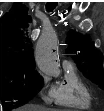
In case 4, 90 mL of non-ionic contrast were administered intravenously through the left arm at a rate of 2 mL/s using a scan delay of 65 seconds. Scans were obtained using 1.25 mm collimation at 0.9 mm intervals through the thorax in cases 1-3; in case 4, the study was obtained with 5 mm collimation at 4 mm intervals and reconstructed at 1.25 x Fig. 1. CT angiogram of 70 year old male with metastatic renal cell carcinoma and obstruction of superior vena cava.
A. Transverse image through thorax at level of upper limit of pericardium shows occlusion of superior vena cava by metastatic adenopathy (*) and extensive network of venous collaterals (long white arrows) in mediastinum. Note early opacification of pulmonary vein (curved white arrow) in left lung through systemic-to-pulmonary venous anastomoses (not shown) across pleural thickening (short white arrow) and probable adhesions from remote left lower lobectomy. A, B. Pericardial veins (arrowheads in A) at anterior aspect of ascending aorta and pulmonary trunk are continuous with small vessels (arrows in B) along intrapericardial portions of great arteries. C. At root of ascending aorta and base of pulmonary trunk, collateral vessels (black arrows) spread along left coronary artery (arrowhead) and its branches and drain into anterior interventricular vein (white arrows). Latter also receives collateral vessel (curved arrow) directly from left lateral wall of pulmonary trunk. D. Slightly more inferiorly, collateral vessel (long white arrow) drains into left coronary vein (short white arrow). On right, collateral branches (black arrows) pass anteriorl or posteriorl to right coronary artery (arrowhead) on their way to right atrial appendage (RAA). E. At lower level, there is early opacification of left coronary vein (short white arrow), and focal collections of contrast are present in wall of right atrial appendage (arrowheads). Collateral vessel (black arrow) posterior to right coronary artery (curved arrow) made loop inferiorly (not shown) before emptying into right auricular wall (black arrowhead). Collateral vessel situated anterior to coronary artery in D drained into wall of appendage more anteriorly (white arrowhead). P
= pericardium
B
E
C A
D
Casullo et al.
1.25 mm.
The CT scans of the first three patients showed an extensive network of venous collaterals in the upper mediastinum, particularly around the aortic arch and pericardium. These collateral vessels included pericardial, esophageal, bronchial and mediastinal branches of the left brachiocephalic, left vertebral, left pericardiacophrenic, left internal thoracic or superior intercostal veins. The collateral vessels also communicated with the azygos-hemiazygos,
phrenic and, in two cases, pulmonary veins. In the fourth patient, the scan showed a collateral venous network joining the left and right inferior thyroid veins in the upper mediastinum and lower neck that communicated with the paravertebral venous plexus and gave rise to several small mediastinal and pericardial branches.
At the union of the pericardium with the ascending aorta and pulmonary trunk, pericardial branches were noted to be in continuity with small vessels distributed along the walls Fig. 2. CT angiogram of 71 year old male with metastatic lung carcinoma and obstruction of superior vena cava.
A. Transverse image through thorax at level of upper limit of pericardium (P) shows almost complete occlusion of superior vena cava (curved white arrow) by metastatic adenopathy (*) and associated venous collaterals (straight white arrows) in anterior mediastinum. A, B. Pericardial branches (arrowheads in A) at anterior aspect of ascending aorta are continuous with small epicardial vessels in aortopulmonary groove (straight black arrow in B) and along pulmonary trunk anteriorly (curved black arrow in B). C. At level of left coronary artery (black arrowhead), collateral vessels from lateral wall of pulmonary trunk (curved black arrow) and within posterior aspect of aortopulmonary groove (feathered black arrow) could not be followed further due to image-degrading pulsation artifacts. Vessel (straight black arrow) in anterior aspect of aortopulmonary groove descends towards right atrium. D. More inferiorly, collateral vessel (black arrows) from anterior aspect of aortopulmonary groove (straight black arrow C) passes posterior to calcified right coronary artery (white arrowhead) before connecting with right atrial appendage (RAA).
A
C
B
D
Fig. 3. CT angiogram of 70 year old male with acute chest pain and remote aortocoronary bypass and surgical ligation of left brachiocephalic vein.
A. Transverse image through thorax at level of aortic arch shows narrowed end of left brachiocephalic vein (curved white arrow) just before occlusion by two surgical clips (curved black arrow). There is extensive network of venous collaterals (straight white arrows) in mediastinum.
B, C. Pericardial veins (arrowheads in B) around great arteries are continuous with small vessels (black arrows in C) along intrapericardial segments of ascending aorta and pulmonary trunk. Pericardium (P) was not seen anteriorly and was thin laterally. D, E. At base of great arteries, there is early opacification of left coronary vein (short white arrow in E) by collateral vessels (black arrows) from lateral wall of pulmonary trunk and posterior wall of ascending aorta. Collateral vessel from anterior wall of aorta gives off branches that accompany right coronary artery (curved white arrow) on their way to right atrial appendage (RAA). Black arrowhead in D points to left coronary artery. F. More inferiorly, collateral vessel (upper black arrow) contacts anterior surface of right atrial appendage. F, G. Collateral vessel along anterior wall of ascending aorta (lower black arrow in F and arrow in G) descends to aortic root where it forms loop posteriorly and connects with anterosuperior wall of left atrium (arrowheads).
Image in G is reformatted CT image of chest in oblique sagittal plane. RA = right atrium, LA = left atrium, Ao = ascending aorta
B C
A
E F
D
G
Casullo et al.
of the intrapericardial segments of the great arteries in all patients (Figs. 1-4). These vessels measured 1.5-2.5 mm in diameter and extended to the root of the ascending aorta and base of the pulmonary trunk where they crossed the coronary arteries. The vessels crossing the right coronary artery drained into the right atrial appendage (all cases), and those crossing the left coronary artery drained into the anterior interventricular and left coronary veins (cases 1 and 3). In the second case, tiny collateral vessels on the left could only be traced to the level of the left coronary artery due to image-degrading pulsation artifacts; we assume that these veins also drained into the great cardiac vein (Fig. 2C). The collateral vessels that crossed the right coronary artery joined the anterior aspect of the right atrial appendage. In case 1, focal collections of contrast measuring about 5 x 8 mm were noted in the wall of the appendage where the collateral vessels fended (Fig. 1E).
The unusual course of a collateral vessel from the anterior wall of the ascending aorta is demonstrated in Figure 3F, G.
After descending along the wall of the aorta to the aortic root, the vessel made a loop posteriorly and connected with the anterosuperior wall of the left atrium.
Fewer than four or five intrapericardial vessels
communicated with pericardial veins. The vessels, except for a single channel in case 4, ramified and intercommunicated as they descended towards the base of the great arteries.
Figure 4 shows the single vessel in case 4; it coursed along the left lateral wall of the ascending aorta and drained into the right atrial appendage after passing under the right coronary artery. A common finding among all patients was the presence of at least one collateral channel along the wall of the ascending aorta located close to the aortopulmonary groove.
DISCUSSION
The various collateral venous pathways associated with SVC obstruction have been reported by several authors (1, 2). The findings from this study demonstrate yet another collateral pathway through anastomoses between extracardiac and cardiac veins at the pericardial attachment to the great arteries. While the existence of widespread anastamoses of extracardiac arteries with the coronary arterial bed has been recognized since 1786 (3), little could be found in the English literature on systemic-cardiac connections on the venous side of the circulation. Baroldi and Scomazzoni (4), in their monograph on the coronary circulation published in 1965, briefly mention the presence of very small anastomoses between coronary and systemic veins in normal hearts. According to these investigators, the anastomoses are found at the pericardial reflections around the pulmonary veins, the venae cavae, and the ascending aorta and pulmonary trunk, similar to the extracardiac anastomoses of the coronary arteries.
In 1965, Clarke (5, 6) obtained microradiographs of the mural vessels of the normal human ascending aorta and pulmonary trunk following the injection of micropaque into the coronary sinus post mortem. He found a network of longitudinal venous channels, measuring 100-140 µm in diameter, in the adventitial layers of both arteries that received branches from the media and drained into tributaries of the ventricular coronary veins. In addition, cross anastomoses between the adventitial veins of the ascending aorta and pulmonary trunk were observed across the aortopulmonary groove.
The adventitia of the ascending aorta and pulmonary Fig. 4. Reformatted CT image of chest, in oblique coronal
plane, of 66 year old male with catheter-related thrombosis (*) of left brachiocephalic vein (short white arrow). Level shown is through ascending aorta and innominate artery. At pericardial attachment to ascending aorta (black arrowhead), there is continuity between pericardial vein (long white arrow) and longitudinal vessel (long black arrow) along intrapericardial portion of ascending aorta near aortopulmonary groove (white arrowhead). Vessel passes posterior to right coronary artery (curved black arrow) before draining into right atrial appendage (not shown). Pericardial vein in question was branch of venous plexus joining left (curved white arrow) and right inferior thyroid veins. P = pericardium
trunk is fused with the fibrous pericardium whose venous drainage is via the pericardiacophrenic veins and the pericardial branches of mediastinal, bronchial and esophageal veins. Therefore, the adventitial layer of the great arteries at the level of the pericardial reflections serves as an entry-point to the coronary circulation for extracardiac veins. The collateral vessels observed along the intrapericardial segments of the ascending aorta and pulmonary trunk in the present cases most likely correspond to the adventitial venous channels described by Clarke, albeit significantly enlarged from collateral flow.
The collateral vessels crossing the left coronary artery are then tributaries of the anterior interventricular and left coronary veins (7). More inferiorly, the left coronary vein then joins the oblique vein of the left atrium to mark the beginning of the coronary sinus. Likewise, the collateral vessels crossing the right coronary artery probably represent tributaries of the anterior cardiac veins that drain the inter-aortopulmonary region and the conus arteriosus. The anterior cardiac veins have been shown to drain directly into the right atrium or indirectly, through intramural tunnels that open into the right atrium (8, 9). We could not determine whether these presumed anterior cardiac veins emptied directly into the lumen of the right atrial appendage or whether they entered vascular channels within its walls. The focal mural collections of contrast observed in the right atrial appendage in one patient are at least suggestive of these intramural vascular tunnels (Fig.
1E).
The unusual course of a collateral vessel from the anterior wall of the ascending aorta to the anterosuperior wall of the left atrium, observed in one patient (Fig. 3F, G), suggests a possible connection between adventitial veins (and the vasa vasorum) of the great arteries and the left atrium.
Whether this vein drained directly into the left atrial lumen or connected with the epicardial veins of the left atrium (9) remains unknown.
It is conceivable that in some of the present cases, anastomoses between systemic and cardiac veins had developed through adhesions around the large arteries near the pericardial attachments. However, there was no radiological evidence of pericardial thickening in any patient, and only a small pericardial effusion was present in cases 1 and 2.
Natural systemic-cardiac venous anastomoses in normal subjects are unimportant and virtually closed their
function is to enlarge in systemic venous obstruction. The extent and capacity of natural anastomoses in patients with SVC obstruction would depend on their number (likely determined at birth) and on the relative resistance to collateral blood flow. As long as the larger venous collaterals remain patent, the amount of blood flow redirected to the right atrium through these extracardiac anastomoses would most likely be relatively small, as demonstrated in our patients. In certain types of Fontan procedures for congenital heart disease, such as the total cavopulmonary connection for tricuspid atresia, systemic veins could conceivably decompress through systemic- cardiac anastomoses into the common atrium. However, the resulting right-to-left shunt might not be insignificant in patients who have persistent cyanosis despite prior ligation or embolization of other more common venous collaterals that are known to develop in these patients (10).
REFERENCES
1. Cihangiroglu M, Lin BH, Dachman AH. Collateral pathways in superior vena caval obstruction as seen on CT. J Comput Assist Tomogr 2001;25:1-8
2. Kapur S, Paik E, Rezaei A, Vu DN. Where there is blood, there is a way: unusual collateral vessels in superior and inferior vena cava obstruction. Radiographics 2010;30:67-78 3. Haller A von. First lines of physiology. A reprint of the 1786
edition. Sources of science; series no. 32. New York: Johnson Reprint Corp, 1966:49
4. Baroldi G, Scomazzoni G. Coronary circulation in the normal and the pathologic heart. Washington, DC: Armed Forces Institute of Pathology, 1965:79
5. Clarke JA. An X-ray microscopic study of the vasa vasorum of the normal human ascending aorta. Br Heart J 1965;27:99-104 6. Clarke JA. An x-ray microscopic study of the vasa vasorum
of the normal human pulmonary trunk. Acta Anat (Basel) 1965;61:6-14
7. Schaffler GJ, Groell R, Peichel KH, Rienmüller R. Imaging the coronary venous drainage system using electron-beam CT.
Surg Radiol Anat 2000;22:35-39
8. Ortale JR, Marquez CQ. Anatomy of the intramural venous sinuses of the right atrium and their tributaries. Surg Radiol Anat 1998;20:23-29
9. von Lüdinghausen M, Ohmachi N, Besch S, Mettenleiter A.
Atrial veins of the human heart. Clin Anat 1995;8:169-189 10. Sugiyama H, Yoo SJ, Williams W, Benson LN. Characterization
and treatment of systemic venous to pulmonary venous collaterals seen after the Fontan operation. Cardiol Young 2003;13:424-430