교신저자 : 고우신, 울산광역시 중구 신정2동 동의대 울산한방병원 (Tel : 052-256-8101, E-mail;
[email protected])
∙
접수 2009/03/09∙
수정 2009/03/24∙
채택 2009/04/03 The Journal of Korean Oriental Medical Ophthalmology &Otolaryngology & Dermatology 2009;22(1) : 89-99
The Anti-allergy Effects of Injinho-tang on the RBL-2H3 cells
Kyeung-Jeong Eo ․ Ji-Hyo Lyu ․ Sun-Ae Lyu ․ Hwa-Jung Yoon ․ Woo-Shin Ko 1. Clinical Research Center Of Oriental Medicine; Department of Oriental Medicine,
College of Oriental Medicine, Dongeui University
RBL-2H3 cells에서의 茵蔯蒿湯의 항 알레르기 효과
어경정 ․ 류지효 ․ 유선애 ․ 윤화정 ․ 고우신
茵蔯蒿湯은 한의학에서 여러 가지 질환에 사용되어 왔다. 특히 간경의 濕熱로 인한 질환에 습열을 제거함으로써 證 의 완화에 많은 응용이 되외 왔다. 본 연구는 濕熱로 인한 질환중의 하나인 아토피 피부염에 그동안 응용이 적은 茵 蔯蒿湯을 실험적으로 응용함으로써 임상적인 가치를 가질 수 있는 지에 대한 기초연구를 진행하였다. 본 실험에 사용 된 세포주는 rat leukemia (RBL-2H3) cells로 茵蔯蒿湯은 알레르기와 관련된 사이토카인인 tumor necrosis factor (TNF)-α, interleukin (IL)-4를 용량의존적으로 억제하였지만 세포독성은 일으키지 않았다. 그리고 동물실험에서 茵蔯 蒿湯은 PCA반응에서 충분한 억제효과를 나타내었다. 또한 Compound 48/80으로 유도된 anaphylaxis shock도 용량 의존적으로 억제함이 밝혀졌다.
Key words : Injinho-tang, Allergy, RBl-2H3 cell, cytokine, TNF-a
Introduction
Mast cells and basophils are critical participants in allergic diseases 1) . There cells express surface membrane receptors with high affinity and specificity for immunoglobulin (Ig)E, which is induced by IL-4 2) . The interaction
of antigen-bound IgE with surface membrane receptors causes the release of histamine, prostaglandins, leukotrienes, and cytokines 1) . These cytokine-induced reactions cause tissue inflammation, anaphylaxis, and scratching behaviors 3,4) .
Injinho-tang (IJHT) has been used as a
traditional Oriental prescribable medicine. It is
widely used as a medication for jaundice
associated with liver inflammation 5) . This is a
medicine preparation consisting of three herbs:
Artemisia capillaris Thunberg (茵蔯), Rheum rhabarbarum (大黃), Gardenia jasminoides Ellis (梔子). Recent studies have reported that IJHT, A. capillaris extract (ACE), and scoparone isolated from A. capillaris have anti- inflammatory and analgesic activities 5-9) . However, study on the anti-allergic effect of IJHT in the mast cells has not been identified.
Here, we investigated the anti-allergic activities of the IJHT on rat basophilic leukemia (RBL)-2H3 cells and allergic models.
We found that IJHT inhibited degranulation, production and expression of pro-inflammatory cytokines from PMA and (or) A23187-induced RBL-2H3 cells. Therefore, Compound 48/80- induced systemic anaphylaxis and passive cutaneous anaphylaxis were inhibited by IJHT.
Materials and Methods
1. Reagents
Phorbol 12-myristate 13-acetate (PMA), calcium ionophore A23187, 3-(4,5-dimethylthiazol-2-yl) -2,5-diphenyltetrazolium bromide (MTT), ρ - n i t r o - p h e n y l - N - β - D - g l u c o s a m i n i d e , anti-dinitrophenyl (DNP) IgE and DNP-human serum albumin (HSA) were purchased from Sigma Chemical Co. (St. Louis, MO).
Dulbecco's Modified Eagle's Medium (DMEM) containing L-glutamine (200 ㎎/ℓ) and FBS were purchased from Hyclone (Logan, UT).
TNF ELISA set (BD OptEIA TM Rat TNF ELISA Set), IL-4 ELISA set (BD OptEIA TM Rat IL-4 ELISA Set) were purchased from BD
Biosciences (Franklin Lakes, NJ).
2. Preparation of Injinho-tang (IJHT) Each of the IJHT was identified and authenticated by Professor , College of Oriental Medicine, Dongeui University (Busan, Korea) (Table 1). IJHT, a one day dose for human adults were boiled with distilled water at 100
℃, and the whole mixture is decocted until the volume is reduced by half. The extract water (400 ㎖) was filtered through 0.22 ㎛ filter and the filtrate was freeze-dried (yield, 13.85 g) and kept at 4 ℃. The dried filtrate was dissolved in phosphate buffered saline (PBS) and filtered through 0.22 ㎛ filter before use.
Table 1. Prescription of Injinho-tang (IJHT)
Herbs Dose
(One day) Artemisia capillaris Thunberg (茵蔯) 40 g
Rheum rhabarbarum (大黃) 20 g Gardenia jasminoides Ellis (梔子) 8 g
3. Animals
We purchased original stock of male ICR mice (5 weeks old) from Samtaco Bio Korea Inc. (Osan, Gyeonggi-do, Republic of Korea).
The mice were housed six to eight per cage in a laminar air-flow room maintained at a temperature of 22 ± 1 ℃ and relative humidity of 55 ± 10 ℃ throughout the study.
4. Cells culture
RBL-2H3 cells were cultured in Dulbecco's
Modified Eagle's Medium (DMEM) with 10 % (v/v) heat-inactivated fetal bovine serum (FBS), 100 U/㎖ penicillin and 100 ㎍/㎖ streptomycin in a humidified incubator with 5 % CO 2 . In all experiments, RBL-2H3 cells were treated for 1 h with the presence of the indicated concentrations of GST-G prior to stimulation with 50 nM PMA plus 1 μM A23187 in serum-free DMEM.
5. MTT assay
The cell viability of GST-G was assessed using the MTT assay 10) in the remaining cells after Griess reaction. The MTT solution (0.5
㎎/㎖) was added to each well. After incubation for 2 h at 37 ℃ and 5 % CO 2 , the supernatant were removed and formed fornazan crystals in viable cells were measured at 540 nm with a microplate reader. The percentage of cell viability was calculated against untreated cells. All experiments were performed in triplicate well.
6. β -hexosaminidase assay
β-hexosaminidase was measured in both supernatant and pellet fractions using a previously reported method 11) . Briefly, RBL-2H3 cells (3 × 10 5 cells) were treated for 1 h with the presence of the indicated concentrations of GST-G prior to stimulation with 50 nM PMA plus 1 μM A23187 and incubated at 37 ℃ for 50 min. After stimulation, 50 ㎕ of each sample was incubated with 50 ㎕ of 1 mM ρ -nitro-phenyl-N-β-D-glucosaminide dissolved in
0.1 M citrate buffer, pH 5, in 96 well microtiter plate at 37 ℃ for 1 h. The reaction was terminated with 200 ㎕/well of 0.1 M carbonate buffer, pH 10.5. The plate was read at 405 ㎚ in an ELISA reader. The inhibition percentage of β-hexosaminidase release was calculated using the following equation :
β-hexosaminidase release (%) =
A 405 of Sup
× 100 A 405 of Sup + A 405 of pellet
where is A 405 is absorption of measured at 405
㎚ and sup. is supernatant.
7. Enzyme-linked immunosorbent assay for pro-inflammatory cytokines (TNF- α , IL-4)
Each cytokines concentration in RBL-2H3 cells were measured with commercially available Rat TNF, IL-4 ELISA kit (BD Biosciences), according to the manufacture's protocol. Color development was measured at 450 ㎚ using an automated microplate ELISA reader.
8. Isolation of total RNA from cells and Reverse Transcriptase-Polymerase Chain Reaction (RT-PCR)
Total RNA was isolated as per the
manufacture's instructions. Briefly, cells were
lysed additional Trizol reagent (Invitrogen,
Carlsbad, CA) and the cell lysate was passed
through the pipette several times. 0.2 ㎖ of
chloroform was added per 1 ㎖ of Trizol
reagent. The tubes were shaken vigorously and incubated at room temperature for 2-3 min.
The samples were centrifuged at 14,000 g for 20 min. The aqueous phase was transferred to a fresh tube and RNA was precipitated by the addition of 0.5 ㎖ isopropanol. The RNA pellet was air-dried and resuspended in nuclease-free water. The concentration of RNA was estimated spectrophotometrically. Three microgram RNAs were reverse-transcribed using M-MLV reverse transcriptase (Promega, Madison, WI). Single stranded cDNA was amplified by PCR with primers (Table 2).
PCR amplifications were done in a 20 ㎕ PCR PreMix (Bioneer Co., Korea) containing 10 mM Tris-HCl, 40 mM KCl, 1.5 mM MgCl 2 , 250 μM dNTP, 1 unit of Taq polymerase.
Amplifications were carried out in a PCR machine (ASTEC PC802) using an initial denaturation at 95 ℃ for 5 min followed by 30 cycles (TNF-α : 35 cycles) of denaturation for 60 sec at 95 ℃, annealing for 60 sec at 52
℃ and extension for 60 sec at 72 ℃. This was concluded with a final extension for 7 min at 72 ℃. Amplicons were separated in 1% agarose gels in 0.5× TBE buffer at 100 V for 30 min, stained with ethidium bromide
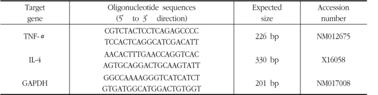
Target gene
Oligonucleotide sequences (5’ to 3’ direction)
Expected size
Accession number
TNF-α CGTCTACTCCTCAGAGCCCC
TCCACTCAGGCATCGACATT 226 bp NM012675
IL-4 AACACTTTGAACCAGGTCAC
AGTGCAGGACTGCAAGTATT 330 bp X16058
GAPDH GGCCAAAAGGGTCATCATCT
GTGATGGCATGGACTGTGGT 201 bp NM017008
Table 2. Oligonucleotide primers used for PCR in this study.
and visualised under UV light. GAPDH (Glyceraldehyde-3-phosphate dehydrogenase) was used as an internal control to evaluate relative expressions of TNF-α and IL-4.
9. Compound 48/80-induced systemic anaphylactic shock
Mice (n=8) were given an intraperitoneal injection of the mast cells degranulator Compound 48/80 (8.0 ㎎/㎏). IJHT was dissolved in saline and administered orally with a sonde 2 h before the injection of Compound 48/80. Mortality was monitored for 30 min after induction of anaphylactic shock.
10. Passive cutaneous anaphylaxis (PCA)
IgE-dependent cutaneous reaction was
generated by sensitizing the skin with the
intradermal injection of anti-DNP IgE followed
48 h later with an injection of DNP-HSA into
the mice tail vein. the DNP-HSA was diluted
in PBS. The mice were injected intradermally
with 300 ng of anti-DNP IgE into each of
three dorsal skin sites that had been shaved
48 h earlier. The sites were outlined with a
water-in-soluble red marker. Forty-eight hours
later, each mouse received an injection of 300
㎕ of 1 : 9 mixture of 10 ㎎/㎖ DNP-HSA in PBS and 4 % Evans blue via the tail vein.
Two hours before this injenction, IJHT was administered orally with a sonde. The mice were sacrificed 30 min after the intravenous challenge. The dorsal skin of the mouse was removed for measurement of the pigment area.
The amount of dye was then determined colorimetrically after extraction with 0.5 ㎖ of 1 N KOH and 4.5 ㎖ of a mixture of acetone and phosphoric acid (in a ratio of 5 : 13, v/v), based on the method of Katayama et al . 12) . The absorbance of the extract was measured at 620 ㎚ in a spectrophotometer, and the amount of dye was calculated using an Evans blue calibration curve.
11. Statistical analysis
Data is presented as the mean ± SE (standard error) of at least three separate experiments. Comparisons between two groups were analyzed using Student’s t-test. P values less than 0.05 considered be statistically significant.
Results
1. Effect of IJHT on degranulation and cell viability in RBL-2H3 cells
Inhibitory effects of IJHT on the release of β-hexosaminidase from RBL-2H3 cells were evaluated by the methods, as described in Materials and Methods. The release of β -hexosaminiase decreased significantly with all
concentrations of IJHT. The inhibition rate of β-hexosaminidase release were 66.2 % with a dose of 0.5 ㎎/㎖, 80.26 % with a dose of 1.0
㎎/㎖, and 92.23 % with a dose of 2.0 ㎎/㎖.
(Fig. 1).
β-hexosaminidaserelease(%)
***
***
0 5 10 15 20 25 30 35 40 45
***
0 0 1.0 2.0
- + + +
IJHT (mg/ml) PMA+A23187
0.5 +
β-hexosaminidaserelease(%)
***
***
0 5 10 15 20 25 30 35 40 45
***
0 0 1.0 2.0
- + + +
IJHT (mg/ml) PMA+A23187
0.5 +
Fig. 1. Effect of IJHT on degranulation in RBL-2H3 cells.
Cells were treated with the indicated concentration of IJHT. Degranulation was assessed by β -hexosaminidase release into the supernatant. β -hexosaminidase released into the medium is presented as mean ± SE ( n =4). *** P < 0.005;
significantly different from the stimulated group.
IJHT (mg/ml) 0 1.0 2.0
Cell viability(%)
0 20 40 60 80 100 120
0.5
IJHT (mg/ml) 0 1.0 2.0
Cell viability(%)
0 20 40 60 80 100 120
0 20 40 60 80 100 120
0.5
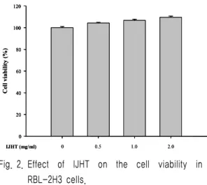
Fig. 2. Effect of IJHT on the cell viability in RBL-2H3 cells.
Cell viability was evaluated by MTT assay. Data
represent the mean ± SE of three independent
experiments.
2. Effect of IJHT on the cell viability The cell viability effect of IJHT on RBL-2H3 cells was evaluated by MTT assay.
IJHT concentrations from 1.0 ㎎/㎖ to 2.0 ㎎/
㎖ had no effect on cell survival (Fig. 2).
These results suggest IJHT inhibits A23187-induced TNF-α and IL-4 production in RBL-2H3 cells without effect on the cell viability in each condition.
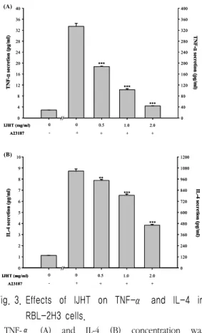
3. Effect of IJHT on secretion of TNF-α and IL-4 in RBL-2H3 cells
To assess the effect of IJHT on the secretion of pro-inflammatory cytokines, RBL-2H3 cells were treated with various concentrations of IJHT for 8 h. The levels of TNF-α and IL-4 were analyzed by ELISA.
Treatment with IJHT significantly inhibited cytokines secretion in RBL-2H3 cells (Fig. 3A, 3B). When RBL-2H3 cells were treated only A23187 the cells secreted 334.02 ± 10.78 pg/
㎖ TNF-α and 1046.54 ± 22.11 pg/㎖ IL-4.
Whereas, RBL-2H3 cells were pre-treated with 2.0 ㎎/㎖ IJHT, the cells secreted 43.28 ± 0.56 pg/㎖ TNF-α and 460.75 ± 9.54 pg/㎖ IL-4.
4. Effect of IJHT on expression of TNF- α and IL-4 in RBL-2H3 cells
Also to assess the effect of IJHT on the pro-inflammatory cytokines (TNF-α and IL-4) expression. These cytokines expression decreased significantly by IJHT in dose-dependent manner (Fig. 4). In contrast to TNF-α and IL-4, the level of GAPDH mRNA expression remained the same under these conditions.
TNF-αsecretion(pg/ml)
IJHT (mg/ml) 0 0 0.5
A23187 - + + + +
2.0 1.0
TNF-αsecretion(pg/ml)
0 4 8 12 16 20 24 28 32 36 40
0 40 80 120 160 200 240 280 320 360 400
//
***
***
***
TNF-αsecretion(pg/ml)
IJHT (mg/ml) 0 0 0.5
A23187 - + + + +
2.0 1.0
TNF-αsecretion(pg/ml)
0 4 8 12 16 20 24 28 32 36 40
0 40 80 120 160 200 240 280 320 360 400
//
***
***
***
0 1 2 3 4 5 6 7 8 9 10
0 120 240 360 480 600 720 840 960 1080 1200
//
IJHT (mg/ml) 0 0 0.5
A23187 - + + + +
2.0 1.0
**
***
***
IL-4secretion(pg/ml) IL-4secretion(pg/ml)
0 1 2 3 4 5 6 7 8 9 10
0 120 240 360 480 600 720 840 960 1080 1200
//
IJHT (mg/ml) 0 0 0.5
A23187 - + + + +
2.0 1.0
**
***
***
IL-4secretion(pg/ml) IL-4secretion(pg/ml)
(A)
(B)
Fig. 3. Effects of IJHT on TNF-α and IL-4 in RBL-2H3 cells.
TNF-α (A) and IL-4 (B) concentration was measured from cell supernatants using ELISA method. Vertical bars represent as the mean ± SE from 4 wells. ** P < 0.01, *** P < 0.005;
significantly different from the stimulated group.
1 2 3 4 5 TNF-α
IL-4
GAPDH
330 bp
201 bp 226 bp 1 2 3 4 5
TNF-α
IL-4
GAPDH
330 bp
201 bp 226 bp
Fig. 4. Effects of IJHT in the expression of pro-inflammatory cytokines in RBL-2H3 cells.
Total RNA was isolated, TNF-α and IL-4 mRNA expression was detected by RT-PCR analysis. Lane 1. negative control group; lane 2. positive control group (only treated A23187); lane 3. IJHT 0.5 ㎎/㎖
+ A23187; lane 4. IJHT 1.0 ㎎/㎖ + A23187; lane 5.
IJHT 2.0 ㎎/㎖ + A23187.
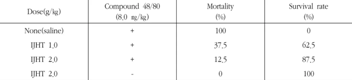
5. Effect of IJHT on Compound 48/80- induced systemic anaphylaxis
To determine the contribution of IJHT in anaphylactic reaction, we used an in vivo model of systemic anaphylaxis. As a mast cell degranulator, Compound 48/80 (8.0 ㎎/㎖) was used. After the injection of Compound 48/80, the mice monitored for 30 min, after which the mortality rate was determined. The period for observation of mortality was based on the control mice that had died in 30 min by Compound 48/80. An oral administration of saline as a control resulted in a fatal reaction in 100 % of group. When IJHT was orally administered at the dosed of 1.0 and 2.0 g/㎏
2 h before Compound 48/80 injection, the mortality was inhibited (Table 3).
6. Effect of IJHT on PCA
Local injection of anti-DNP IgE followed by an intravenous antigenic challenge was performed. Anti-DNP IgE was injected into dorsal skin sites. After 48 h, all animals were injected intravenously with DNP-HSA containing
Table 3. Effect of IJHT on Compound 48/80-induced anaphylaxis shock Dose(g/㎏) Compound 48/80
(8.0 ㎎/㎏)
Mortality (%)
Survival rate (%)
None(saline) + 100 0
IJHT 1.0 + 37.5 62.5
IJHT 2.0 + 12.5 87.5
IJHT 2.0 - 0 100
Note. Group of mice (n=8/group) were orally pre-treated with 500 ㎕ of saline of IJHT at various doses 2 h before the intraperitoneal (i.p.) injection of Compound 48/80. Mortality (%) within 30 min following Compound 48/80 i.p. injection is represented as :
(number of dead mice ÷ total number of experimental mice) × 100
Saline IJHT 1.0 g/kg IJHT 2.0 g/kg 0.0
0.5 1.0 1.5 2.0 2.5 3.0
***
Amounts of dye (μg/site) ***
Saline IJHT 1.0 g/kg IJHT 2.0 g/kg 0.0
0.5 1.0 1.5 2.0 2.5 3.0
***
Amounts of dye (μg/site) ***