교신저자 : 김원일, 동의대학교 한의과대학 내과학교실 (Tel: 051-850-7444, E-mail : [email protected].)
∙접수 200811/03 ∙수정 2008/11/28 ∙채택 2008/12/09 Otolaryngology & Dermatology 2008;21(3) : 94-103
Anti-allergic Effect of Gyokeisamultang-gagam (艽桂四物湯-加減) in the RBL-2H3 Mast Cells
Pei-Yun Tsung
1‧ Woo-Jin Shin
1‧ Ji-Hyo Lyu
2‧ Seong-Hui Kim
2Hwa-Jung Yoon
3‧ Won-Il Kim
1,*1. Department of Internal Medicine, College of Oriental Medicine, Dong-eui University
2. Clinical Research Center of Oriental Medicine, Dong-eui University 3. Department of Ophthalmology & Otolaryngology & Dermatology, College of Oriental Medicine, Dong-eui University
RBL-2H3 cells에서 Gyokeisamultang-gagam의 항알레르기 효과
총배빈
1‧ 신우진
1‧ 류지효
2‧ 김성희
2‧ 윤화정
3‧ 김원일
1,*비만세포의 한 종류인 rat basophilic leukemia (RBL-2H3) 세포를 이용하여 교계사물탕-가감의 항알레르기 효과를 확인하고자 하였다. Phorbol 12-myristate 13-acetate (PMA)와 calcium ionophore A23187을 이용하여 RBL-2H3 세 포를 자극한 후 세포의 탈과립 정도를 β-hexosaminidase assay로 확인한 결과, 전 처리한 교계사물탕-가감의 농도 의존적으로 탈과립을 억제하였다. Pro-inflammatory cytokines인 tumor necrosis factor (TNF)-alpha와 interleukin (IL)-4의 분비량을 enzyme-linked immunosorbent assay (ELISA)로 확인한 결과 교계사물탕-가감의 농도 의존적으로 감소하였으며, 이들 cytokines와 염증 반응에 주요한 인자인 cyclooxygenase (COX)-2의 mRNA 발현 정도 역시 교 계사물탕-가감에 의해 감소함을 확인할 수 있었다. 이러한 실험 결과로 보아 교계사물탕-가감은 알레르기 관련 질환의 치료에 응용될 수 있을 것으로 사료된다.
Key words : Gyokeisamultang-gagam , Mast cell, Degranulation, Pro-inflammatory cytokine, Cyclooxygenase-2
Introduction
Basophils as well as mast cells play important
roles in both immediate and late-phase reactions
of type Ⅰ allergy
1). Mast cells are a major
source of inflammatory mediators, some of which are preformed and stored in secretory granules and others (such as cytokines and lipid-derived eicosanoids)
2). It is well known that immunoglobulin E (IgE)-dependent mast cell activation is associated with allergic diseases
3). Activated mast cells release inflammatory mediators (histamine), both preformed cytokines and newly synthesized cytokines
4,5). β-hexosaminidase is stored in secretion granules of mast cells, and is released concomitantly with histamine when mast cells are immunologically activated, and β -hexosaminidase activity in the medium is used as a marker of mast cell degranulation
6). Tumor necrosis factor (TNF)-α, a kind of cytokine which mediates the inflammation pathway, is major target of therapeutic strategy in many chronic inflammatory conditions
7-9). T-helper 2 (Th2) cells produce interleukin (IL)-4, -5, -10, and -13 and the cytokines function as important factors in atopic dermatitis and asthma processes. IL-4 plays a major role in B-cell activation and isotype switching, resulting in generation of IgE antibodies
10-13). Cyclooxygenase (COX)-2, one of the major mediators of the inflammatory reaction, is also strongly induced in activated monocytes/
macrophages. Several recent studies demonstrated that prostaglandin (PG)D
2, which is the COX-2 metabolite released from activated mast cells, is also essential for the pathgenesis of eosinophilic airway inflammation
14,15).
Gyokeisamultang-gagam (GST-G) is a therapy made according to the book titled,
『Experience of Senior Oriental Medical Docto
r』. This therapy is used to nourish blood, warm meridian, expel dampness, and remove obstructions in meridian. This theory is mainly used to cure cold-dampness, arthrodynia of extremities, numbness and unconsciousness of muscular and skins, limited movement of extremities, difficulties in moving about, white-coated tongue fur, deep-moderate or deep-thready or soft-thready pulses, blue and cyanotic complexion, noted pain points, and heel pains
16). Samul-tang gagam can be found in the theses on allergic rhinitis
17), contact dermatitis
18), rheumatoid arthritis, allergic dermatitis
19), and some other allergic diseases;
however, no research has ever been conducted on Gyokeisamul-tang. To elucidate the anti- allergic effect of GST-G, we examined the effect of GST-G on degranulation, pro- inflammatory cytokines secretion and expression from the phorbol 12-myristate 13-acetate (PMA) plus calcium ionophore A23187-induced rat basophilic leukemia (RBL-2H3) mast cells.
Materials and Methods
1. Preparation of Gyokeisamultang-gagam (GST-G)
Each of the GST-G was identified and
authenticated by Professor W.I. Kim, College
of Oriental Medicine, Dongeui University
(Busan, Korea) (Table 1). GST-G, a one day
dose for human adults were boiled with
distilled water at 100 ℃, and the whole
mixture is decocted until the volume is
reduced by half. The extract water (400 ml)
was filtered through 0.22 μm filter and the filtrate was freeze-dried (yield, 33.84 g) and kept at 4 ℃. The dried filtrate was dissolved in phosphate buffered saline (PBS) and filtered through 0.22 μm filter before use.
Table 1. Prescription of Gyokeisamultang-gagam (GST-G)
Herbs Dose
Angelica gigas Nakai (當歸) Cnidium officinale Makino (川芎)
Paeonia lactiflora Pallas (赤芍藥) Rehmannia glutinosa Liboschitz var.
purpurea Makino (熟地黃) Cinnamomum cassia Blume (桂枝) Gentiana macrophylla Pallas (秦艽) Acyranthes bidentata Blume (牛膝)
Phlomis umbrosa (續斷) Aralia contientalis (獨活) Caesalpina asppan Linne (蘇木)
Glycyrrhizae radix (甘草)
12 g 6 g 12 g 12 g 6 g 9 g 12 g 12 g 12 g 6 g 6 g
2. Reagents
Phorbol 12-myristate 13-acetate (PMA), calcium ionophore A23187, 3-(4,5-dimethylthiazol- 2-yl)-2,5-diphenyltetrazolium bromide (MTT) and ρ-nitro-phenyl-N-β-D-glucosaminide were purchased from Sigma Chemical Co. (St. Louis, MO). Dulbecco's Modified Eagle's Medium (DMEM) containing L-glutamine (200 mg/L) and FBS were purchased from Hyclone (Logan, UT). TNF ELISA kit (BD OptEIA
TMRat TNF ELISA Set), IL-4 ELISA kit (BD OptEIA
TMRat IL-4 ELISA Set) were purchased from BD Biosciences (Franklin Lakes, NJ).
3. Cells culture
RBL-2H3 cells were cultured in Dulbecco's Modified Eagle's Medium (DMEM) with 10 % (v/v) heat-inactivated fetal bovine serum (FBS), 100 U/ml penicillin and 100 ㎍/ml streptomycin in a humidified incubator with 5 % CO
2. In all experiments, RBL-2H3 cells were treated for 1 h with the presence of the indicated concentrations of GST-G prior to stimulation with 50 nM PMA plus 1 μM A23187 in serum-free DMEM.
4. MTT assay
The cell viability of GST-G was assessed using the MTT assay
20)in the remaining cells after Griess reaction. The MTT solution (0.5 mg/ml) was added to each well. After incubation for 2 h at 37 ℃ and 5 % CO
2, the supernatant were removed and formed fornazan crystals in viable cells were measured at 540 nm with a microplate reader. The percentage of cell viability was calculated against untreated cells. All experiments were performed in triplicate well.
5. β-hexosaminidase assay
β-hexosaminidase was measured in both supernatant and pellet fractions using a previously reported method
21). Briefly, RBL- 2H3 cells (3 × 10
5cells) were treated for 1 h with the presence of the indicated concentrations of GST-G prior to stimulation with 50 nM PMA plus 1 μM A23187 and incubated at 37
℃ for 50 min. After stimulation, 50 ㎕ of
each sample was incubated with 50 ㎕ of 1
mM ρ-nitro-phenyl-N-β-D-glucosaminide dissolved
in 0.1 M citrate buffer, pH 5, in 96 well microtiter plate at 37 ℃ for 1 h. The reaction was terminated with 200 ㎕/well of 0.1 M carbonate buffer, pH 10.5. The plate was read at 405 nm in an ELISA reader. The inhibition percentage of β-hexosaminidase release was calculated using the following equation : β-hexosaminidase release (%) =
(A
405of sup.) / (A
405of sup. + A
405of pellet)
× 100
where is A
405is absorption of measured at 405 nm and sup. is supernatant.
6. Enzyme-linked immunosorbent assay for pro-inflammatory cytokines (TNF-α, IL-4) Each cytokines concentration in RBL-2H3 cells were measured with commercially available Rat TNF, IL-4 ELISA kit (BD Biosciences), according to the manufacture's protocol. Color development was measured at 450 nm using an automated microplate ELISA reader.
7. Isolation of total RNA from cells and Reverse Transcriptase-Polymerase Chain Reaction (RT-PCR)
Total RNA was isolated as per the manufacture's instructions. Briefly, cells were lysed additional Trizol reagent (Invitrogen, Carlsbad, CA) and the cell lysate was passed through the pipette several times. 0.2 ml of chloroform was added per 1 ml of Trizol reagent. The tubes were shaken vigorously and incubated at room temperature for 2-3 min.
The samples were centrifuged at 14,000 g for
20 min. The aqueous phase was transferred to a fresh tube and RNA was precipitated by the addition of 0.5 ml isopropanol. The RNA pellet was air-dried and resuspended in nuclease-free water. The concentration of RNA was estimated spectrophotometrically. Three microgram RNAs were reverse-transcribed using M-MLV reverse transcriptase (Promega, Madison, WI). Single stranded cDNA was amplified by PCR with primers (Table 2).
PCR amplifications were done in a 20 ㎕ PCR PreMix (Bioneer Co., Korea) containing 10 mM Tris-HCl, 40 mM KCl, 1.5 mM MgCl
2, 250 μM dNTP, 1 unit of Taq polymerase.
Amplifications were carried out in a PCR machine (ASTEC PC802) using an initial denaturation at 95 ℃ for 5 min followed by 30 cycles (TNF-α, COX-1 : 35 cycles) of denaturation for 60 sec at 95 ℃, annealing for 60 sec at 52 ℃ (COX-1, COX-2 : 55 ℃) and extension for 60 sec at 72 ℃. This was concluded with a final extension for 7 min at 72 ℃. Amplicons were separated in 1 % agarose gels in 0.5× TBE buffer at 100 V for 30 min, stained with ethidium bromide and visualised under UV light. GAPDH (Glyceraldehyde-3-phosphate dehydrogenase) was used as an internal control to evaluate relative expressions of TNF-α, IL-4 and COX-2.
8. Statistical analysis
Data is presented as the mean ± SE (standard error) of at least three separate experiments.
Comparisons between two groups were
analyzed using Student’s t-test. P values less
than 0.05 considered be statistically significant.
Target gene
Oligonucleotide sequences (5’ to 3’ direction)
Expected size
Accession number
TNF-α CGTCTACTCCTCAGAGCCCC
TCCACTCAGGCATCGACATT 226 bp NM012675
IL-4 AACACTTTGAACCAGGTCAC
AGTGCAGGACTGCAAGTATT 330 bp X16058
COX-1 ACTGGTCTGCCTCAACACCA
CAAGGGTGAGACCCCAAGTT 223 bp S67721
COX-2 TGACCAGAGCAGAGAGATGA
CATAAGGCCTTTCAAGGAGA 250 bp S67722
GAPDH GGCCAAAAGGGTCATCATCT
GTGATGGCATGGACTGTGGT 201 bp NM017008
Table 2. Oligonucleotide primers used for PCR in this study.
0 5 10 15 20 25 30 35 40 45 50
0 5 10 15 20 25 30 35 40 45 50
0 5 10 15 20 25 30 35 40 45 50
0 5 10 15 20 25 30 35 40 45 50
β-hexosaminidaserelease (%) β-hexosaminidaserelease (%)
GST-G (mg/ml) 0 0 0.5
PMA+A23187 - + + + +
2.0 1.0
***
*** ***
Fig. 1. Effect of GST-G on degranulation in RBL-2H3 cells.
Cells were treated with the indicated concentration of GST-G. Degranulation was assessed by β -hexosaminidase release into the supernatant. β -hexosaminidase released into the medium is presented as mean ± SE (n=3). ***
P< 0.005;
significantly different from the stimulated group.
Results
1. Effect of GST-G on degranulation in RBL-2H3 cells
Inhibitory effects of GST-G on the release of β-hexosaminidase from RBL-2H3 cells were
evaluated by the methods, as described in Materials and Methods. The release of β -hexosaminiase decreased significantly with all concentrations of GST-G. The inhibition rate of β-hexosaminidase release were 17.97 % with a dose of 0.5 mg/ml, 31.26 % with a dose of 1.0 mg/ml, and 33.5 % with a dose of 2.0 mg/ml (Fig. 1).
2. Effect of GST-G on the cell viability The cell viability effect of GST-G on RBL-2H3 cells was evaluated by MTT assay.
As shown in Fig. 2, SGT concentrations from 0.5 mg/ml to 2.0 mg/ml had no effect on cell survival. These results suggest GST-G inhibits PMA plus A23187-induced TNF-α, IL-4, and COX-2 production in RBL-2H3 cells without effect on the cell viability in each condition.
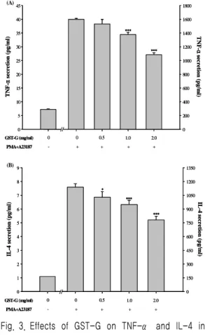
3. Effect of GST-G on secretion of TNF-α and IL-4 in RBL-2H3 cells
To determine whether GST-G can modulate
pro-inflammatory cytokines (TNF-α and IL-4) secretion. Cells were pre-treated with various concentration of GST-G and then PMA plus A23187 challenge for 10 h. Treatment with GST-G significantly and dose-dependently inhibited TNF-α and IL-4 secretion in RBL-2H3 cells (Fig. 3A, 3B). The inhibitory rate : 32.23 % with a dose of 2.0 mg/ml (TNF-α) and 31.42 % with a dose of 2.0 mg/ml (IL-4).
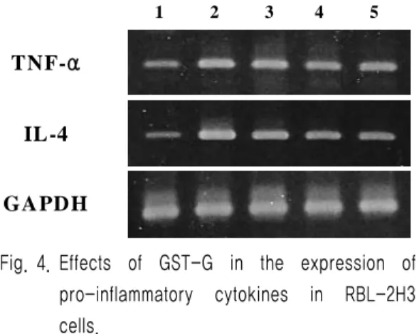
4. Effect of GST-G on expression of TNF- α and IL-4 in RBL-2H3 cells
Also to assess the effect of GST-G on the pro-inflammatory cytokines (TNF-α and IL-4) expression. These cytokines expression decreased significantly by GST-G in dose-dependent manner (Fig. 4). In contrast to TNF-α and IL-4, the level of GAPDH mRNA expression remained the same under these conditions.
Cell viability (%)
0 10 20 30 40 50 60 70 80 90 100 110
GST-G (mg/ml) 0 0.5 1.0 2.0
Fig. 2. Effect of GST-G on the cell viability in RBL-2H3 cells.
Cell viability was evaluated by MTT assay. Data represent the mean ± SE of three independent experiments.
0 5 10 15 20 25 30 35 40 45
0 200 400 600 800 1000 1200 1400 1600 1800
//
TNF-αsecretion(pg/ml) TNF-αsecretion(pg/ml)
***
***
GST-G (mg/ml) 0 0 0.5
PMA+A23187 - + + + +
2.0 1.0 0
5 10 15 20 25 30 35 40 45
0 200 400 600 800 1000 1200 1400 1600 1800
//
TNF-αsecretion(pg/ml) TNF-αsecretion(pg/ml)
***
***
GST-G (mg/ml) 0 0 0.5
PMA+A23187 - + + + +
2.0 1.0
0 1 2 3 4 5 6 7 8 9
0 150 300 450 600 750 900 1050 1200 1350
*
***
***
IL-4secretion(pg/ml) IL-4secretion(pg/ml)
//
GST-G (mg/ml) 0 0 0.5
PMA+A23187 - + + + +
2.0 1.0 0
1 2 3 4 5 6 7 8 9
0 150 300 450 600 750 900 1050 1200 1350
*
***
***
IL-4secretion(pg/ml) IL-4secretion(pg/ml)
//
GST-G (mg/ml) 0 0 0.5
PMA+A23187 - + + + +
2.0 1.0 (B)
(A)