Kor. J. Pharmacogn.
45(2) : 174∼ 180 (2014)
174
D-Pinitol의 유방암 증식 및 재발 억제 효능
김윤섭·박지성·김민지·황방연·이종길·송석길*
충북대학교 약학대학
Inhibitory Effect of D-pinitol on Both Growth and Recurrence of Breast Tumor from MDA-MB-231 Cancer Cells
Yoon-seob Kim, Ji-sung Park, Minji Kim, Bang Yeon Hwang, Chong-Kil Lee and Sukgil Song*
College of Pharmacy and Medical Research Center, Chungbuk National University, 52, Naesudong-ro, Heungdeok-gu, Cheongju, Chungbuk 361-763, Korea
Abstract − D-Pinitol, an anti-diabetic substance, is a naturally occurring compound found in legumes. In this study, we inves- tigated the inhibitory effect of D-pinitol on growth and recurrence of breast cancer. When D-pinitol was treated on MDA-MB- 231 or MCF-7 breast cancer cells, it was observed that the viability of the two cancer cell lines was reduced in MTT assay.
In order to examine the effect on the growth of breast tumor, mouse xenograft assay was carried out. On day 0, nine millions cells of MDA-MB-231 were injected subcutaneously into nude mouse and D-pinitol was administered orally at the dose of 500 mg/kg or 1000 mg/kg body weight for consecutive 45 days. Tumor size was reduced in dose-dependent manner upto 95.4%
in 1000 mpk-treated group, compared with the non-treated control group. When D-pinitol was co-administrated with 4µg of doxorubicin, recurrence of breast tumor was delayed by two weeks, compared with the mouse group of doxorubicin mono- therapy. Consistent with this data, it was observed that the population of cancer stem cells (CSCs), responsible for recurrence of cancer, within tumor mass was significantly reduced. Taken together, D-pinitol inhibits the growth of breast cancer and relapse of the tumor by suppressing the proliferation of CSCs.
Key words − D-pinitol, Breast cancer, Cancer stem cells
D-pinitol은 D-chiro-inositol(DCI)의 3번 탄소에 methoxy 기를 가진 분자 구조이며 콩과 식물 carob(학명: Ceratonia siliqu)에서 추출한 천연 물질로 혈당강하 효과가 있는 것으 로 알려져 있다1). 섭취한 D-pinitol은 위장에서 탈메틸화에 의해 D-chiro-inositol(DCI)로 변형되어 원활하지 못한 인슐 린 신호전달을 보완해주는 기능을 하는 것으로 알려져 왔 으며2), 특히 DCI는 인슐린 저항성으로 인한 2형 당뇨병 환 자에게 효능이 있는 것으로 보고되어 있다3).또한 제2형 당 뇨병의 일차치료제인 metformin은 biguanide계 경구형 치료 제로서 AMP-activated protein kinase (AMPK)를 활성화하 여 유방암의 증식을 억제하고 재발을 지연시키는 효능이 있 는 것으로 알려져 있다5).
유방암은 선진국형 질병으로서 미국 같은 경우 가장 흔하 게 발병되고 있는 여성암이며, 유방암으로 죽는 여성이 매
12분당 1명으로, 두 번째 흔한 암 사망이 관찰되고 있다4). 우리나라는 여성암 중 2위를 차지하고 있고, 최근 20년간 유방암으로 인한 사망과 발생은 점차 증가되고 있는 추세 이며, 2015년에 달하면 1995년 유방암 사망률의 2.3배로 인 구증가율보다 2배나 높은 수치로 증가될 것이 예측되고 있 다5). 그 이유로는 서구화된 식생활로 인한 비만, 늦은 결혼 과 출산, 빠른 초경과 늦은 폐경 등으로 에스트로겐에 노출 되는 총 기간의 증가를 지목하고 있다6). 유방암의 재발은 치료 후 여러 해에 걸쳐 나타나는데, 지금까지 보고된 유방 암 재발 시기에 관한 여러 연구들을 살펴보면, 재발 환자의 70%는 수술 후 3년 이내에 재발하며, 유방암 수술 후 재발 의 연 위험도는 수술 후 2~3년째 가장 높은 수치을 보이고, 다음으로 4~7년 사이에 두 번째로 높은 수치를 나타내는 경 우가 많았다고 보고된다.7-9) 유방암은 암 줄기세포, EMT (Epithelial mesenchymal transition), β1-integrin, Notch signaling, Wnt signaling, Hedgehog signaling 등에 의해서 재발이 일어난다고 알려져 있으며10), 이러한 재발 원인 중
*교신저자(E-mail): songs@chungbuk.ac.kr (Tel): +82-43-261-2817
하나인 암 줄기세포(Cancer stem cell, CSCs)는 혈액암11-12), 유방암13), 뇌암14), 전립선암15), 흑색종16), 자궁암17), 대장암18), 간암19), 폐암20), 췌장암21), 위장암22)에 존재하는 것으로 보 고되었다.
유방암 줄기세포는 CD44+, CD24− 표현형을 가지고 있으 며 종양 덩어리 내에 1%정도 존재하는 것으로 알려져 있지 만 암 줄기세포가 남아 있을 경우 재발을 유발하는 것으로 알려져 있어 암 줄기세포의 제거가 항암치료의 중요한 요 인으로 지목되고 있다24). 현재까지 D-pinitol에 대해 암과 관 련된 논문은 NF-κB pathway activation으로 발현되는 proliferation, apoptosis, invasion, angiogenesis 관련 유전자 의 발현을 억제 시킨다는 내용이 보고된 바 있으며25), 2012 년 rat에 발암물질인 DMBA을 주입 후 형성된 breast carcinoma가 D-pinitol에 의하여 억제되며, Bcl-2의 발현이 억제되고 p53과 caspase-3의 발현이 유도된다고 보고된 바 있다26).
본 연구에서는 D-pinitol에 의한 유방암의 성장억제와 재 발지연을 확인하고 재발에 관련된 암 줄기세포의 증식에 미 치는 영향을 평가하였다.
재료 및 방법
실험 재료 − 시험물질로서 D-pinitol과 대조군 물질인 doxorubicin hydrochloride와 metformin은 Sigma(Sigma, St.
Louis, MO, USA)사에서 구입하였으며 충북대학교 약학대 학 약품 미생물학 연구실에 보관하였다.
시약 및 기기 − 세포배양에 필요한 RPMI, DMEM, FBS, penicillin/streptomycin, PBS는 Gibco(Gibco, Grand Island, NY, USA)사에서 구입하였으며, 배양기는 Sanyo(Sanyo, Moriguchi, Osaka, Japan)사의 CO2 배양기를 사용하였다.
MTT assay에 사용한 MTT는 Amresco(Amresco, Solon, OHIO, USA)사에서 구입하였으며, MTT solvent에 들어가 는 HCl과 isopropanol은 Deajung(Daejung, Siheung-si, Gyeonggi-do, Korea)사에서, triton X-100은 USB(USB, Clevelanf, OHIO, USA)사에서 구입하였다. Formazan을 녹이는데 사용한 obital shaker는 Heidolph(Heidolph, Walpersdorfer, Schwabach, Germany)사의 vibramax 100을 사용하였고, 흡광도를 측정하는 microplate reader기는 Molecular devices(Molecular devices, Silicon Valley, CA, United States)사의 spectramax M2를 사용하였다. 암줄기세 포의 비율을 측정하기 위해 사용되는 anti-CD44 항체와 anti- CD24 항체는 BD(BD, San Diego, CA, USA)사에서 구입 하였으며, BSA는 Sigma사에서 구입하였다. 암줄기세포의 비율을 확인하는데 사용한 유세포 분석기는 BD사의 canto
Ⅱ를 사용하였다. 96-well plate는 SPL(SPL, Pocheon-si, Gyeonggi-do, Korea)사에서 구입하였으며, 1 ml syringe와
1 ml insulin syringe, cell strainer는 BD사에서 구입하였다.
종양의 크기는 Mitutoyo(Mitutoyo, Kawasaki, Kanagawa, Japan)사의 vernier caliper를 구입하여 측정하였다.
세포 배양 − 인체유래 유방암 세포주인 MDA-MB-231 세 포는 10% FBS와 1% penicillin/streptomycin을 첨가한 RPMI 1640 배지를 사용하여 배양 하였으며, 인체유래 유 방암 세포주인 MCF-7 세포는 10% FBS와 1% penicillin/
streptomycin을 첨가한 DMEM 배지를 사용하여 37oC, 5%
CO2 조건에서 배양 하였다.
세포 성장 측정 − 세포의 성장을 측정하기 위해 MTT assay를 실시하였다. MDA-MB-231 세포주와 MCF-7 세포 주를 각각 배지 200 µl에 5×10⁴ cell씩 포함되도록 하여 96- well plate에 분주하여 24시간 동안 배양하였다. 배지를 제 거한 뒤 배양된 세포에 doxorubicin hydrochloride(최종농도 :1µM), metformin(최종농도:5 mM)과 D-pinitol(최종농도:1, 5, 10 mM)이 첨가된 배지를 200 µl씩 넣어준 후 48시간 배 양하였다. 48시간 후 MTT solution(5 mg/ml)을 넣어준 뒤 2 시간 동안 차광된 37oC, 5% CO2 배양기에서 배양하였다.
배지를 제거한 후 MTT solvent(HCl 50 µl, isopropanol 15 ml, 10% triton X-100)를 100 µl 넣어준 뒤 obital shaker 에서 300 rpm으로 20분간 생성된 formazan을 용해시켜 주 었다. Microplate reader를 이용하여 측정 파장 570 nm으로 흡광도를 측정하였다.
실험 동물 − SPF인 4주령 BALB/c nu mouse를 Orient bio(Orient bio, Seongnam-si, Gyeonggi-do, Korea)사에서 구입 후 semi-SPF 동물실에서 1주간의 적응기간을 거친 뒤 실험에 사용했다. 동물 사육실은 온도 23±1oC, 습도 50%, 12시간씩 밤낮을 유지하였다. 실험동물은 격리용 마우스 케 이지에서 사육하였고, 사료와 식수는 자유로이 섭취하도록 하였다.
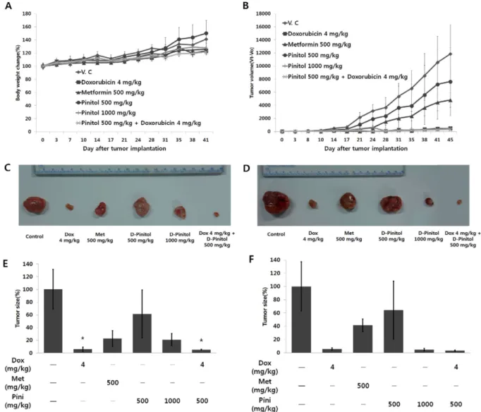
이종 이식 마우스 모델(Nude mouse xenograft assay) − 종양 성장 관련 실험: BALB/c nu mouse에 MDA- MB-231세포(9×10⁶cell/mouse)를 PBS 100 µl에 부유시킨 뒤 1 ml syringe로 우측의 견갑부와 흉벽 사이의 액와 부위 피 하에 이식하였다. 종양의 크기가 일정수준에 도달하였을 때, 약물 투여군 별로 7개체씩 무작위로 분류하였다. D-pinitol (최종 투여용량: 500, 1000 mg/kg)과 metformin(최종 투여용 량: 500 mg/kg)은 증류수를 사용하여 50 mg/ml의 농도로 용 해한 후 마우스에 10 ml/kg 액량으로 반복하여 45일 동안 경구 투여하였다. Doxorubicin hydrochloride은 PBS를 이용 하여 0.4 mg/ml의 농도로 용해한 후 마우스에 20 µl씩 1 ml insulin syringe를 이용하여 반복적으로 종양 내(intratumoral) 에 총 4회(0, 5, 10, 15일) 투여하였다. 형성된 종양의 크기 가 53.0 mm3 도달 시 부터 45일째까지 총 14회 개체별로 vernier caliper를 이용하여 3-방향을 측정한 후 길이(length)
× 너비(width) × 높이(height)/2의 계산식에 의해 종양크기
를 측정하였다. 모든 동물에 대하여 약물 투여 개시 후 일 반증상 관찰 및 체중의 변화를 측정하였다.
− 재발 및 암 줄기세포 관련 실험: 종양 성장 관련 실 험과 달라진 조건은 metformin 투여군은 제외하였으며, D- pinitol 투여군은 300 mg/kg 투여군을 추가 시켰다. 또한 형 성된 종양의 크기가 53.0 mm3 도달 시 부터 63일째까지 총 28회 개체별로 vernier caliper를 이용하여 3-방향을 측정한 후 길이(length) × 너비(width) × 높이(height)/2의 계산식에 의해 종양크기를 측정하였다. 그 외 나머지 조건은 종양 성 장 관련 실험과 동일하다.
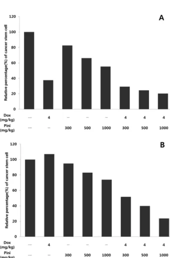
암줄기세포의 비율 측정 − 유방암의 암줄기세포 marker 인 CD44분자와 CD24분자를 유세포분석기를 이용하여 암 줄기세포의 비율을 측정하였다. 형성된 종양을 분리하여 cell strainer를 이용하여 단일세포화 한 뒤 ack 시약을 이용하여 적혈구를 제거해 주었다. 적혈구가 제거된 약 1×10⁶ cell을 1% BSA을 첨가한 PBS(BSA/PBS)로 한번 세척한 후, 50 µl 의 PBS/BSA 안에 부유시켰다. 세포에 anti-CD44 항체와 anti-CD24 항체를 첨가한 후 4oC에서 20분 동안 반응시키 고, PBS로 두 번 세척하여, 500 µl의 PBS로 다시 부유시켜 유세포분석기로 CD44+, CD24− 표현형인 세포 비율을 분석 하였다.
통계분석 − 모든 실험값은 임의대로 100%로 표시한 대조 군에 대하여 백분율로 나타내었다. 실험 결과는 T-test(Micro office, Excel)로 검정하였으며, p값이 0.05 미만일 때 통계 적으로 유의성이 있다고 판정하였다.
결과 및 고찰
D-pinitol이 유방암 세포주 성장에 미치는 영향을 평가하 기 위하여 MTT assay를 수행하였다. MDA-MB-231 세포 주와 MCF-7 세포주를 24시간 배양한 뒤, 1 µM doxorubicin, 5 mM metformin 그리고 1, 5, 10 mM D-pinitol을 48시간 동안 처리하여 MTT assay를 수행하였다. MDA-MB-231 세 포주에서는 1 µM doxorubicin을 처리하였을 때 21.7%
(p<0.05) 감소하였고 5 mM metformin을 처리하였을 때는 12.7%가 감소하였다. 또한 1, 5, 10 mM D-pinitol을 처리하 였을 때는 농도별로 19% (p<0.05), 25% (p<0.01), 28.1%
(p<0.05)가 감소하여 농도 의존적으로 성장이 억제되는 것 을 확인하였다(Fig. 1A). MCF-7 세포주에서는 1 µM doxorubicin을 처리하였을 때 41.1% (p<0.05) 감소하였고 5 mM metformin을 처리하였을 때는 14.9% (p<0.01)가 감 소하였으며 1, 5, 10 mM D-pinitol을 처리하였을 때는 농도 별로 20.7% (p<0.05), 21.8% (p<0.05), 22% (p<0.05)가 감 소한 것을 확인하였다(Fig. 1B). MTT assay에 사용한 MDA-MB-231 세포주는 MCF-7 세포주에 비해서 약물 내 성관련 유전자인 p130cas의 발현이 높아 항암제로 알려진
doxorubicin에 대한 내성이 높은 것으로 보고되어 있는데27), MTT assay를 한 결과에서도 MDA-MB-231 세포주가 MCF-7 세포주에 비해서 doxorubicin의 감수성이 낮은 것으 로 확인되었다. 그러나 D-pinitol은 doxorubicin과는 다르게 MCF-7 세포주에 비해서 MDA-MB-231 세포주에 대한 증 식 억제 효능이 높은 것을 확인할 수 있었다.
인체유래 암세포주를 nude mouse에 이식하여 항암효능 을 평가하는 mouse xenograft asssy는 시험물질의 효능을 in vivo상에서 확인하기에 적합한 동물시험계이다. 따라서 MTT assay에서 확인하였던 두 가지의 세포주 중 doxorubicin에 내성이 있으며 D-pinitol에는 감수성인 MDA-MB-231 세포 주를 mouse에 이식한 xenograft model을 이용하여 생체 내 에서 종양 성장에 어떠한 영향을 미치는지 확인하였다.
MDA-MB-231세포(9×10⁶cell/mouse)를 BALB/c nude mouse 우측의 견갑부와 흉벽 사이의 액와 부위 피하에 이식하였 고 형성된 종양의 크기가 53.0 mm3 도달 시 부터 D-pinitol (최종 투여용량: 500, 1000 mg/kg)을 반복하여 경구 투여하 였으며, doxorubicin hydrochloride은 PBS를 이용하여 0.4 mg/ml의 농도로 용해한 후 마우스에 20 µl씩 반복적으 Fig. 1. Growth inhibition of D-pinitol on breast cancer cell lines. MDA-MB-231 and MCF-7 cell lines were treated with 1-10 mM D-pinitol for 48 hour. Doxorubicin (1µM) and met- formin (5 mM) were used as comparators. Cell viability of (A) MDA-MB-231 and (B) MCF-7 cell lines were estimated by MTT assay (*p<0.05, **p<0.01, ***p<0.001).
로 종양 내에 투여하였다. 투여기간 동안 약물의 독성 정도 를 알아보기 위해 동물의 일반증상 및 체중변화를 관찰하 였으나, 약물 투여 후 일반적인 증상이나 유의적인 체중감 소가 없는 것으로 보아 전체적으로 약물에 대한 독성은 나 타나지 않은 것으로 판단하였다(Fig. 2A). 시험물질 투여 후 25일째에 대조군은 종양의 크기가 3243.7±1010.3 mm3으로 성장하였으나, D-pinitol 500 mg/kg 투여군에서는 종양크기 가 1993.5±1216.6 mm3으로 38.5%의 종양성장 억제효과를, D-pinitol 1000 mg/kg 투여군에서는 종양크기가 681.1±
310.4 mm3으로 79%의 종양성장 억제효과를 나타내었다. 또 한, doxorubicin 4 mg/kg과 병용 투여한 D-pinitol 500 mg/
kg 투여군에서는 종양크기가 171.6±51.2 mm3 (p<0.05)으로
94.7%의 종양성장 억제효과를 나타내었다. Doxorubicin 4 mg/kg 투여군에서는 190.8±105.2 mm3 (p<0.05)으로 94.1%의 종양성장 억제효과가 있었다(Fig. 2B, E). 시험물 질 투여 후 45일째에 대조군은 종양의 크기가 11960.9±
4445.1 mm3으로 성장하였으나, D-pinitol 500 mg/kg 투여군 에서는 종양크기가 7693.5±5231 mm3으로 35.7%의 종양성 장 억제효과를, D-pinitol 1000 mg/kg 투여군에서는 종양크 기가 552.7±234.9 mm3으로 95.4%의 종양성장 억제효과를 나타내었다. 또한, doxorubicin 4 mg/kg과 병용 투여한 D- pinitol 500 mg/kg 투여군에서는 종양크기가 383.5±31.97 mm3 으로 96.8%의 종양성장 억제효과를 나타내었다. Doxorubicin 4 mg/kg 투여군에서는 종양크기가 648.1±194.5 mm3으로
Fig. 2. Inhibition of breast tumor growth by D-pinitol in nude mouse xenograft assay. On day 0, nine million MDA-MB-231 cell were implanted subcutaneously into nude mouse. D-pinitol at the dose of 500 mg/kg or 1000 mg/kg body weight was administered orally for consecutive 45 days. (A) The body weights of the tumor-bearing nude mice were measured to evaluate overall toxicity of D-pinitol. (B) Tumor volumes were estimated by the formula: length (mm) × width (mm) × height (mm)/2. Photographs of rep- resentative MDA-MB-231 tumor mass at (C) 25 day and (D) 45 day were shown. At (E) 25 day and (F) 45 day, relative tumor size was measured (*p<0.05).
94.58%의 종양성장 억제효과가 있었다(Fig. 2D, F). D- pinitol에 의한 인체 유방암 종양 성장 억제가 투여량을 증 가시킬수록 종양의 크기가 감소하였다.
유방암 치료 후에도 재발이 일어나는 이유 중 하나는 암 줄기세포에 의한 새로운 종양의 증식에서 기인하는 것으로 알려져 왔다. 유방암 재발에 영향을 미치는 암줄기세포는 CD44+, CD24−의 표현형을 지닌다. 따라서 우리는 MDA- MB-231 세포주를 BALB/c nude mouse에 이식한 mouse xenograft assay를 수행하여 D-pinitol이 암줄기세포의 증식 에 미치는 영향 분석하였다. D-pinitol 투여 후 25일과 46일 째의 종양덩어리를 단일세포화 시킨 뒤, anti-CD44 항체와 anti-CD24 항체를 이용하여 암줄기세포의 비율을 유세포분 석기을 통해 분석하였다. 25일째의 경우 대조군 대비 doxorubicin 4 mg/kg 투여군에서 62.4% 감소하였으며 D- pinitol 300, 500, 1000 mg/kg 투여군 각각에서 17.4%, 33.7%, 44.8% 감소하였고 doxorubicin 4 mg/kg과 병용 투 여한 D-pinitol 300, 500, 1000 mg/kg 투여군 각각에서는 각
각 70.7%, 75.6%, 79.7% 감소하는 것을 확인하였다(Fig.
3A). 또한 46일째의 경우 대조군 대비 doxorubicin 4 mg/kg 투여군에서 7.1% 증가하였으며 D-pinitol 300, 500, 1000 mg/kg 투여군 각각에서 5.2%, 17.1%, 26.1% 감소하 였고 doxorubicin 4 mg/kg과 병용 투여한 D-pinitol 300, 500, 1000 mg/kg 투여군 각각에서는 각각 48.3%, 60.2%, 76.3% 감소하는 것을 확인하였다(Fig. 3B). D-pinitol은 종 양 내에 암줄기세포의 비율을 감소시키는 효능이 있는 것 으로 확인되었으나, 46일째 종양에서 doxorubicin 단독투여 군의 종양 내 암줄기세포 비율은 vehicle control 투여군 보 다 높게 측정되어 암줄기세포 억제에 영향을 주지 않는 것 으로 확인 되었다. 또한, doxorubicin과 D-pinitol의 단독 투 여군과 doxorubicin과 D-pinitol의 병용투여군의 암줄기세포 비율을 비교해 보았을 때 병용 투여군에서 암줄기세포의 비 율은 현저히 감소하여 시너지 효과가 나타나는 것을 확인 할 수 있었다.
D-pinitol은 암세포주로부터 발생하는 CSCs를 비율을 감 소시키고 종양 내 CSCs의 증식을 억제하였다. 이는 D-pinitol 이 암의 재발을 억제할 수 있음을 시사한다. 따라서 D-pinitol 에 의해 유방암 재발이 지연되는지를 확인하기 위해 MDA- MB-231 세포주를 BALB/c nude mouse에 이식하고 종양재 발을 분석하였다. Doxorubicin 4 mg/kg과 병용 투여한 D- pinitol 300, 500 mg/kg 투여군에서는 35일 동안은 종양재발 이 관찰되지 않았으며, doxorubicin 4 mg/kg과 병용 투여한 D-pinitol 1000 mg/kg 투여군에서는 37일 동안 종양재발이 관찰되지 않았다. 또한 doxorubicin 4 mg/kg 단독 투여군에 서는 30일 동안 종양재발이 나타나지 않음을 확인하였다 (Fig. 4). 특히 44일째에 관찰된 doxorubicin 단독 투여군의 종양크기는 D-pinitol 1000 mg/Kg 병용 투여군에서 2주 후 관찰되었다. 따라서 D-pinitol은 암 성장 억제와 더불어 종 Fig. 3. Inhibition of the growth of the cancer stem cells within
tumor mass by D-pinitol. On (A) 25 day and (B) 46 day, the mice were sacrificed and CSCs population of the breast tumors were measured by flow cytometry analysis after double stain- ing with anti-CD44 and anti-CD24 antibodies.
Fig. 4. Delay of breast tumor relapse in mouse xenograft assay by D-pinitol. D-pinitol at the dose of 300 mg/kg, 500 mg/kg and 1000 mg/kg body weight was administered orally for con- secutive 63 days. Doxorubicin at the dose of 4 mg/kg was injected within breast tumor four times for 15 day.
양 내의 암 줄기세포 비율을 감소시켜 암 재발을 지연시키 는 효능이 있음을 말해준다.
결 론
본 연구는 D-pinitol이 유방암 성장 억제와 재발 지연에 대한 효능이 있음을 확인하였고, 이는 암 줄기세포의 증식 억제에서 기인함을 규명하였다. 이와 같은 연구결과는 천연 식물에서 추출한 안전한 물질인 D-pinitol이 항암 보조제로 사용될 수 있음을 시사한다.
사 사
이 논문은 2011년도 충북대학교 학술연구지원사업의 연 구비 지원에 의하여 연구되었음.
인용문헌
1. Narayanan, C. R., Joshi, D. D., Mudjumdar, A. M. and Dhekne, V.V. (1987) Pinitol, a new anti-diabetic compound from the leaves of Bougainvillea spectabilis. Curr. Sci. 56:
139-141.
2. Larner, J. (2002) D-chiro-inositol - its functional role in insu- lin action and its deficit in insulin resistance. Int. J. Exp. Dia- betes Res. 3: 47-60.
3. Koo, B. J., Kim, H. J. and Park, K. S. (2007) The clinical study to evaluate the safety and efficacy of D-chiro-inositol in patients with type 2 diabetes. Korean J. med. 72: 29-36.
4. Witters, L. A. (2001) The blooming of the French lilac. J.
Clin. Invest. 108: 1105-1107.
5. Zakikhani, M., Dowling, R., Fantus, I. G., Sonenberg, N. and Pollak, M. (2006) Metformin is an AMP kinase-Dependent growth inhibitor for breast cancer cells. Cancer Res. 66:
10269-10273.
6. Euhus, D. M. (2001) Understanding mathematical models for breast cancer risk assessment and counseling. Breast J. 7:
224-232.
7. Park, S. K., Lim, Y. K. and Yoo, K. Y. (1998) Prediction of future breast cancer mortality in Korea. Korean J. Epidemiol.
20: 267-274.
8. Park, B., Choi, K. S., Lee, Y. Y., Jun, J. K. and Seo, H. G.
(2012) Cancer screening status in Korea, 2011: results from the Korean National Cancer Screening Survey. Asian Pac. J.
Cancer Prev. 13: 1187-1191.
9. Bland, K. I., Copeland, E. M. III., Breast. In: Schwartz, S. I., Shires, G. T. and Spencer, F. C. (1994) Principles of surgery, 6th ed. NewYork: McGraw-Hill 554-557.
10. Jatoi, I., Tsimelzon, A., Weiss, H., Clark, G. M. and Hilsen- beck, S. G. (2005) Hazard rates of recurrence following diag- nosis of primary breast cancer. Breast Cancer Res. Treat. 89:
173-178.
11. Demicheli, R., Abbattista, A., Miceli, R., Valagussa, P. and Bonadonna, G. (1996) Time distribution of the recurrence risk for breast cancer patients undergoing mastectomy: further support about the concept of tumor dormancy. Breast Cancer Res. Treat. 41: 177-185.
12. Ahmad, A. (2012) Pathway to breast cancer recurrence. ISRN Oncol. 2013: Article ID 290568, 16.
13. Lapidot, T., Sirard, C., Vormoor, J., Tsvee, L., Christian, A., Josef, V., Barbara, M., Trang, H., Julio, C. C., Mark, M., Bruce, P., Michael, A. C. and John, E. D. (1994) A cell ini- tiating human acute myeloid leukaemia after transplantation into SCID mice. Nature 367: 645-648.
14. Bonnet, D. and Dick, J. (1997) Human acute myeloid leu- kemia is organized as a hierarchy that originates from a prim- itive hematopoietic cell. Nat. Med. 3: 730-737.
15. Al-Hajj, M., Wicha, M.S., Benito-Hernandez, A., Morrison, S.J. and Clarke, M.F. (2003) Prospective identification of tumorigenic breast cancer stem cells. Proc. Natl. Acad. Sci.
100: 3983-3988.
16. Hemmati, H. D., Nakano, I., Lazareff, J. A., Masterman- Smith, M., Geschwind, D.H., Bronner-Fraser, M. and Korn- blum, H.I. (2003) Cancerous stem cells can arise from pedi- atric brain tumors. Proc. Natl. Acad. Sci. 100: 15178-15183.
17. Collins, A.T., Berry, P.A., Hyde, C., Stower, M.J. and Mait- land, N.J. (2005) Prospective identification of tumorigenic prostate cancer stem cells. Cancer Res. 65: 10946-10951.
18. Fang, D., Nguyen, T. K., Leishear, K., Finko, R., Kulp, A. N., Hotz, S., Van Belle, P. A., Xu, X., Elder, D. E. and Herlyn, M. (2005) A tumorigenic subpopulation with stem cell prop- erties in melanomas. Cancer Res. 65: 9328-9337.
19. Bapat, S. A., Mali, A. M., Koppikar, C. B. and Kurrey, N. K.
(2005) Stem and progenitor like cells contribute to the aggressive behaviour of human epithelial ovarian cancer.
Cancer Res. 65: 3025-3029.
20. O’Brien, C. A., Pollett, A., Gallinger, S. and Dick, J. E.
(2007) A human colon cancer cell capable of initiating tumor growth in immunodeficient mice. Nature 445: 106-110.
21. Ma, S., Chan, K. W., Hu, L., Lee, T. K., Wo, J. Y., Ng, I. O., Zheng, B. J. and Guan, X. Y. (2007) Identification and char- acterisation of tumorigenic liver cancer stem/progenitor cells.
Gastroenterology 132: 2542-2556.
22. Ho, M. M., Ng, A. V., Lam, S. and Hung, J. Y. (2007) Side population in human lung cancer cell lines and tumors is enriched with stem-like cancer cells. Cancer Res. 67: 4827- 4833.
23. Hermann, P., Huber, S., Herrler, T., Aicher, A., Ellwart, J. W., Guba, M., Bruns, C. J. and Heeschen, C. (2007) Distinct pop- ulations of cancer stem cells determine tumour growth and metastatic activity in human pancreatic cancer. Cell Stem Cell 1: 313-323.
24. Fukuda, K., Siakawa, Y., Ohashi, M., Kumagai, K., Kitajima,
M., Okano, H., Matsuzaki, Y. and Kitagawa, Y. (2009) Tumor initiating potential of side population cells in human gastric cancer. Int. J. Oncol. 34: 1201-1207.
25. Lagadec, C., Vlashi, E., Donna, L. D., Dekmezian, C. and Pajonk, F. (2012) Radiation-induced reprogramming of breast cancer cells. Stem Cells 30: 833-844.
26. Al-Hajj, M., Becker, M. W., Wicha, M., Weissman, I. and Clarke, M. F. (2004) Therapeutic implications of cancer stem cells. Curr. Opin. Genet. Dev. 14: 43-47.
27. Sethi, G., Ahn, K. S., Sung, B. K. and Aggarwal, B. B. (2008) Pinitol targets nuclear factor-kB activation pathway leading to inhibition of gene products associated with proliferation, apoptosis, invasion, and angiogenesis. Mol. Cancer Ther. 7:
1604-1614.
28. Thamaraiselvan, R., Natarajan, N. and Maruthaiveeran, P. B.
(2013) D-pinitol prevents rat breast carcinogenesis induced by 7, 12-dimethylbenz [a] anthracene through inhibition of bcl-2 and induction of p53, caspase-3 proteins and modu- lation of hepatic biotransformation enzymes and antioxidants.
Biomed. Prev. Nutr. 3: 31-41.
29. Ta, H. Q., Thomas, K. S., Schrecengost, R. S. and Bouton, A.
H. (2008) A novel association between p130Cas and resis- tance to the chemotherapeutic drug adriamycin in human breast cancer cells. Cancer Res. 68: 8796-8804.
(2014. 6. 12 접수; 2014. 6. 16 심사; 2014. 6. 18 게재확정)