대한내과학회지: 제 75 권 제 3 호 2008
원발성 흉선 MALT 림프종 1예
계명대학교 의과대학 내과학교실
박우영 ․ 성희진 ․ 이상민 ․ 김주연 ․ 김진영 ․ 도영록 ․ 송홍석
A case of primary thymic MALT lymphoma
Woo Young Park, M.D., Hee Jin Seong, M.D., Sang Min Lee, M.D., Ju Youn Kim, M.D., Jin Young Kim, M.D., Young Rok Do, M.D. and Hong Suk Song, M.D.
Department of Internal Medicine, Keimyung University School of Medicine, Dongsan Medical Center, Daegu, Korea
Primary thymic MALT lymphoma is a rare thymic tumor, with only eight previous cases having been described worldwide to date.
We report a case of a 60-year-old Korean woman diagnosed as primary thymic MALT lymphoma. She was found to have an anterior mediastinal tumor during a medical check-up in 2006 and was referred to our hospital for further examination and treatment. The thymus was resected through a median sternotomy and pathology revealed primary thymic MALT lymphoma. Two months later, a follow-up chest CT showed a residual mediastinal soft tissue mass and increased FDG uptake was detected on PET CT scan.
The patient was irradiated with 4,140 cGy. After radiation therapy, no evidence of residual soft tissue was found in follow-up chest CT scan and the patient is alive and well 15 months after treatment.
We report the details of this case of primary thymic MALT lymphoma treated with irradiation and also offer a review of the literature. (Korean J Med 75:343-348, 2008)
Key Words: Thymus; Lymphoma; MALT
∙Received: 2007. 5. 30
∙Accepted: 2007. 7. 18
∙Correspondence to: Hong Suk Song, M.D., Department of Internal Medicine, Keimyung University School of Medicine, 194 Dongsan-dong, Jung-gu, Daegu 700-712, Korea E-mail: [email protected]
서 론
MALT 림프종은 정상적으로 위장관 조직을 구성하는 MALT에서 발생하는 B세포 림프종이다1). 비호지킨림프종 의 8%를 차지하며 위장관은 MALT 림프종의 호발 부위로 위 MALT 림프종에 대해서는 많은 연구가 이루어져 있다.
그러나 MALT 림프종은 위장관 외 안와, 폐, 갑상선, 침샘, 피부, 연부조직, 방광, 신장, 중추신경 등 어느 조직에서든 발생 가능하며 위장관외 MALT 림프종은 대부분 병기가 낮 아 국소 치료에 반응이 좋은 것으로 알려져 있다2). 조직학
적으로는 중심세포양세포(centrocyte-like cell)의 침윤 및 림 프상피병변(lymphoepithelial lesion)의 형성이 특징이다.
발생학적으로 흉선은 3번째와 4번째 인두주머니에서 기 원하는데 크게 림프조직과 상피조직으로 구성되어 있다. 흉 선에서 발생하는 림프종으로 호지킨병, 림프모구림프종, 미 만성 대형 B세포 림프종 등은 잘 알려져 있으나 원발성 MALT 림프종은 드문 예로 1990년에 Isaacson 등5)이 2예를 보고한 이래 현재까지 8예가 보고되고 있다3-9).
저자들은 흉선에서 MALT 림프종이 발생한 1예를 경험 하였기에 문헌고찰과 함께 보고하는 바이다.
- The Korean Journal of Medicine: Vol. 75, No. 3, 2008 -
Figure 1. Chest PA showing enlarged right cardiopericardial silhouette border.
Figure 2. Chest CT showing a well defined homogeneous anterior mediastinal tumor and there was no evidence of invasion to surrounding tissue and LN enlargement.
Figure 3. Pathologic finding of Thymic MALT-type lymp- homa. Microscopic finding of the tumor consists of mono- tonous centrocyte-like cells and lymphoepithelial lesion surrounding Hassall's corpuscles (H&E stain, ×400).
증 례
환 자: 김○○, 여자 61세 주 소: 특이 증상 없음.
현병력: 특별한 증상 없이 일차의료기관에서 촬영한 흉 부전산화단층촬영상 흉선에 종양이 발견되어 내원하였다.
과거력 및 가족력: 9년 전 제2형 당뇨병 진단 받았고, 30 년 전 자궁근종으로 수술 받았으며 가족력에서 특이 사항은 없었다.
이학적 소견: 입원 당시 혈압은 120/80 mmHg, 맥박수 70 회/분, 호흡수 22회/분, 체온 36.6℃였고, 공막의 황달 소견 및 결막의 창백 소견은 관찰되지 않았으며, 두경부에 촉지 되는 림프절은 없었다. 폐와 심음은 정상 소견이었고, 복부 압통도 없었다.
검사 소견: 입원 당시 말초혈액검사에서 백혈구 4,610/
mm3, 혈색소 15.1 g/dL, 헤마토크리트 44.9%, 혈소판 246,000 /mm3, 뇨 검사상 뇨비중 1.010, 뇨단백 음성, 뇨당 음성, 고 배율 시야에서 백혈구 11-15개가 보였고, 뇨침 검사상 원주 는 없었다. 생화학 검사상 칼슘 10.2 mg/dL, 혈액 뇨소질소 10 mg/dL, 크레아티닌 0.8 mg/dL, 총 단백 8.1 g/dL, 알부민
4.0 g/dL, 총 빌리루빈 0.7 mg/dL, ALP 48 U/L, AST 25 U/L, ALT 33 U/L, 총 콜레스테롤 189 mg/dL, LDH 337.4 U/L, 요 산 3.8 mg/dL, 프로트롬빈시간 10.2초, 활성부분트롬보플라 스틴시간 25.4초였다.
방사선학적 소견: 흉부 단순촬영상 우측 심연이 커져 있 고(그림 1), 흉부전산화단층촬영에서 전 종격 흉선에 난형의
- Woo Young Park, et al: Primary thymic MALT lymphoma -
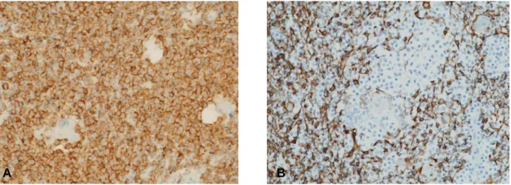
Figure 4. (A) Pathologic finding of thymic MALT lymphoma. The centrocyte-like cells are diffuse positive for CD20 (IH stain,
×200). (B) The thymic epithelial cells are positive for CK only in medulla portion (IH stain, ×200).
Figure 5. Two months after surgical excision, there was residual anterior mediastinal soft tissue mass in follow up chest CT scan.
종양이 발견되었고, 주위 조직 침윤이나 림프절 전이 소견 은 없었다(그림 2).
조직학적 소견: 수술 소견상 종양의 우측 흉막과 심낭 침 윤이 관찰되었고, 상대정맥 림프절 종대가 관찰되었다. 흉 선 절제와 함께 우측 흉막과 심낭 절제술을 시행하였고, 절 제된 검체 조직 표본은 육안적으로 8.0×6.5×4.0 cm 크기의 황갈색 덩어리로 무게는 62 gm이었으며 우측 흉막과 심낭 침윤이 관찰되었다. 광학현미경 하에서 피질과 수질의 기본
적인 구조는 보존되어 있었고, 중심세포양세포의 증식, 흉 선소체(Hassall's corpuscles) 주위로 림프상피병변이 현저하 여 흉선 조직의 소실을 보였다(그림 3). 면역조직화학검사상 에서 중심세포양세포는 CD20 미만성 양성(그림 4), CD79 양성, Bcl-2 양성, CD3 음성, CD10 음성, CD23 음성, CyclinD1 음성, Bcl-6 음성으로 B세포 표현형을 보였고, 흉 선상피세포는 CK 수질 부분에만 양성 소견을 보였으며(그 림 4), 같이 절제된 상대정맥 림프절 전이는 없었다.
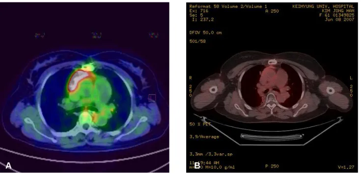
치료 및 경과: 이상 소견으로 원발성 흉선 MALT 림프종 (Ann Arbor stage IE)으로 진단하였고, 수술로 절제를 하였으 나 2개월 후 추적 흉부전산화단층촬영상 종격에 잔존 조직 이 발견되었고(그림 5), 추가로 검사한 PET-CT에서 FDG uptake 증가가 관찰 되었다(그림 7). 수술 4개월 후 시행한 추적 흉부전산화단층촬영상 잔존 종격 조직 크기의 증가가 관찰되어(그림 6), 4,140 cGy의 선량으로 국소 방사선 치료 를 시행하였으며 현재까지 15개월간 무병 생존을 보여 외래 추적관찰 중이다(그림 6, 7).
고 찰
1983년 Isaacson 등1)이 처음으로 MALT 림프종에 대해 기 술한 이후 1994년에 Harris 등10)이 처음으로 B세포 림프종의 외투층 B세포 림프종(Marginal Zone B-cell lymphoma; MZL) 으로 분류하였고, 최근 WHO 분류 체계에 의해 외투층 B세 포 림프종의 하나로 분류되었다. MALT 림프종은 비호지킨 림프종의 7.6~8% 정도로 흔하지 않으며 호발 연령은 60대 이고, 남녀 비율은 1:1.5로 여자에서 좀 더 발생하는 것으로 알려져 있다11, 13). 대부분 원발 부위는 위장관이며 그 외 안
A B
- 대한내과학회지: 제 75 권 제 3 호 통권 제 577 호 2008 -
Figure 6. (A) Four months after surgical excision, follow up chest CT scan showing increased size of residual anterior mediastinal soft tissue mass. (B) Three months after radiation therapy, there was no evidence of residual soft tissue mass in follow up chest CT scan.
Figure 7. (A) Four months after surgical excision, follow up PET-CT scan showing increased FDG uptake of residual soft tissue.
(B) Nine months after radiation therapy, there was no evidence of FDG uptake in follow up PET-CT scan.
와, 폐, 갑상선, 침샘, 피부, 연부조직, 방광, 신장, 중추신경 등 림프절외 어느 조직에서도 발생 가능하나 원발성 흉선 MALT 림프종은 드물어 현재까지 8예가 보고된 것으로 알 려져 있다3, 5-9).
위 MALT 림프종 발생과 헬리코박터 감염 및 자가면역질 환과의 연관성이 알려지면서 MALT 림프종 인식에 하나의 전기가 마련되었고, 항원의 지속적인 자극에 의한 점막 림 프 조직의 장기간 증식 과정이 MALT 림프종의 병태생리로
A B
A B
- 박우영 외 6인: 원발성 흉선 MALT 림프종 1예 -
알려져 있다12).
조직학적으로 MALT 림프종은 중심세포양세포의 침윤 및 림프상피병변이 특징적이나 면역조직화학염색상 CD20, CD79a, bcl-2, IgM 양성, CD5, CD10, bcl-6, cyclinD1, IgG, IgA 음성 및 세포유전학적 분석으로 진단이 가능하다14).
임상적으로 위장관외 MALT 림프종은 대개 증상 없이 통 상적인 방사선 검사에서 우연히 발견되거나 종양에 의한 발 생 부위의 국소 증상으로 발견되게 된다. 위장관외 MALT 림프종의 74%는 원발 장기 및 국소 림프절에 국한된 경우 이고 26%에서 병기 3~4를 보이는데 이러한 경우에는 미만 성 대형 B세포 림프종으로 전환되어 병발하는 경우가 많다12). 대부분 진단 시 병기가 낮아 완전 관해율은 76%, 10년 생존 율은 75% 이상되어 예후는 양호하며 IPI score가 낮은 환자 의 경우 5년 생존율이 99%까지 이른다13, 15).
치료로는 원발 부위에 국한된 경우 국소 치료인 방사선 또는 수술적 제거로 완치가 가능하며 헬리코박터 감염이 있 는 위 MALT 림프종은 항생제 치료로도 관해율이 70% 이상 된다16). 병기가 더 진행한 경우에는 항암 치료가 주된 치료 이고, 미만성 대형 B세포 림프종으로 전환되거나 병발한 경 우에는 복합 항암 화학 요법이 필요하다.
저자들의 증례도 특별한 증상 없이 흉부전산화단층촬영에 서 종격내 흉선 종양이 발견되어 수술적 제거를 시행하고 추 적 검사에서 발견된 잔존 종양에 대해서는 국소 방사선 치료 를 시행한 후 현재까지 15개월간 무병 생존을 보이는 원발성 흉선 MALT 림프종 증례이며 아직 이에 대한 병태 생리, 임 상적 특징 및 치료가 정립되지 않은 상태이다. 본 저자들은 앞으로 원발성 흉선 MALT 림프종에 대한 증례 발표, 정보 수집 등 더 많은 연구가 필요함을 고찰 할 수 있었다.
요 약
저자들은 특별한 증상 없이 흉부전산화단층촬영에서 발 견된 종격동내 흉선 종양으로 내원하여 수술적 제거 후 추 적 검사상 발견된 잔존 종양에 대해 국소 방사선 치료를 시 행한 후 현재까지 15개월간 무병 생존을 보인 원발성 흉선 MALT 림프종 1예를 경험 하였기에 문헌고찰과 함께 보고 하는 바이다.
중심 단어: 흉선; 림프종; MALT
REFERENCES
1) Isaacson P, Wright DH. Malignant lymphoma of mucosa-
associated lymphoid tissue: a distinctive type of B-cell lymphoma. Cancer 52:1410-1416, 1983
2) Isaacson PG, Spencer J. Malignant lymphoma of mucosa- associated lymphoid tissue. Histopathology 11:445-462, 1987 3) Yamasaki S, Matsushita H, Tanimura S, Nakatani T, Hara S,
Endou Y, Hara M. B-cell lymphoma of mucosa-associated lymphoid tissue of the thymus: a report of two cases with a background of Sjogren's syndrome and monoclonal gammopathy. Hum Pathol 29:1021-1024, 1998
4) Nakagawa A, Nakamura S, Koshikawa T, Nakayama A, Nagasaka T, Motoori T, Kojima M, Hosomura Y, Ueda R, Mori S, Asai J, Suchi T. Clinicopathologic study of primary mediastinal non-lymphoblastic non-Hodgkin's lymphomas among the Japanese. Acta Pathol Jpn 43:44-54, 1993
5) Isaacson PG, Chan JK, Tang C, Addis BJ. Low-grade B-cell lymphoma of mucosa-associated lymphoid tissue arising in the thymus: a thymic lymphoma mimicking myoepithelial sialad- enitis. Am J Surg Pathol 14:342-351, 1990
6) Takagi N, Nakamura S, Yamamoto K, Kunishima K, Tkagi I, Suyama M, Shinoda M, Sugiura T, Oyama A, Hisamitsu S, Takashi K, Keniichi K, Ryuzo U, Toshitada T, Yutaka A, Taizan S. Malignant lymphoma of mucosa-associated lymphoid tissue arising in the thymus of a patient with Sjogren's symdrome: a morphologic, phenotypic and genotypic study.
Cancer 69:1347-1355, 1992
7) Yokose T, Kodama T, Matsuno Y, Shimosato Y, Nishimura M, Mukai K. Low grade B cell lymphoma of mucosa- associasted lymphoid tissue in the thymus of a patient with rheumatoid arthritis. Patholo Int 48:74-81, 1998
8) Yi JG, Kim DH, Choi CS. Malignant lymphoma of mucosa-associated lymphoid tissue (MALT lymphoma) arising in the thymus: radiologic findings. AJR Am J Roentrenol 171:899-900, 1998
9) Moriyama E, Yokose T, Kodama T, Matsuno Y, Hojo F, Takahashi K, Nagai K, Nishiwaki Y, Ochiai A. Low-grade B-cell lymphoma of mucosa-associated lymphoid tissue in the thymus of a patient with pulmonary amyloid nodules. Jpn J Clin Oncol 30:349-353, 2000
10) Harris NL, Jaffe ES, Stein H, Banks PM, Chan JK, Cleary ML, Delsol G, De Wolf-Peeters C, Falini B, Gatter KC. A revised European-American classification of lymphoid neoplasms:
a proposal from the International Lymphoma Study Group.
Blood 84:1361-1392, 1994
11) The Non-Hodgkin's Lymphoma Classification Project. A clinical evaluation of the International Lymphoma Study Group Classification of non-Hodgkin's lymphoma. Blood 89:
3909-3918, 1997
12) Cohen SM, Petryk M, Varma M, Kozuch PS, Ames ED, Grossbard ML. Non-Hodgkin's lymphoma of mucosa-associated lymphoid tissue. Oncologist 11:1100-1117, 2006
- The Korean Journal of Medicine: Vol. 75, No. 3, 2008 -
13) Zucca E, Conconi A, Pedrinis E, Cortelazzo S, Motta T, Gospodarowicz MK, Patterson BJ, Ferreri AJ, Ponzoni M, Devizzi L, Giardini R, Pinotti G, Capella C, Zinzani PL, Pileri S, Lopez-Guillermo A, Campo E, Ambrosetti A. Baldini L, Cavalli F. Nongastric marginal zone B-cell lymphoma of mucosa-associated lymphoid tissue. Blood 101:2489-2495, 2003
14) Wotherspoon AC, Dogan A, Du MQ. Mucosa-associated lymphoid tissue lymphoma. Curr Opin Hematol 9:50-55, 2002
15) Thieblemont C, Bastion Y, Berger F, Rieux C, Salles G, Pomontet C, Felman P, Coiffier B. Mucosa-associated lymphoid tissue gastrointestinal and nongastrointestinal lymphoma behavior: analysis of 108 patients. J Clin Oncol 15:1624-1630, 1997
16) Hunt RH. Peptic ulcer disease: defining the treatment strate- gies in the era of Helicobacter pylori. Am J Gastroentrol 92(4 Suppl):36S-40S, 1997