https://doi.org/10.5468/ogs.2019.62.3.149 pISSN 2287-8572 · eISSN 2287-8580
Robotic single-site staging operation for early-stage endometrial cancer: initial experience at a single institution
Hyewon Chung * , Tae-Kyu Jang * , Seung Hyub Nam, Sang-Hoon Kwon, So-Jin Shin, Chi-Heum Cho
Department of Obstetrics and Gynecology, Keimyung University School of Medicine, Daegu, Korea
Objective
The aims of this study were to introduce surgical guidelines, and to evaluate the feasibility and safety of a robotic single-site staging (RSSS) operation for early-stage endometrial cancer.
Methods
Patients with a preoperative diagnosis of endometrial cancer (International Federation of Gynecology and Obstetrics stages IA to IB) from endometrial curettage and preoperative imaging studies were selected at Dongsan Medical Center from March 2014 to November 2015. All surgical procedures, including hysterectomy, salpingo-oophorectomy, bilateral pelvic node dissection, and cytology aspiration, were performed by robotic single-site instruments (da Vinci Si
®surgical system; Intuitive Surgical, Sunnyvale, CA, USA).
Results
A total of 15 women with early-stage endometrial cancer underwent the RSSS operation. The median patient age and body mass index were 53 years (range, 37–70 years) and 25.4 kg/m
2(range, 18.3–46.4 kg/m
2). The median docking time, console time, and total operative time were 8 minutes (range, 4–15 minutes), 75 minutes (range, 55–115 minutes), and 155 minutes (range, 125–190 minutes), respectively. The median retrieval of both pelvic lymph nodes was 9 (range, 6–15). There were no conversions to laparoscopy or laparotomy.
Conclusion
The RSSS operation is feasible and safe in patients with early-stage endometrial cancer. In this study, operative times were reasonable, and the surgical procedure was well-tolerated by the patients. Further evaluation of patients with early-stage endometrial cancer should be performed in large-scale comparative studies using the laparoendoscopic, single-site staging operation to confirm the safety and benefits of the RSSS operation for early-stage endometrial cancer.
Keywords: Single site; Single port; Robotic; Staging operation; Endometrial cancer
Co-Corresponding author: So-Jin Shin
Department of Obstetrics and Gynecology, Keimyung University School of Medicine, 56 Dalseong-ro, Jung-gu, Daegu, Korea E-mail: [email protected]
https://orcid.org/0000-0002-7432-6025
*
These authors contributed equally to this work.
Articles published in Obstet Gynecol Sci are open-access, distributed under the terms of the Creative Commons Attribution Non-Commercial License (http://creativecommons.
org/licenses/by-nc/3.0/) which permits unrestricted non-commercial use, distribution, and reproduction in any medium, provided the original work is properly cited.
Copyright © 2019 Korean Society of Obstetrics and Gynecology Received: 2017.12.29. Revised: 2018.05.28. Accepted: 2018.11.20.
Corresponding author: Chi-Heum Cho
Department of Obstetrics and Gynecology, Keimyung University School of Medicine, 56 Dalseong-ro, Jung-gu, Daegu, Korea E-mail: [email protected]
https://orcid.org/0000-0002-0437-4099
Introduction
Current trends in minimally invasive surgery are focused on decreasing surgical trauma through the elimination of incisions, which results in less postoperative discomfort, de- creased hospital stays, improved cosmetic results, and less wound-related complications [1,2]. Laparoendoscopic single- site surgery (LESS) utilizes a minimal number of skin incisions to gain access to the abdominal or pelvic cavity. There has been an increase in the use of LESS for the management of gynecologic disease, even for the most advanced oncologic procedures. Park et al. [3] described the feasibility and effi- cacy of LESS on patients with early-stage endometrial cancer.
In addition, some retrospective, multi-institutional studies de- scribed the use of single-incision laparoscopy for endometrial cancer staging and early-stage cervical cancer [4,5]. Advanc- es in instrumentation are providing solutions to the technical challenges of LESS and encouraging reconsideration of the use of a single incision for laparoscopic surgery. However, de- spite being a more advanced surgical method, LESS presents various surgical challenges, including a limited range of mo- tion due to the parallel angle of the surgical instruments, and the difficulty in manipulating a flexible camera and surgical instruments in a limited space through a small skin incision [6-8]. Therefore, robotic technology applied to LESS has been postulated to overcome these limitations [9,10]. In 2009, Kaouk et al. [11] reported the first successful series of single- site robotic procedures in humans; they noted an improved ability in intracorporeal dissecting and suturing because of the robotic semi-flexible instrument and the triangulation achieved by crossing the curved cannulas. A less-recognized benefit was the reduction in fatigue and strain for the oper- ating surgeon [12]. These studies show that advanced tech- nology used to perform operations in various surgical fields can be successful.
Many studies have reported the feasibility, safety, and ef- ficacy of robotic-assisted, single-site surgery in benign gy- necologic diseases [13-15]. However, few studies have used robotic-assisted, single-site surgery in gynecologic oncology.
Preliminary studies that have evaluated the use of this tech- nique for the management of malignant disorders in gyne- cology have demonstrated the feasibility of this approach [16].
Endometrial cancer is the most common malignancy of the female genital tract in the United States [17] and the
third most common malignancy of the female genital tract in South Korea, and its incidence is rapidly increasing [18]. The standard surgical management for early-stage endometrial cancer is surgery, including a total hysterectomy, bilateral salpingo-oophorectomy, and pelvic and/or para-aortic lymph node dissection [19]. As mentioned previously, only a few re- ports about robot-assisted, single-site surgery in gynecologic oncology have been published. This study is a preliminary evaluation of a robotic single-site staging (RSSS) operation including pelvic node dissection at a single institution by a single surgeon. The aims of our preliminary study are to introduce surgical guidelines, and to evaluate the feasibility and safety of the RSSS operation on patients with early-stage endometrial cancer.
Materials and methods
1. Patients and basic characteristics
A total of 15 patients who underwent the RSSS operation between March 2014 and November 2015 in the Depart- ment of Obstetrics and Gynecology at the Keimyung Univer- sity Dongsan Medical Center (Daegu, Korea) were included in this study. Prior to their operations, all patients were informed about the RSSS techniques, benefits, and related risks of possible laparoscopic or laparotomic conversion, and signed a written consent form.
Patients with a preoperative diagnosis of endometrial can- cer (International Federation of Gynecology and Obstetrics stage IA and IB) by endometrial curettage or biopsy were selected. Inclusion criteria were as follows: 1) No evidence of metastasis to other organs in the preoperative imaging, 2) a uterus size smaller than 14 gestational weeks, and 3) well (G1) and moderately (G2) differentiated endometrioid endometrial cancer diagnosed by preoperative endometrial curettage or biopsy. The standard exclusion criteria used for any laparoscopic cancer surgery were applied, but there were no restrictions related to body mass index (BMI) or previous abdominal surgeries.
Perioperative and postoperative data were collected pro-
spectively. Operative times were recorded electronically and
were defined as the interval between the start of the incision
to closure. In addition, the time it took to perform the fol-
lowing was recorded: 1) umbilical incision and single-port
placement, 2) robotic docking, 3) surgeon console time, and
4) vaginal cuff closure. The total operation time was calculat- ed from setting time to console time. Intraoperative param- eters included estimated blood loss, requirement for blood transfusion, conversion to multiport laparoscopy or lapa- rotomy, and presence of drainage. Postoperative parameters included length of hospital stay, postoperative hemoglobin changes after 4 hours, and complications and presence of postoperative therapy according to the permanent biopsy. All patients were followed up at the outpatient clinic 2 weeks and 6 weeks after discharge.
2. Surgical technique
All RSSS operations were performed using a da Vinci Si
®Surgical System (Intuitive Surgical, Sunnyvale, CA, USA). The surgical team consisted of the primary surgeon, the bedside assistant, and a robot system-dedicated scrub technician and circulating nurse. This single-site instrument is a multiple- channel single port composed of: a robotic, 8.5-mm, high- definition, and 3-dimensional (3D) endoscope; 2 types of curved robotic cannulas; and one 5-mm accessory cannula.
The patient was placed in the typical low-lithotomy posi- tion after induction of general anesthesia. The body of the patient was then positioned in the Trendelenburg position (at a 30-degree angle). A single 2.5-cm vertical periumbilical incision was usually made to the left of the umbilicus using an open Hasson approach. The left periumbilical incision pro- vided an easier approach and resulted in less postoperative scarring. The lubricated single-site port was inserted into the abdominal cavity, and the lower rim of the single-site port was clamped using a traumatic Kelly forceps (AliMed, Ded- ham, MA, USA). After checking the other organs, a pneumo- peritoneum was made with carbon dioxide at a pressure of 12 mmHg. A trocar for the camera and a 3D, 8.5-mm endo- scope (30 degrees) were inserted carefully along the endo- scopic cannula. The abdominal cavity was inspected to con- firm the feasibility of the RSSS operation, and to verify any adhesion and/or obstacle for node dissection. The operator coagulated both salpinges before the uterine manipulation device was inserted to prevent the possibility of metastasis to other organs. After coagulating both salpinges, the 3D, 8.5- mm endoscope was removed. A Rumi
®uterine manipulation device (Cooper Surgical, Trumbull, Connecticut, USA) was in- serted to hold the cervix tight and enable efficient movement during the operation.
One 5×250-mm curved cannula (arm 2) was inserted
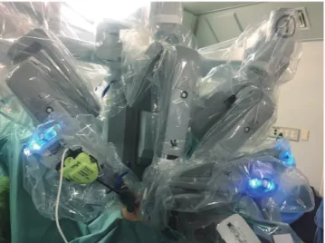
through the designated lumen until the end of the cannula was visible in the visible field of the endoscope. While the other cannula (arm 1) was inserted using the same method as arm 2, the already inserted cannula was held by the as- sistant to prevent displacement. Lastly, the 2 curved cannulas were positioned in a cross position to avoid collision, and a monopolar hook (arm 2) and fenestrated bipolar grasper (arm 1) were placed in each arm of the cannulas for the right- handed surgeon (Fig. 1).
The assistant’s 5-mm accessory cannula was inserted to perform several functions in the procedure: 1) suction and irrigation, 2) coagulation and cutting simultaneously by the LigaSure 5-mm blunt tip (Covidien, Minneapolis, MN, USA), and 3) insertion of V-loc™ 2-0 sutures (Covidien), which are unidirectional barbed sutures used exclusively with a straight- ened needle.
All steps of the RSSS operation were performed sequential- ly, from cytology aspiration to bilateral pelvic node dissection, type I hysterectomy (classification of radical hysterectomy by the Surgeons Committee of the Gynecologic Cancer Group, which was part of the European Organization of Research and Treatment of Cancer in 2007), and salpingo-oophorec- tomy. For the pelvic lymphadenectomy, the peritoneal space, located between the external iliac arteries and the round ligament and infundibulopelvic ligament, was opened by an incision along the external iliac vessel. After dissection of the internal and external iliac artery bifurcations, the ureter was retracted to the infundibulopelvic ligament site to prevent ureter injury. The external pelvic lymph nodes parallel to the
Fig. 1. Full view after completion of docking.
external iliac artery were carefully removed, avoiding injury to the external iliac vessel and the genitofemoral nerve beside the psoas muscle. For the internal pelvic lymphadenectomy, the obturator area was opened by retraction of the external iliac vein and medial dissection of the lymphatic tissues. The obturator nerve was identified downward of the lymphatic tissue, and the internal pelvic lymph nodes were carefully re- moved, avoiding obturator nerve injury. In the retroperitoneal space, careful dissection of the lymph nodes away from the ureter and vessels was accomplished using the monopolar hook and the fenestrated bipolar grasper (Fig. 2A). At this point in the operation, energy was used sparingly to avoid thermal injury of the ureter, vessels, and nerves. All retrieved pelvic lymph nodes remained in the ipsilateral retroperitoneal space until the hysterectomy was finished. The paravesical and pararectal spaces were gently opened. The round and infundibulopelvic ligaments were cut and coagulated with the 5-mm LigaSure via the assistant port. The uterine artery was ligated and secured by an endoclip. A colpotomy was circumferentially performed with the monopolar hook, and the specimen was delivered vaginally. After the hysterectomy, the remaining nodal tissue was placed in a sterile endo- scopic bag through the vagina by the second assistant and extracted. Robotic instruments were then exchanged with the fenestrated bipolar grasper placed on the left arm (arm 1) and the needle driver on the right arm (arm 2), which was replaced with a long cannula (5×300 mm) for a stronger and more rigid suture. The vagina cuff was repaired with a con- tinuous suture by V-Loc™ (Covidien), which is a unidirection- al barbed suture, with a straightened needle in all patients (Fig. 2B and C). After all steps of the RSSS operation were completed, the peritoneum and fascia were repaired using
absorbable sutures. The skin was closed using a liquid topical skin adhesive agent to lessen the scarring.
3. Statistical analyses
Statistics were primarily descriptive. The median and range were utilized for skewed data. Categorical data were pre- sented as the number of patients and a percentage.
Results
A cytology aspiration, bilateral pelvic lymph node dissection, type I hysterectomy, and salpingo-oophorectomy were per- formed on all patients who underwent the RSSS operation.
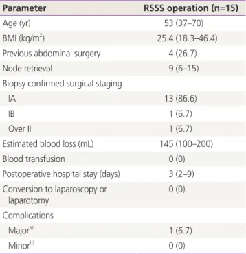
The basic characteristics of the patients, and the periop- erative and postoperative parameters are shown in Table 1.
The median age of the women was 53 years (range, 37–70 years), and the median BMI was 25.4 kg/m
2(range, 18.3–
46.4 kg/m
2). Four of 15 (27%) patients had a history of pre- vious abdominal surgery, which included cesarean sections, but there were no conversions to laparoscopy or laparotomy.
In this study, no additional ports were used for the RSSS op- eration.
The median total operation time was 155 minutes (range, 125–190 minutes), and the total operation time was mea- sured separately according to the time it took to perform each procedure. Table 2 shows each procedural time. The median console time was 75 minutes (range, 55–115 min- utes), and the console time gradually shortened as the RSSS operations were performed (Fig. 3).
The median retrieval of both pelvic lymph nodes was 9 nodes (range, 6–15 nodes), and estimated blood loss was
A B C
Fig. 2. (A) Careful dissection of the lymph nodes away from the ureter and vessels accomplished by the monopolar hook and the fenes-
trated bipolar grasper at the retroperitoneal space. (B) Completion of vagina cuff repair by continuous suture of V-Loc™ (Covidien, Min-
neapolis, MN, USA). (C) Full view after completion of full staging operation.
145 mL (range, 100–200 mL). Drainage was inserted into the pelvic cavity for the first 3 patients due to the possibility of postoperative bleeding. However, for the remaining patients, drainage was not inserted, and there were no complications associated with postoperative bleeding irrespective of the drainage insertion.
The median postoperative hospital stay was 3 days (range, 2–9 days). None of the patients required a transfusion. One patient had an incisional hernia diagnosed by a physical exam and computed tomography 5 months after the surgery.
This patient was transferred to the general surgery depart- ment and received surgical treatment using bilayer mesh. The patient is now free of any symptoms regarding the incisional hernia.
According to the histological biopsy results, 4 patients required adjuvant therapy due to risk factors for recurrence and upstaging after the surgery. Among these, 3 patients received concurrent chemoradiation therapy with 6 cycles of cisplatin, and 1 patient received radiation therapy. These 4 patients finished all adjuvant therapy and continue to be dis- ease free (Table 3).
Discussion
The RSSS operation is a new platform that has been used for benign gynecologic diseases. Compared with laparoscopic single-site surgery, the RSSS operation provides easier ma- nipulation and makes an enhanced approach possible for the operator.
We performed the RSSS operation with pelvic node dissec- tion on patients with gynecologic malignancies, especially those with low-risk early-stage endometrial cancer. Sinno et al. [20] reported a single-site robotic sentinel lymph node bi- opsy and hysterectomy in endometrial cancer and discussed the possibility of using the RSSS operation in this type of cancer. In a large-scale study, Vizza et al. [21] described the Table 1. Basic characteristic of patients and operative parameters
of the study population
Parameter RSSS operation (n=15)
Age (yr) 53 (37–70)
BMI (kg/m
2) 25.4 (18.3–46.4)
Previous abdominal surgery 4 (26.7)
Node retrieval 9 (6–15)
Biopsy confirmed surgical staging
IA 13 (86.6)
IB 1 (6.7)
Over II 1 (6.7)
Estimated blood loss (mL) 145 (100–200)
Blood transfusion 0 (0)
Postoperative hospital stay (days) 3 (2–9) Conversion to laparoscopy or
laparotomy 0 (0)
Complications
Major
a)1 (6.7)
Minor
b)0 (0)
Data are presented as medians (ranges) or number (%) unless other- wise specified.
BMI, body mass index; RSSS, robotic single-site staging.
a)
Major complications include hernia, bowel injury or ileus, vaginal cuff dehiscence, vaginal cuff infection, and vaginal bleeding and require surgical intervention or hospital readmission;
b)Minor compli- cations include any event other than the major complications listed above.
Table 2. Procedural times
Duration of procedures (min) RSSS operation (n=15)
Setting time 25 (15–35)
Preparation time 10 (4–20)
Docking time 8 (4–15)
Console time 75 (55–115)
Vaginal cuff closure time 11 (8–18)
Total operation time 155 (125–190)
Data are presented as medians (ranges).
RSSS, robotic single-site staging.
Fig. 3. Total operative time by chronological procedure number.
RSSS, robotic single-site staging.
Procedure number
RSSS operation 300
250 200 150 100 50
0 5 10 15
Total operative time (min)
feasibility of a robotic single-site hysterectomy in 15 patients with low-risk endometrial cancer. However, their internal protocol showed that a lymph node dissection was not per- formed. The aims of our study were to evaluate the feasibility of the RSSS operation with lymph node dissection in patients with early-stage endometrial cancer, and to suggest surgical guidelines for node dissection using a robotic single-site plat- form.
All RSSS operations were accomplished successfully with- out additional port insertions or conversions to a laparotomy or laparoscopy. There was no exclusion criterion related to
BMI in the selection of patients, since the robotic surgical platform was developed for obese patients. There were no perioperative complications regarding patient 12, who had a BMI of 46.44 kg/m
2.
We believe that there are 2 surgical difficulties in perform- ing the RSSS operation on patients with endometrial cancer.
The first is efficient node dissection. In this study, a bilateral pelvic lymphadenectomy was performed without the firefly system for sentinel node mapping. The median number of nodes retrieved was 9 (range, 6–15). This may appear to be a small number, but considering that node involvement and
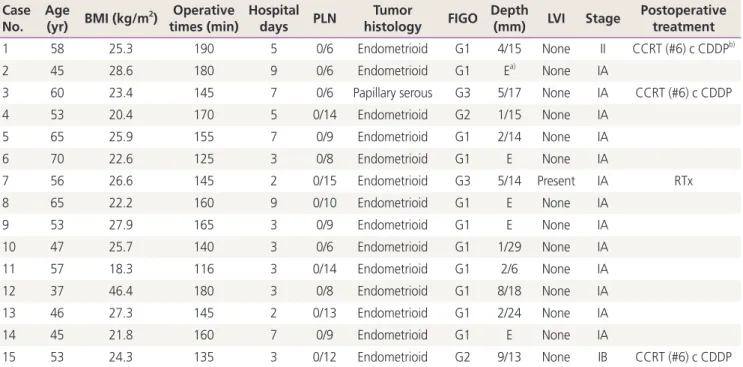
Table 3. Case series of robotic single-site staging operation Case
No.
Age
(yr) BMI (kg/m
2) Operative times (min)
Hospital
days PLN Tumor
histology FIGO Depth
(mm) LVI Stage Postoperative treatment
1 58 25.3 190 5 0/6 Endometrioid G1 4/15 None II CCRT (#6) c CDDP
b)2 45 28.6 180 9 0/6 Endometrioid G1 E
a)None IA
3 60 23.4 145 7 0/6 Papillary serous G3 5/17 None IA CCRT (#6) c CDDP
4 53 20.4 170 5 0/14 Endometrioid G2 1/15 None IA
5 65 25.9 155 7 0/9 Endometrioid G1 2/14 None IA
6 70 22.6 125 3 0/8 Endometrioid G1 E None IA
7 56 26.6 145 2 0/15 Endometrioid G3 5/14 Present IA RTx
8 65 22.2 160 9 0/10 Endometrioid G1 E None IA
9 53 27.9 165 3 0/9 Endometrioid G1 E None IA
10 47 25.7 140 3 0/6 Endometrioid G1 1/29 None IA
11 57 18.3 116 3 0/14 Endometrioid G1 2/6 None IA
12 37 46.4 180 3 0/8 Endometrioid G1 8/18 None IA
13 46 27.3 145 2 0/13 Endometrioid G1 2/24 None IA
14 45 21.8 160 7 0/9 Endometrioid G1 E None IA
15 53 24.3 135 3 0/12 Endometrioid G2 9/13 None IB CCRT (#6) c CDDP
BMI, body mass index; PLN, pelvic lymphnode dissection; FIGO, International Federation of Gynecology and Obstetrics; LVI, lymphovascular in- vasion; CCRT, concurrent chemoradiotherapy; CDDP, cisplatin; RTx, radiotherapy.
a)