MicroRNA 155 Expression Pattern and its Clinic-pathologic Implication in Human Lung Cancer
Mi Kyeong Kim1,4, Dong Chul Moon2, Hye Jin Hyun1, Jong-Sik Kim3, Choi Tae Jin4 and Sang Bong Jung5*
1Department of Clinical Laboratory Science, Gimhae College, Gimhae 50811, Korea
2Department of Physical Therapy, Gimhae College, Gimhae 50811, Korea
3Department of Anatomy, Kosin University College of Medicine, Busan 49267, Korea
4Department of Microbiology, Pukyong National University, Busan 48513, Korea
5Department of Clinical Laboratory Science, Dong-Eui Institute of Technology, Busan 47230, Korea
Received March 7, 2016 /Revised August 29, 2016 /Accepted September 22, 2016
Lung cancer is currently the most common malignant disease and the leading cause of mortality in the world and non-small cell lung cancer (NSCLC) accounts for 75-80% of lung cancer cases. miR-155 gene was found to be over expressed in several solid tumors, such as thyroid carcinoma, breast can- cer, colon cancer, cervical cancer, pancreatic ductal adenocarcinoma (PDAC) and lung cancer. The aims of this study were to define the expression of miR-155 in lung cancer and its associated clin- ic-pathologic characteristics. Total RNA was purified from formalin-fixed, paraffin-embedded NSCLC tissues and benign lung tissues. Expression of miR-155 in human lung cancer tissues were evaluated as mean fold changes of miR-155 in cancer tissues compared to benign lung tissues by quantitative real-time reverse transcriptase polymerase chain reaction (real-time qRT-PCR) and associations of miR- 155 expression with clinic-pathologic findings of cancer. Compared with the benign control group, miR-155 expression was significantly overexpressed in NSCLCs (p=<0.001). miR-155 was more overex- pressed in squamous cell carcinoma than in adenocarcinoma. Poorly differentiated tumors showed sig- nificantly overexpression of miR-155 than well-differentiated tumors (p=<0.001). Overexpression of miR-155 was significantly associated with lymph node metastasis (p=<0.05). In survival analysis for all NSCLC patients, high miR-155 expression was significantly correlated with worse overall survival (p=<0.05). These results suggested that miR-155 might play an important role in lung cancer pro- gression and metastasis.
Key words : Lung cancer, metastasis, MicroRNA, miR-155
*Corresponding author
*Tel : +82-51-860-3520, Fax : +82-51-860-3450
*E-mail : [email protected]
This is an Open-Access article distributed under the terms of the Creative Commons Attribution Non-Commercial License (http://creativecommons.org/licenses/by-nc/3.0) which permits unrestricted non-commercial use, distribution, and reproduction in any medium, provided the original work is properly cited.
Journal of Life Science 2016 Vol. 26. No. 9. 1056~1062 DOI : http://dx.doi.org/10.5352/JLS.2016.26.9.1056
서 론
폐암은 우리나라뿐만 아니라 전 세계적으로 악성 종양 중 가장 높은 암사망율을 보이고 있으며, 여러 연구를 통해 치료 성적을 향상시키려는 노력이 진행되어 왔으나 생존율에 있어 서는 유의한 향상을 가져오지는 못했다. 폐암은 병리학적으로 소세포암(small cell carcinoma)과 비소세포암(non-small cell carcinoma)으로 분류되며, 비소세포암은 편평상피암(squ- amous cell carcinoma), 선암(adenocarcinoma) 등으로 구분된 다. 폐암의 발생은 암유전자(oncogene)의 활성화와 암 억제 유전자(tumor suppressor gene)의 불활성화에 따른 유전적 불
안정성의 증가 등이 관련된다고 알려져 있다[35]. 최근 연구에 서 암 유전자(oncogene)나 암 억제 유전자(tumor suppressor gene)를 포함하여 많은 단백질을 지정하는 유전자를 조절하는 것으로 알려진 다양한 종류의 microRNA (miRNA)가 발견되 어서 암 발생에 관여하는 유전자가 매우 복잡하고 다양한 것 으로 밝혀지고 있다[5, 10, 11, 17, 21, 22, 29].
miRNA는 21~25개의 nucleotide로 이루어진 small non- coding, single-stranded RNA로서 각각의 miRNA는 수백 가 지의 표적 유전자를 조절할 수 있다고 알려져 있으며, 표적 유전자는 다수의 miRNA에 의해 조절될 수 있다고 알려져 있 다[14]. 또한 폐암을 포함한 다양한 암에서 miRNA 의 발현 변화가 관찰되며, 일부의 암에서는 miRNA의 발현이 연관이 있다고 알려져 있다[1, 3, 8, 16, 24, 25, 32]. miR-155는 림프종 (lymphoma), 자궁경부암(cervical cancer), 유방암(breast can- cer) 등에서 발현이 증가되고[5, 17, 31], miR-17-92 family는 림프종, 폐암 등에서 발현이 증가된다고 보고되었으며[10, 22], miR-221, miR-222 등은 갑상선 유두암 등에서 발현이 증가된 다고 보고되었다[11, 21]. miR-15, miR-16은 만성 림프구성 백
Journal of Life Science 2016, Vol. 26. No. 9 1057
Table 1. Clinicopathological characteristics of lung cancer pa- tients
Variables Case No.
Total cases Gender
Male Female Age
<65
≥65
Histological type
aSCC
bAC
Tumor size (cm)
<5
≥5
cLN metastasis No
Yes Differentiation
Well Moderate Poor Smoking
No Yes Stage
Ⅰ/Ⅱ
Ⅲ/Ⅳ Recurrence
No Yes Survival status
Dead Alive
47 34 13 25 22 27 20 20 27 26 21 20 17 10 14 33 23 24 36 11 24 23
aSCC Squamous cell carcinoma
bAC Adenocarcinoma
cLN Lymph node
Table 2. Clinicopathological characteristics of Control group
Total cases 10
Gender (mean age±SD) Male
Female Diagnosis
Anthracofibrosis
7 (50.90±12.27) 3 (50.42±12.28)
10 혈병(chronic lymphocytic leukemia) 등에서 발현이 억제되고
[2], let-7 family는 폐암, 난소암(ovarian cancer), 췌장암(pan- creatic cancer), 유방암 등에서 발현이 억제되며[13, 14, 19, 29], miR-143, miR-145는 직장암(rectal cancer), 대장암에서 발현 이 억제된다고 알려져 있다[7, 23, 36].
따라서 종양에 따라 특이적으로 변화된 miRNA의 발현 양 상을 파악하는 것은 암을 조기에 진단하는데 도움이 될 수 있으며, miRNA를 대상으로 한 항암치료는 새로운 표적치료 의 가능성을 제시하고 있다. 폐암과 miRNA의 발현 양상에 대한 국내연구는 세포주를 대상으로 한 연구가 대부분이고 조직을 대상으로 한 연구는 미비한 실정이다. 이에 본 연구에 서는 한국인 폐암 환자의 조직에서 특이적으로 발현되는 miRNA의 양상을 양성 폐질환자 와 비교 분석하고, 폐암환자 의 임상병리학적 특성과의 상관성을 분석하여 miRNA가 암 진단의 생물표지자로서의 가능성을 조사하여 향후 암의 조기 진단 및 치료, 예후 연구에 기초 자료를 제공하고자 하였다.
재료 및 방법 연구대상
본 연구는 성균관대학교 창원 삼성병원에 내원하여 생검이 나 근치수술을 시행하여 조직병리학적으로 폐암으로 진단을 받은 47명의 환자를 대상으로 하였다. 본 연구는 성균관대학 교 창원 삼성병원의 IRB 심의(심의번호 2012-scmc-044-00)를 거쳐 승인을 획득하였다. 그리고 성별, 연령, 종양의 위치와 크기, 전이 유무, 흡연 유무, 병기, 재발 유무 등 각 환자들의 임상병리학적 특성을 조사하였다. 폐암환자군은 편평상피암 (squamous cell carcinoma)이 27명, 선암(adenocarcinoma)이 20명이었으며, 분화도에 따라서는 저등도, 중등도, 고등도가 각각 10명, 17명, 20명이었다. 대조군으로는 양성 폐 질환인 탄분섬유증(Anthracofibrosis) 환자 10명의 파라핀 포매 조직 을 재료로 사용하였다. 폐암 유형을 확인하기 위하여 hema- toxylin and eosin 염색을 한 후 현미경으로 관찰하여 WHO (World Health Organization)분류에 따라 폐암 유형을 결정하 였다. 실험에 사용된 폐암환자군과 양성폐질환자군의 임상적 특징은 Table 1, 2에 표시하였다.
Total RNA 분리
파라핀 조직으로부터 total RNA를 분리하기 위하여 RNA paraffin kit (Applied Roche Science, Indianapolis, IN, USA) 를 사용하였다. RNA의 추출은 제조사의 매뉴얼에 따라서 다 음과 같이 수행하였다. 파라핀 조직을 RNase-free tube에 넣고 melting buffer 300 μl를 tube에 넣은 다음 원심 시켜 72℃ 10분 간 반응을 시킨 후 Proteinase K를 20 μl첨가하였다. 60℃에서 추출물에 binding buffer 400 μl와 100% ethanol 800 μl를 넣고 mix후 collection tube에 cartridge를 넣고 binding buffer와
ethanol을 첨가된 추출물 700 μl를 옮겨 넣은 후 원심을 2회 반복 실시하였다. 마지막 용액을 버리고 Wash buffer와 100%
ethanol 500 μl를 cartridge에 넣고 원심 시킨 후 용액을 제거하 는데 과정을 2회 더 반복하고 마지막 용액을 제거 하였다. car- tridge에 65℃ RNase-free DW 50 μl를 첨가한 후 1분간 반응
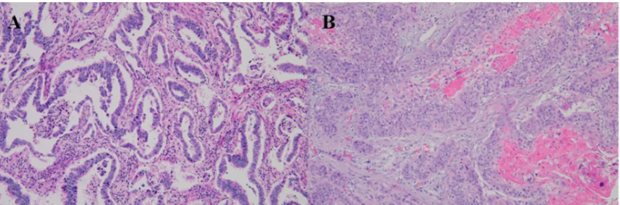
Fig. 1. Comparison of Adenocarcinoma (A) and squamous cell carcinoma (B) by H&E staining. Adenocarcinoma consists of malignant glands invading a fibrous stroma. Squamous cell carcinoma resembles stratified squamous epithelium of the upper airway and shows obvious keratinization.
Quantitative real time PCR (qPCR)
TaqMan MicroRNA Reverse Transcription Kit (Applied Biosystems, Foster City, CA, USA)를 사용하여 RNA 100 ng 에 stem loop RT-primer (5X) 3 μl와 RT Master mix 7 μl, RNase inhibitor (20U/μl) 0.2 μl, RNase-free water 2.8 μl를 혼합한 용액 15 μl를 Gene Amp PCR system 9600을 사용하여 16℃에서 30분, 42℃에서 30분, 85℃에서 5분간 반응시켜 cDNA를 합성하였다. 합성된 cDNA는 5배 희석하여 Quanti- tative real time PCR에 사용하였다. 합성된 cDNA 2 μl에 TaqMan 2X universal PCR master mix (Applied Biosystems, Foster City, CA, USA) 10 μl, Primer and Probe mix (20X) 1 μl, RNase free water 7 μl를 혼합한 용액 20 μl를 ABI PRISM 7000 sequence detection system (Applied Biosystems, Foster City, CA, USA)을 사용하여 Quantitative real time PCR을 수 행하였고 2중 분석으로 miRNA의 발현 정도를 측정하였다.
표적 miRNA의 표준화 대조군으로 U6 small RNA (RNU6B) 를 정량 하였고 동일 시료로 Quantitative real time PCR를 3회 반복 시행하여 평균값을 구하여 발현량을 정량화 하였다.
U6B발현을 기준으로 하여 생성된 각각의 역치 주기 값(thres- hold cycle value, CT value)들을 표준화 하였으며 이후 표준 화된 이 값을 상대치로 환산하였다.
Data Normalization
Control 에 비해 sample의 유전자 발현량이 얼마나 증감하 고 있는 지를 분석하기 위해 ΔCt값을 산출하였다. ΔCt값은 sample 의 Ct값에서 internal control사용된 RNU6B의 Ct값을 빼서 △Ct을 측정하여 Normalization하였으며, △Ct값이 음 의 값이면 증폭되는 cycle수가 빠르기 때문에 유전자 발현량의 증가를 의미하고 △Ct값이 양의 값이면 증폭되는 cycle수가 느리기 때문에 유전자 발현량의 감소를 의미한다.
여 측정하였고, 양성 폐 질환자의 △△Ct값을 ‘1’로 기준하여 폐암 환자의 ratio값을 산출하였다(Ratio = 2-△△Ct). Ratio값 이 ‘1’보다 낮으면 증폭산물이 일정량으로 증폭되는 cycle수가 느리므로 발현율이 낮은 ‘down’를 의미하고 ‘1’보다 높으면 증폭산물이 일정량으로 증폭되는 cycle수가 빠르므로 발현율 이 높은 ‘over’를 의미한다.
통계분석
MicroRNA발현 변화는 평균 ± 표준편차로 나타내었고 각 임상병리학적 특성 에 따른 miRNA의 발현을 보기 위해 두 그룹간의 비교는 independent t-test를 이용하였고 세 그룹간 의 비교는 one-way ANOVA를 이용하여 분석하였다. 통계학 적 분석은 PASW version 19.0을 이용하였고 유의수준은 p<
0.05로 하였다.
생존분석
MicoRNA-155의 발현에 따른 생존분석은 사망이 발생한 시점마다 구간생존율을 구하여 이들의 누적으로서 누적생존 율을 구하는 Kaplan-Meier모형을 사용하였으며, 기준값은 양 성폐질환자의 평균값으로 하였다. 기준값 보다 낮은 △Ct값은 높은 발현으로 분류하였고, 높은 △Ct값은 낮은 발현으로 분 류하였다.
결 과
Hematoxylin-eosin 염색을 이용한 폐암의 분류
조직학적으로 WHO분류 기준에 따라 각질(keratin)을 형성 하고, 중층편평상피와 유사할 경우 편평세포암으로 분류하였 으며, 선조직(gland)을 형성하여 섬유기질을 침범하는 특징을 보일 경우 선암종으로 분류하였다(Fig. 1).
Journal of Life Science 2016, Vol. 26. No. 9 1059
Table 3. Relationship between the expression of miR-155 in lung cancer and benign lung disease miR-155
Sample Benign Lung cancer p value
RNU6B 27.34 30.47
miR-155 Ct 29.05 30.51
a△Ct 1.71 0.04
<0.001
b△△Ct
0.00 -1.67
cRatio 1.00 3.18
a△Ct = MicroRNA Ct - RNU6B Ct
b△△Ct = lung cancer △Ct - benign △Ct
cRatio = 2-△△Ct
Quantitative real-time PCR을 이용한 miR-155의 발 현 양상
양성 폐 질환자와 폐암 환자군에서 miR-155의 발현양상을 비교하기 위하여 RNU6B, miR-155의 probe를 이용하여 정량 실시간연쇄중합반응을 수행한 결과 양성 폐질환자에 비해 비 소세포페암환자에서 miR-155발현율이 높음을 확인하였다.
Table 3에서 보듯이 폐암 환자군과 양성 폐질환자의 miR-155 의 △Ct값을 분석한 결과 양성폐질환자의 miR-155의 값은 1.71, 폐암환자의 miR-155의 값은 0.04로 유의성 있는 결과가 나왔다(p<0.001).
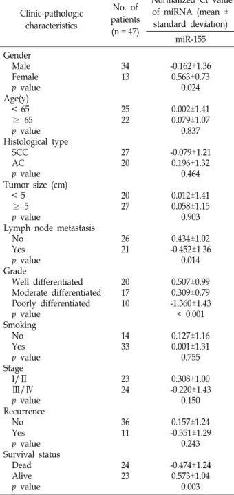
폐암 환자의 임상병리학적 특성과 miRNA와의 연관성 폐암 환자군의 임상병리학적 특성과 miRNA의 발현율과의 상관관계를 분석한 결과 성별에 따라서 miR-155는 여성에서 남성에 비해 유의하게 높게 발현되었다(p=0.024). 병리조직학 적 분류에 따라서는 편평상피세포암종에서 선암종에 비해 높 게 발현되어 종양의 병리조직학적 유형에 따른 차이를 나타내 었으나 통계적 유의성은 없었다. 종양 크기에 따른 발현은 ≥5 에서〈5에 비해 높게 발현되었지만 통계적 유의성은 없었다.
림프절 전이에 따른 발현은 전이된 조직이 전이되지 않은 조 직이 보다 발현이 높은 경향을 보였고 통계적 유의성이 있었 다(p=0.014). 분화도에 따라서는 분화도가 좋을수록 낮게 발현 되었고 분화도가 나쁠수록 높게 발현되었으나 통계적 유의성 이 있었다(p<0.001).
흡연 유무에 따라서는 흡연한 환자에서 높은 발현을 보였으 나 통계적 유의성은 없었다. 병기가 진행됨에 따라 높은 발현 을 보였으나 통계적 유의성은 없었다. 재발 유무에 따라서 miR-155는 재발한 환자에서 높은 발현을 보였으나 통계적 유 의성은 없었다(Table 4).
miR-155 발현에 따른 생존률
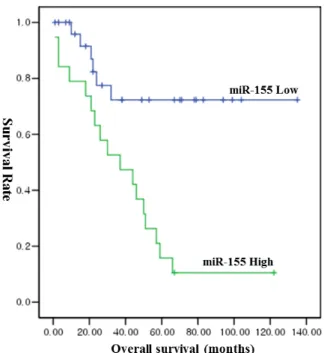
생존률 분석에서 중앙값 보다 낮은 △Ct값은 높은 발현으로 분류하였고 높은 △Ct값은 낮은 발현으로 분류하였다. Fig. 2 에서 보듯이 Kaplan-Meier모형의 생존분석결과 폐암환자의 5년 생존률은 miR-155의 과 발현이 관찰된 환자에서는 15.8%
였으며, 과 발현이 관찰되지 않은 환자에서는 77.8%로 miR- 155의 과 발현이 관찰된 환자와 비교하여 유의하게 낮은 생존
Table 4. Relationship between the expression of microRNA and clinic-pathologic characteristics in lung cancer
Clinic-pathologic characteristics
No. of patients (n = 47)
aNormalized Ct value of miRNA (mean ±
standard deviation) miR-155 Gender
Male Female p value Age(y) < 65 ≥ 65 p value Histological type SCC
AC p value Tumor size (cm) < 5
≥ 5 p value
Lymph node metastasis No
Yes p value Grade
Well differentiated Moderate differentiated Poorly differentiated p value
Smoking No Yes p value Stage I/Ⅱ Ⅲ/Ⅳ p value Recurrence No Yes p value Survival status Dead Alive p value
34 13
25 22
27 20
20 27
26 21
20 17 10
14 33
23 24
36 11
24 23
-0.162±1.36 0.563±0.73
0.024 0.002±1.41 0.079±1.07
0.837 -0.079±1.21
0.196±1.32 0.464 0.012±1.41 0.058±1.15
0.903 0.434±1.02 -0.452±1.36
0.014 0.507±0.99 0.309±0.79 -1.360±1.43
< 0.001 0.127±1.16 0.001±1.31
0.755 0.308±1.00 -0.220±1.43
0.150 0.157±1.24 -0.351±1.29
0.243 -0.474±1.24
0.573±1.04 0.003
aNormalized Ct = △Ct
Fig. 2. Survival analysis using the Kaplan-Meier method: overall survival (OS) according to the expression levels of miR- 155 in patients with lung cancer. high miR-155 expre- ssion was significantly correlated with worse OS (p=
<0.05).
률을 보였다(p=<0.001).
고 찰
폐암은 매년 사망률이 증가하고 있으나 조기 진단이 어렵고 림프절이나 혈액을 통해 전신으로 전이할 수 있으며, 5년 생존 율 또한 낮다[26, 28]. 최근 연구에서는 암 발생에 있어 miRNA 의 발현 변화가 관찰되며, miRNA의 발현이 암과 연관이 있다 고 알려져 있다[6, 9, 20, 29]. 폐암의 생성과정에서도 miRNA 는 표적으로 하는 mRNA유전자에 따라 종양유전자 또는 종양 억제유전자로 작용할 수 있으므로 종양에 따라 특이적으로 변화된 miRNA의 발현 양상을 파악하는 것은 암을 조기에 진 단하는데 도움이 될 수 있다고 사료된다. 따라서 본 연구에서 는 폐암 환자 조직 47예와 양성 폐질환자 조직 10예를 대상으 로 특징적으로 발현하는 miR-155를 선정하여 폐암 환자와 양 성 폐질환자에서의 miRNA발현을 비교 분석하였으며, 폐암 환자의 임상병리학적 특성에 따른 miRNA발현의 상관성을 비 교 분석하였다.
Quantitative real-time PCR을 통해 정량 분석한 결과 miR- 155가 양성 폐 질환자에 비해 발현율이 증가한 것을 확인하였 다. miR-155는 염색체 21q21.3에 위치하고 갑상선암, 대장암, 췌장암등에서 발현이 증가된다고 보고되었다[6, 9, 20, 29, 30].
본 연구에서도 miR-155는 폐암에서 양성 폐질환자에 비해 발 현이 증가되어 이전 연구와 동일한 결과를 나타내었다.
에 비해 높게 발현되어 miRNA의 발현은 성별에 따라 차이가 있을 것으로 사료된다. 연령에서는 <65에서 ≥65에 비해 높게 발현이 되었는데 연령에 대한 miRNA에 대한 연구는 미흡하 지만 본 연구에서는 연령층이 낮을수록 높게 발현되는 경향을 보였다. 병리조직학적 분류에 따라서 miR-155는 편평상피세 포암종에서 높게 발현되어 종양간의 발현차이를 나타내었다.
이러한 결과로 볼 때 암종의 병리조직학적 유형에 따라 miRNA의 작용기전이 다를 것으로 생각되어지며 이와 관련된 연구가 진행되어야 할 것으로 사료된다. 림프절 전이에 따른 발현은 Kono, Zhang 등의 연구에 따르면 miR-155의 과 발현 이 다양한 암에서 전이를 촉진시킨다고 보고하였다[4, 18, 35].
본 연구에서 miR-155는 전이된 조직이 전이되지 않은 조직보 다 높게 발현되어 이전 연구와 비슷한 결과를 나타내었다. 종 양의 분화도에 따라서는 분화도가 낮을수록 높은 발현을 보였 고 이에 대한 연구는 아직 미흡하기 때문에 이에 대한 연구가 지속적으로 이루어져야 될 것이다.
miR-155는 종양에 따라 침투와 증식을 촉진시킨다고 보고 되었다[31, 33]. 종양의 증식과 관련된 miRNA들의 목표 유전 자와 그 작용기전에 대한 연구가 이루어진다면 종양의 치료에 도움이 될 것으로 사료되며 앞으로 여기에 대한 연구가 더 이루어져 될 것이다.
담배 흡연은 체내에 benzo[a]pyrene이나 nicotine-derived nitroso-amineketon 등의 발암물질을 생성하여 폐암을 일으킨 다고 보고되었다[12]. 본 연구에서도 폐암환자와 miR-155의 발 현빈도에 대한 통계적 유의성은 없었으나 발현 정도는 높게 나 타나 흡연이 miR-155의 발현과도 관련이 있을 것으로 사료된다.
폐암의 발생병기에 따라서는 병기가 진행됨에 따라 miR- 155의 발현이 증가되어 miR-155는 폐암의 진행과 관련이 있을 것으로 사료되며, 그 작용기전에 대한 연구가 더 이루어져야 할 것으로 사료된다. 생존 상태에 따라서는 사망한 환자에서 생존중인 환자에 비해 과 발현 양상을 보였다. 이러한 결과는 miR-155가 과 발현이 환자의 병기 및 분화도와 상관성을 가지 기 때문으로 사료된다.
현재까지 우리나라에서 폐암 환자의 임상병리학적 특성과 miRNA와의 연관성에 대한 분석한 연구는 미비한 실정이다.
우리나라에서 폐암과 양성 폐질환자의 miRNA에 대한 연구는 2009년 Son 등의 연구 이후 처음이라 할 수 있다[27]. 하지만 Son 등의 연구에서는 폐암환자의 임상병리학적 특성과 miRNA 에 대한 연구는 없었고 본 연구에서는 이러한 폐암 조직에서 특이적으로 발현되는 miRNA를 조사하고 그 중 폐암 진단에 가능성을 줄 수 있는 표적 miRNA를 선별하였다.
References
1. Bloomston, M., Frankel, W. L., Petrocca, F., Volinia, S.,
Journal of Life Science 2016, Vol. 26. No. 9 1061
Alder, H., Hagen, J. P., Liu, C. G., Bhatt, D., Taccioli, C.
and Croce, C. M. 2007. MicroRNA expression patterns to differentiate pancreatic adenocarcinoma from normal pan- creas and chronic pancreatitis. JAMA. 297, 1901-1908.
2. Calin, G. A., Dumitru, C. D., Shimizu, M., Bichi, R., Zupo, S., Noch, E., Aldler, H., Rattan, S., Keating, M., Rai, K., Rassenti, L., Kipps, T., Negrini, M., Bullrich, F. and Croce, C. M. 2002. Frequent deletions and down-regulation of mi- cro- RNA genes miR15 and miR16 at 13q14 in chronic lym- phocytic leukemia. Proc. Natl. Acad. Sci. USA 99, 15524-15529.
3. Calin, G. A., Sevignani, C., Dumitru, C. D., Hyslop, T., Noch, E., Yendamuri, S., Shimizu, M., Rattan, S., Bullrich, F., Negrini, M. and Croce, C. M. 2004. Human microRNA genes are frequently located at fragile sites and genomic re- gions involved in cancers. Proc. Natl. Acad. Sci. USA 101, 2999-3004.
4. Donnem, T., Lonvik, K., Eklo, K., Berg, T., Sorbye, S, W., Al-Shibli, K., Al-Saad, S., Andersen, S., Stenvold, H., Bremnes, R, M. and Busund, L. T. 2011. Independent and tissue-specific prognostic impact of miR-126 in nonsmall cell lung cancer: coexpression with vascular endothelial growth factor-A predicts poor survival. Cancer 117, 3193-3200.
5. Eis, P. S., Tam, W., Sun, L., Chadburn, A., Li, Z., Gomez, M. F., Lund, E. and Dahlberg, J. E. 2005. Accumulation of miR-155 and BIC RNA in human B cell lymphomas. Proc.
Natl. Acad. Sci. USA 102, 3627-3632.
6. Faraoni, I., Antonetti, F, R., Cardone, J. and Bonmassar, E.
2009. miR-155 gene: a typical multifunctional microRNA.
Biochim. Biophys. Acta. 1792, 497-505.
7. Fuse, M., Nohata, N., Kojima, S., Sakamoto, S., Chiyomaru, T., Kawakami, K., Enokida, H., Nakagawa, M., Naya, Y., Ichikawa, T. and Seki, M. 2011. Restoration of miR-145 ex- pression suppresses cell proliferation, migration and in- vasion in prostate cancer by targeting FSCN1. Int. J. Oncol.
38, 1093-1101.
8. Garzon, R., Fabbri, M., Cimmino, A., Calin, G, A. and Croce, C, M. 2006. MicroRNA expression and function in cancer.
Trends. Mol. Med. 12, 580-587.
9. Gironella, M., Seux, M., Xie, M. J., Cano, C., Tomasini, R., Gommeaux, I., Garcia, S., Nowak, J., Yeung, M. L., Jeang, K. T., Chaix, A., Fazli, L., Motoo, Y., Wang, Q., Rocchi, P., Russo, A., Gleave, M., Dagorn, J. C., Iovanna, J. L., Carrier, A., Pebuseque, M. J. and Dusetti, N. J. 2007. Tumor protein 53-induced nuclear protein 1 expression is repressed by miR-155, and its restoration inhibits pancreatic tumor development. Proc. Natl. Acad. Sci. USA 104, 16170-16175.
10. Hayashita, Y., Osada, H., Tatematsu, Y., Yamada, H., Yana- gisawa, K., Tomida, S., Yatabe, Y., Kawahara, K., Sekido, Y. and Takhashi, T. 2005. A polycistronic microRNA cluster, miR-17-92, is overexpressed in human lung cancers and en- hances cell proliferation. Cancer Res. 65, 9628-9632.
11. He, H., Jazdzewski, K., Li, W., Liyanarachchi, S., Nagy, R., Volinia, S., Calin, G. A., Liu, C. G., Franssila, K., Suster, S., kloos, R. T., Croce, C. M. and Chapelle, P. 2005. The role of microRNA genes in papillary thyroid carcinoma.
Proc. Natl. Acad. Sci. USA 102, 19075-19080.
12. Hecht, S. S. 2002. Cigarette smoking and lung cancer: chem- ical mechanisms and approaches to prevention. Lancet Oncol. 8, 461-469.
13. Johnson, S. M., Grosshans, H., Shingara, J., Byrom, M., Jarvis, R., Cheng, A., Labourier, E., Reinert, K. L., Brown, D. and Slack, F. J. 2005. RAS is regulated by the let-7 microRNA family. Cell 120, 635-647.
14. Takamizawa, J., Konishi, H., Yanagisawa, K., Tomida, S., Osada, H., Endoh, H., Harano, T., Yatabe, Y., Nagino, M., Nimura, Y., Mitsudomi, T. and Takahashi, T. 2004. Reduced expression of the let-7 microRNA in human lung cancers in association with shortened postoperative survival. Cancer Res. 64, 3753-3756.
15. Kim, Y. K., Yu, J. and Han, T. S. 2009. Functional links be- tween clustered microRNAs: suppression of cell-cycle in- hibitors by microRNA clusters in gastric cancer. Nucleic.
Acids. Res. 37, 1672-1681.
16. Markou, A., Tsaroucha, E. G., Kaklamanis, L., Fotinou, M., Georgoulias, V. and Lianidou, E. S. 2008. Prognostic value of mature microRNA-21 and microRNA-205 overexpression in non-small cell lung cancer by quantitative real-time RT-PCR. Clin. Chem. 54, 1696-1704.
17. Metzler, M., Wilda, M., Busch, K., Viehmann, S. and Bork- hardt, A. 2004. High expression of precursor microRNA- 155/BIN RNA in children with Burkitt lymphoma. Genes.
Chromosome. Cancer 39, 167-169.
18. Kono, H., Nakamura, M. Ohtsuka, T. Nagayishi, Y., Mori, Y., Takahata, S., Aishima, S. and Tanaka, M. 2013. High ex- pression of microRNA-155 is associated with the aggressive malignant behavior of gallbladder carcinoma. Oncol. Rep. 30, 17-24.
19. Nam, E. J., Yoon, H., Kim, S. W., Kim, H., Kim, Y. T., Kim, J. H., Kim, J. W. and Kim, S. 2008. MicroRNA expression profiles in serous ovarian carcinoma. Clin. Cancer Res. 14, 2690-2695.
20. Nikiforova, M. N., Tseng, G. C., Steward, D., Diorio, D. and Nikiforov, Y. E. 2008. MicroRNA expression profiling of thyroid tumors: biological significance and diagnostic utility.
J. Clin. Endocrinol. Metab. 93, 1600-1608.
21. Pallante, P., Visone, R., Ferracin, M., Ferraro, A., Berlingieri, M. T., Troncone, G., Chiappetta, G., Liu, C. G., Santoro, M., Negrini, M., Croce, C. M. and Fusco, A. 2006. MicroRNA deregulation in human papillary thyroid carcinomas. Endocr.
Relat. Cancer 13, 497-508.
22. Rinaldi, A., Poretti, G., Kwee, I., Zucca, E., Catapano, C.
V., Tibiletti, M. G. and Bertoni, F. 2007. Concomitant MYC and microRNA cluster miR-17-92 (C13orf25) amplification in human mantle cell lymphoma. Leuk. Lymphoma 48, 410- 412.
23. Slaby, O., Svoboda, M., Fabian, P., Smerdova, T., Knoflick- ova, D., Bednarikova, M., Nenutil, R. and Vyzula, R. 2007.
Altered expression of miR-21, miR-31, miR-143 and miR-145 is related to clinicopathologic features of colorectal cancer.
Oncology 72, 397-402.
24. Schetter, A. J., Leung, S. Y., Sohn, J. J., Zanetti, K. A.,
초록:폐암에서 microRNA 155의 발현 양상과 임상병리학적 의의 김미경1,4․문동철2․현혜진1․김종식3․최태진4․정상봉5*
(1김해대학교 임상병리학과, 2김해대학교 물리치료과, 3고신대학교 의과학대학 해부학교실, 4부경대학교 미생물학
과, 5동의과학대학교 임상병리과)
폐암은 전세계적으로 높은 발병율과 사망률을 보이는 암종으로 소세포암종과 비소세포암종으로 구분되어지며, 비소세포암이 75-80%를 차지하고 있다. miR-155의 유전자의 과 발현은 갑상선암, 유방암, 대장암, 자궁 경부암, 췌장선암(PDAC), 폐암 등의 고형암에서 관찰된다. 본 연구에서는 한국인 폐암 환자의 조직에서 특이적으로 발현 되는 miRNA의 양상을 양성 폐질환자 와 비교 분석하고, 폐암환자의 임상병리학적 특성과의 상관성을 분석하여 miRNA가 암 진단의 생물표지자로서의 가능성을 조사하여 향후 암의 조기 진단 및 치료, 예후 연구에 기초 자료 를 제공하고자 하였다. 파라핀 포매 된 비소세포폐암환자 및 양성 폐 질환자의 블록에서 total RN를의 분리하여, 정량 실시간연쇄중합반응을 통해 miR-155의 발현량을 정량 분석을 실시하였으며, miR-155의 발현과 폐암환자의 임상적 특징과의 상관관계를 분석하였다. 폐암 환자군과 양성 폐질환자의 miR-155의 △Ct 값을 분석한 결과 폐암 환자군에서 유의하게 높게 발현되었다(p<0.001). 병리조직학적 분류에 따라서는 편평상피세포암종에서 선암종에 비해 높게 발현되었다. 분화도에 따라서는 저분화 암에서 고분화암에 비해 유의하게 높게 발현되었다(p=<0.001).
또한 miR-155의 과발현은 림프절 전이와도 통계적으로 유의성 나타내었다(p<0.05). 생존분석결과 miR-155의 과발 현은 폐암환자의 생존률과 유의한 상관관계를 나타내었다(p<0.05). 본 연구의 결과로 miR-155의 발현은 폐암의 진행 및 전이에 중요한 역할을 할 것으로 생각되며, 폐암의 조기진단과 예후의 예측을 위하여 보다 다양한 종류의 miRNAs에 대한 연구가 이루어져야 할 것으로 판단된다.
C. M. and Harris, C. C. 2008. MicroRNA expression profiles associated with prognosis and therapeutic outcome in colon adenocarcinoma. JAMA. 299, 425-436.
25. Sevignani, C., Calin, G. A., Siracusa, L. D. and Croce, C.
M. 2006. Mammalian microRNA: a small world for fine-tu- nung gene expression. Mamm. Genome. 17, 189-202.
26. Shayan, R. Achen, M. G. and Stacker, S. A. 2006. Lymphatic vessels in cancer metastasis: bridging the gaps. Carcinogensis 9, 1729-1738.
27. Son, J. W., Kim, Y. J., Cho, H. M., Lee, S. Y., Jang, J. S., Choi, J. E., Lee, J. U., Kang, M. G., Lee, Y. M., Kwon, S.
J., Choi, E., Na, M. J. and Park, J. Y. 2009. MicroRNA ex- pression profiles in Korean non-small cell lung cancer.
Tuberc. Respir. Dis. 67, 413-421
28. Stinchombe, T. E., Lee, C. B. and Socinski, M. A. 2006.
Current approaches to advanced-stage non-small-cell lung cancer: first-line therapy in patients with a good functional status. Clin. Lung Cancer 4, 111-117.
29. Torrisani, J., Bournet, B., Rieu, M. C., Bouisson, M., Souque, A., Escourrou, I., Buscail, L. and Cordelier, P. 2009. Let-7 microRNA transfer in pancreatic cancer-derived cells in- hibits in vitro cell proliferation but fails to alter tumor progression. Hum. Gene Ther. 20, 831-844.
30. Tam, W. 2001. Identification and characterization of human BIC, a gene on chromosome 21 that encodes a noncoding RNA. Gene 274, 157-167.
31. Volinia, S., Calin, G. A., Liu, C. G., Ambs, S., Cimmino, A., Petrocca, F., Visone, R., Iorio, M., Roldo, C., Ferracin, M.,
A microRNA expression signature of human solid tumors defines cancer gene targets. Proc. Natl. Acad. Sci. USA 103, 2257-2261.
32. Yan, L. X., Huang, X. F., Shao, Q., Huang, M. Y., Deng, L., Wu, Q. L., Zeng, Y. X. and Shao, J. Y. 2008. MicroRNA miR-21 overexpression in human breast cancer is associated with advanced clinical stage, lymph node metastasis and patient poor prognosis. RNA. 14, 2348-2360
33. Yu, J., Ohuchida, K., Mizumoto, K., Sato, N., Kayashima, T., Fujita, H., Nakata, K. and Tanaka, M. 2010. MicroRNA, hsa-miR-200c, is an independent prognostic factor in pan- creatic cancer and its upregulation inhibits pancreatic cancer invasion but increases cell proliferation. Mol. Cancer 9, 169.
34. Zhang, B., Pan, X., Cobb, G. P. and Anderson, T. A. 2007.
MicroRNA as oncogenes and tumor suppressor. Dev. Biol.
302, 1-12.
35. Zhang, G. J., Xiao, H. X., Tian, H. P., Liu, Z. L., Xia, S. S.
and Zhou, T. 2013. Upregulation of microRNA-155 pro- motes the migration and invasion of colorectal cancer cells through the regulation of claudin-1 expression. Int. J. Mol.
Med. 31, 1375-1380.
36. Zhu, H., Dougherty, U., Robinson, V., Mustafi, R., Pekow, J., Kupfer, S., Li, Y. C., Hart, J., Goss, K., Fichera, A., Joseph, L. and Bissonnette, M. 2011. EGFR signals downregulate tu- mor suppressors miR-143 and miR-145 in Western diet-pro- moted murine colon cancer: role of G1 regulators. Mol.
Cancer Res. 9, 960-975.