서 론
연교(連翹, Fructus Forsythiae)는 물푸레나무과(Oleaceae)에 속하는 개나리의 열매로 우리나라 전국 각지에서 재배되고 가을 에 성숙한 과실을 채취하여 햇빛에 말려 사용하며1), 한방에서 종 창, 임질, 통경, 이뇨, 치질, 결핵, 나력, 옴, 해독 등의 치료에 널 리 사용되고 있다2). 연교의 함유 성분으로는 lignans, lignan glucosides, flavonoid 및 3,4-dihydroxyphenethyl alcohol의 caffeoyl glycoside 등이 있으며, 약리작용으로 항균3), 항염증4), 혈압강하5), 항산화 작용6-8)이 보고되었고, 저자 등은 연교 메탄올 추출물이 인체 멜라닌세포주인 HM3KO 세포의 tyrosinase 활성 을 억제함을 보고하였다9).
멜라닌은 멜라닌세포에서 합성되는 색소 중합체로서 수상돌 기를 통하여 표피의 각질화세포로 이동되어 피부의 색을 나타내
며10,11), 그 외에도 피부 보호, 독성물질의 흡착, 배아 발생 시 신경
계통의 발달에 관여하는 등 여러 가지 중요한 기능을 하고 있다12).
멜라닌 합성은 여러 단계에 걸친 복잡한 과정으로 많은 유 전자가 관여하며, 포유동물의 경우 약 80개 이상의 유전자가 직 간접적으로 영향을 미치는 것으로 알려져 있는데, 특히 tyrosinase 유전자 family는 멜라닌 합성 조절에 중추적인 역할 을 하고 있다13). 멜라닌은 갈색 또는 검은색을 띄는 eumelanin과 붉은색 또는 노란색을 띄는 pheomelanin의 2종류가 있으며14), tyrosine을 기질로 하여 tyrosinase, tyrosinase-related protein(TRP-1) 및 tyrosinase-related protein(TRP- 2) 등 일련의 효소반응에 의하여 생성된다15).
또한 멜라닌의 합성은 여러가지 사이토카인, basic fibroblast growth factor(bFGF), α-melanocyte stimulating hormone(MSH), cyclic AMP (cAMP) 유도물질인 cholera toxin과 forskolin, melanocyte gene expression factor인 MITF 등에 의해서 조절되 는데, 최근 cAMP pathway는 melanogenic enzymes의 활성과 발현에 중추적인 작용을 하는 것으로 알려져 있다16,17).
따라서 본 연구는 인체 멜라닌 세포주인 HM3KO 세포에서 tyrosinase, TRP-1 단백질 및 tyrosinase 유전자 발현, 세포 내 cAMP 변화 등 연교 메탄올추출물의 멜라닌 생성 억제 기전을 조사하였다.
인체 멜라닌세포주에서 連翹 추출물의 멜라닌생성 억제기전 연구
조미경․문연자*․우원홍
원광대학교 한의학전문대학원 한약자원개발학과
Melanogenesis Inhibition by Forsythiae Fructus Extract in Human Melanoma Cells
Mi Gyeong Jo, Yeun Ja Mun*, Won Hong Woo
Department of Herbal Resources, Professional Graduate School of Oriental Medicine, Wonkwang University
In this study, we have investigated the hypo-pigmentary mechanism of methanol extract of Forsythiae Fructus in human melanocyte cell line, HM3KO. Treatment of HM3KO cells with Forsythiae Fructus extract markedly inhibited melanin biosynthesis in a dose-dependent manner. Decreased melanin contents occurred through the decrease of tyrosinase protein and activity. The mRNA levels of tyrosinase and tyrosinase-related protein 1 (TRP-1) were also reduced by Forsythiae Fructus extract. Moreover, the level of intracellular cyclic AMP (cAMP) was significantly decreased by treatment of Forsythiae Fructus extract. These results suggest that Forsythiae Fructus reduces melanin synthesis by down regulation of tyrosinase mRNA transcription, and this is closely related to the cAMP-dependent pathway.
Key words : Forsythiae Fructus, cAMP, tyrosinase, TRP-1
* 교신저자 : 문연자, 익산시 신용동 344-2, 원광대학교 한의학전문대학원
․E-mail : [email protected], ․Tel : 063-850-6942
․접수 : 2008/03/10 ․채택 : 2008/04/11
재료 및 방법
1. 시료조제
본 실험에 사용한 연교는 시중 건재약국에서 구입하였으며, 연교 100 g에 MeOH 1 ℓ를 혼합하고 6시간 동안 실온에서 초음 파로 분쇄한 후 거즈로 여과하였다. 여과된 연교 추출물은 원심 분리기로 추출액을 분리하여 상온에서 감압농축기(rotary evaporator)로 농축하여 15.78 g(수득률: 15.78%)의 추출물을 얻 었다. 연교 메탄올추출물 시료는 dimethyl sulfoxide (DMSO, Sigma Chemical Co., USA)에 녹인 후 사용하였다.
2. 세포주 배양
인체 멜라닌 세포주(human melanoma cell line)인 HM3KO 세포는 한국 세포주은행에서 분양 받아 CO2 배양기(37℃, 5%)에 서 10% FBS가 포함된 DMEM 배지를 이용하여 배양하였으며, 약 48시간 주기로 배양액을 교체하여 주었다.
3. 세포내 멜라닌 정량(melanin contents) 측정
멜라닌 정량은 Hosoi등18)의 방법을 변형하여 사용하였다.
배양세포를 분리하여 PBS로 세척한 후 각 군당 1×10⁶개 세포를 수확하여 세포용해액(lysis buffer; 1% Triton X-100, 10 mM sodium phosphate, pH 7.0)으로 세포를 용해하고 원심분리하여 멜라닌을 분리하였다. Acid-insoluble material을 얻기 위해 10%
의 dimethyl sulfoxide(DMSO)가 첨가된 1 N NaOH 용액에 8 0℃에서 1시간 동안 처리하여 405 nm에서 흡광도를 측정하였다.
4. 세포내 tyrosinase activity 측정
Tyrosinase 활성은 Martinez-Esparza 등19)의 방법에 의하여 측정하였다. 배양이 끝난 후 세포용해액(lysis buffer; 1% Triton X-100, 10 mM sodium phosphate, pH 7.0, 0.1 mM PMSF)으로 4℃에서 30분간 때때로 흔들어주면서 세포를 파괴시킨 후 이를 원심분리하여 상층액을 취하여 tyrosinase 활성측정용액으로 사용 하였다. 100 mM sodium phosphate(pH 7.0, 100 ㎕)와 100 mM catechol(50 ㎕)를 넣고 온도조절장치가 있는 분광광도계로 37℃, 405 nm에서 1시간 동안 반응시켜 흡광도의 변화를 관찰하였다.
5. Western blot 분석
시료를 농도별로 처리하고 3일 동안 배양한 다음 세포를 회 수하여 단백질을 정제하였다. HM3KO 세포를 PBS로 세척하고, 4℃에서 30분 동안 세포용해액(Nonidet P-40, sodium dodecyl sulphate, 0.1 M Tris-Hcl, pH 7.2, 0.1M PMSF, 10 ㎍/㎖
aprotinin, 10 ㎍/㎖ leupeptin)으로 용해시킨 후 13,000 rpm(4℃, 30분)에서 원심분리하여 상층액을 취하였다. Protein은 Bradford assay로 정량하였고, BSA로 표준곡선을 만들어 단백질 양을 계 산하였다. 50 ㎍ 단백질에 2×sample buffer(1.0 ㎖ glycerol, 0.5
㎖ β-mercaptoethanol, 3.0 ㎖ 10% SDS, 1.25 ㎖ 1.0 M Tris-Hcl, 1 to 2 ㎎ bromophenol blue)를 가하여 7.5% SDS polyacrylamide gel에서 P1, 100 V, 400 mA(Electrophoresis
Power Supply)로 2시간 전기영동하였다. Gel 상에 분리된 단백 질은 60 V에서 3시간 동안 nitroceullose membrane에 전이시키 고, 실온에서 blocking buffer(5% skimmilk/TBST)로 3시간 처리 하였다. Tyrosinase, TRP-1 그리고 TRP-2 antibody(Santa Cruz) 는 각각 1:500으로 희석하여 3시간 처리하여 TBST buffer로 3회 세척하고 1:1,000으로 희석시킨 secondary antibody(Amersham, USA)을 1시간 반 동안 처리하였다. 3회 세척한 다음 Amersham ECL system으로 반응시켜 ChemiDoc에서 band를 확인하였다.
6. 역전사-중합효소 연쇄반응(RT-PCR)
HM3KO 세포 내 전체 RNA는 easy-BlueTM(iNtRON Biotechnology, Korea)를 사용하여 추출하였다. 추출한 RNA를 Oligo d(T)16 (iNtRON Biotechnology, Korea)를 이용하여 역전 사(reverse transcription) 반응으로 cDNA를 합성한 후 i-Master mix PCR kit(iNtRON Biotechnology)를 이용하여 PCR을 시행하 였다. PCR에 사용한 primer는 다음과 같다: tyrosinase forward 5'-CATTTTTGATTTGAGTGTTCT-3'; reverse 5'-TGTGGTAGTC GTCTTTGTCCC-3'; TRP-1 forward 5'-CTTTCTCCCTTCCTTA CTGG-3'; reverse 5'-TGGCTTCATTCTTGGTGCTT-3'; TRP-2 forward 5'-TGAGAAGAAACAAAGTAGGCAGAA-3'; reverse 5'-CAACCCCAAGAGCAAGACGAAAGC-3'; actin forward 5'- TCAGAAGGACTCCTATGTGG-3'; reverse 5'-TCTCTTTGATGT CAGCACG-3'. 각각의 cDNA 증폭은 RNA 2 ㎍ 을 사용하였으 며, 94℃에서 20초, 55℃에서 10초, 72℃에서 50초로 총 35회 증 폭을 시행하였다. 반응액은 2% agarose gel에서 전기영동을 시행 하였으며, ethidium bromide로 염색하였다.
7. cAMP 농도 측정
cAMP 농도는 Cui-X 등20)의 방법에 의하여 측정하였다.
HM3KO 세포에 시료를 150 ㎍/㎖ 농도로 처리한 후 PBS로 세 척하고, 4℃에서 세포용해액(Nonidet P-40, sodium dodecyl sulphate, 0.1 M Tris-Hcl, pH 7.2, 0.1 M PMSF, 10 ㎍/㎖
aprotinin, 10 ㎍/㎖ leupeptin)으로 세포를 용해시킨 후 4℃, 13,000 rpm에서 30분간 원심분리하여 상층액을 취하였다.
Protein은 Bradford assay로 정량하였고, BSA로 표준곡선을 만 들어 단백질 양을 계산하였다. 각 sample(25 ㎍)을 polypropylene tube로 옮겨 water-saturated ether(300 ㎕)로 3번 씻어낸 후 speedVac concentrator로 건조시켰다. Standards와 각 sample에 50 mM sodium acetate buffer(pH 4.8, 100 ㎕)를 첨가 하고 acetic anhydride 와 triethylamine(1:2, 5 ㎕)로 아세틸화시 키고, cAMP antiserum(100 ㎕)과 I125로 표지된 항원(100 ㎕)으 로 4℃에서 24시간 동안 반응시켰다. Charcoal 1 ㎖을 첨가하고 4℃, 3500 rpm에서 20분 동안 원심분리 시킨 후 MINAXI γ에서 측정하였다.
8. 통계학적 분석
실험결과는 mean±S.D.로 표시하였다. 각 군 간의 통계적 유 의성에 대한 검증은 one-way ANOVA test을 이용하였으며, P값
이 0.01 이하인 경우 유의성을 인정하였다.
결 과
1. 멜라닌 합성 억제효과
HM3KO 세포에서 연교 추출물의 멜라닌 합성 억제 작용을 확인하기 위하여 시료를 3일간 처리한 후 세포내 멜라닌 양을 측 정한 결과, 연교 추출물 50, 100, 150 ㎍/㎖ 농도에서 각각 66.7±15%, 56.1±16.3%, 58.8±14.7%로 대조군에 비하여 유의하게 멜라닌 양이 감소되었다(Fig. 1).
또한 멜라닌 합성의 속도조절 효소인 tyrosinase의 활성을 측정한 결과에서도 연교 추출물 50, 100, 150 ㎍/㎖ 처리군에서 각각 대조군의 71.9±8.2%, 65.8±3.3%, 66.9±6.8%로 유의하게 감 소하였다(Fig. 2).
Control 50 100 150
0 20 40 60 80 100 120
∗ ∗
∗
Melanin Contents (% of control)
Concentration (µg/ml)
Fig. 1. Effect of Forsythiae Fructus extract on melanin contents. HM3KO cells were treated with several concentrations of Forsythiae Fructus extract for 3 days. Data are expressed as % of control and each column represents the means±S.D. of five determinations. * p<0.01.
Control 50 100 150
0 20 40 60 80 100 120
∗ ∗
∗
Tyrosinase Activity (% of control)
Concentration (µg/ml)
Fig. 2. Effect of Forsythiae Fructus extract on tyrosinase activity in HM3KO cells. The effect on tyrosinase activity was tested with several concentrations of Forsythiae Fructus in HM3KO cells for 3 days. Data are expressed as % of control and each column represents the means±S.D. of five determinations. * p<0.01
2. 멜라닌합성 관련 효소의 단백질 발현
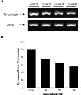
연교 추출물을 처리하고 HM3KO 세포 내 tyrosinase 단백질 발현을 측정한 결과, 50, 100, 150 ㎍/㎖ 군에서 처리 농도에 비 례하여 감소하였으며, 각각 대조군의 77±3.9%, 69.1±0.8%, 65.6±3.2%로 나타났다(Fig. 3).
Control 50 100 150
0 20 40 60 80 100 120
∗ ∗
∗
Tyrosinase/Actin ( % of control )
Concentration (µg/ml) Tyrosinase
Actin
A
B
Control 50 ㎍/㎖ 100 ㎍/㎖ 150 ㎍/㎖
Fig. 3. Effect of Forsythiae Fructus extract on expression level of tyrosinase protein. HM3KO cells were treated with several concentrations of Forsythiae Fructus extract for 3 days. Representative Western blotting (A) analysis densitometric quantification (B) of Forsythiae Fructus extract in HM3KO cells. Data are expressed as % of control and each column represents the means±S.D. of three determinations. * p<0.01
또한 TRP-1 단백질 발현을 조사한 결과 연교 추출물 50 ㎍/
㎖, 100 ㎍/㎖, 150 ㎍/㎖ 농도에서 처리 농도에 비례하여 감소 하였으며, 대조군에 비하여 각각 72.9±8.5%, 63.3±6.2%, 54.1±5.3%로 나타났다(Fig. 4).
TRP-1
Control 50 100 150
0 20 40 60 80 100 120
∗
∗
∗
TRP-1/Actin ( % of control )
Concentration (µg/ml) Actin
A
B
Control 50 ㎍/㎖ 100 ㎍/㎖ 150 ㎍/㎖
Fig. 4. Effect of Forsythiae Fructus extract on expression level of TRP-1 protein. HM3KO cells were treated with several concentrations of Forsythiae Fructus extract for 3 days. Representative Western blotting (A) analysis densitometric quantification (B) of Forsythiae Fructus extract in HM3KO cells. Data are expressed as % of control and each column represents the means±S.D. of three determinations. * p<0.01
3. Tyrosinase mRNA 발현
연교 추출물의 멜라닌 생성 억제 효과는 이들이 tyrosinase 유전자 발현을 조절할 수 있다는 가능성을 제시한다. 따라서 역
전사 중합효소 연쇄반응(RT-PCR)으로 tyrosinase mRNA의 발현을 분석하였다. 그 결과 연교 추출물 50, 100, 150 ㎍/㎖ 농도에서 각각 대조군에 비하여 75.3%, 65%, 56%로 유의하게 감소하였다(Fig. 5).
Control 50 100 150
0 20 40 60 80 100 120
Tyrosinase/Actin ( % of control)
Concentration (µg/ml) Tyrosinase
Actin A
B
Control 50 ㎍/㎖ 100 ㎍/㎖ 150 ㎍/㎖
Fig. 5. Effect of Forsythiae Fructus extract on tyrosinase mRNA gene expression in HM3KO cells. HM3KO cells were treated with several concentrations of Forsythiae Fructus extract for 3 days. Representative RT-PCR (A) analysis densitometric quantification (B) of Forsythiae Fructus extract in HM3KO cells. Data are expressed as % of control.
4. 세포 내 cAMP 변화
HM3KO 세포에 연교 추출물을 150 ㎍/㎖ 농도로 처리하고 3분, 5분, 10분, 20분 후 세포 내 cAMP 농도를 조사하였다. 대조 군의 cAMP 농도는 0.023 ρmol/㎍였으며, 연교 추출물 처리 3분 후에 0.008 ρmol/㎍로 크게 감소하였고, 20분에 0.020 ρmol/㎍
로 cAMP 농도가 다시 회복함을 알 수 있었다(Fig. 6).
Control 3min 5min 10min 20min
0.000 0.005 0.010 0.015 0.020 0.025
∗
∗
∗
cAMP level (pmol/µg of protein)
Incubation Time (minutes)
Fig. 6. Effect of Forsythiae Fructus extract on cAMP efflux. HM3KO cells were treated with 150 ㎍/㎖ concentration of Forsythiae Fructus extract for several minutes. Data are expressed as cAMP efflux and each column represents the means±S.D. of three determinations. * p<0.01
고 찰
연교에 함유되어 있는 성분에는 lignan류(phillygenin,
pinoresinol, arctigenin, matairesinol), lignan glucoside류 (phillyrin, pinoresinol-D-glucose, arctin, matairesinoside), flavonoid(rutin) 및 3,4-dihydroxyphenethyl alcohol의 caffeoyl glycoside류(forsythiaside, acteoside, suspensaside 및 hydroxyacteoside) 등이 있다1-7). 지금까지 연교의 생리활성 작용 에 대한 연구로는 주로 estrogen과 유사하거나 반대되는 성질을 보이며 in vitro에서 항산화활성이 우수한 것으로 보고되었으며, 그 외에 phillyrin은 혈압강하작용, pinoresinol과 pinoresinol-D-glucoside은 cAMP-phosphodiesterase 활성억제 및 혈압강하작용5), phenylpropanoid glycosides는 항균작용, 3β -acetoxy-20,25-ep oxydammarane-24-ol은 항염증 효과가 있는 것 으로 알려져 있다3,4). 연교 추출물을 포함한 생약추출물과 향료를 포함하는 여드름 또는 염증방지용 화장품 조성물이 개시된 바 있으며, 저자 등은 연교 메탄올 추출물이 인체 멜라닌세포주인 HM3KO 세포의 tyrosinase 활성을 억제함을 보고하였다9). 멜라닌 합성 조절에 중추적인 역할을 하고 있는 tyrosinase 유전자 family는 tyrosinase, tyrosinase-related protein(TRP-1) 및 tyrosinase-related protein(TRP- 2)로 이들 중 tyrosinase는 멜라 닌 생성과정의 속도조절 효소로서 처음 두 단계의 반응에 관여 하며21), TRP-2는 DOPAchrome을 DHICA로, TRP-1은 DHICA를 indole -5,6-carboxylic acid로 전환시키는데 관여하고 있다22-24). 본 연구에서 연교 추출물의 멜라닌 생성 억제작용을 확인한 결 과, HM3KO 세포의 tyrosinase 효소활성을 유의하게 억제하였 고, 이와 유사하게 멜라닌의 멜라닌 합성을 효과적으로 감소시켰 다(Fig. 1 & 2).
아직까지 인체 피부에서 TRP-1, TRP-2와 같은 몇몇 다른 단 백질의 발현과 그들의 기능은 명백히 밝혀지지 않았으나25), Commo(2004) 등은 Human eumelanin hair bulbs의 멜라닌세포 에서 tyrosinase와 TRP-1이 검출된 반면에 TRP-2는 검출되지 않 았는데 이는 TRP-2 유전자 발현이 약하기 때문이라고 하였다26). 한편 Wu와 Park(2003) 등은 인체 멜라닌세포에서 PKC-β가 tyrosinase와 TRP-1사이의 복합체 형성을 유도하고 이 복합체에 의해 tyrosinase의 활성화가 유도된 것이라고 하였다27). 또 dbcAMP와 멜라닌생성 유도물질(melanogenic agents)은 tyrosinase 활성과 TRP-1 합성을 증가시키지만28), TRP-2와 멜라 닌합성 사이의 관계는 아직 밝혀지지 않았으며, DHICA의 polymerization이나 decarboxylation, oxidation을 억제하는 차단 인자(blocking factors)가 존재할 것이라고 하였다29).
본 실험 결과에서 tyrosinase와 TRP-1 단백질의 발현은 연교 추출물에 의해 감소하였으나(Fig. 3 & 4), TRP-2는 검출되지 않 았다. 따라서 tyrosinase와 TRP-1 발현의 감소는 tyrosinase와 TRP-1 복합체 형성의 감소를 의미하며, 결과적으로 tyrosinase의 활성 감소로 이어진 것으로 생각된다.
나이에 따른 피부노화로 인해 생긴 색소침착의 가장 공통적 인 유형은 노인흑색점(lentigo senilis)으로, 최근 면역조직화학법 과 역전사중합효소 연쇄반응(RT-PCR)에 의해 노인흑색점 부위 에서 tyrosinase의 단백질과 mRNA 발현이 높게 나타났다30). 따 라서 본 실험에서 연교 추출물이 tyrosinase 유전자의 발현을 조
절하는지 조사하기 위하여 tyrosinase 유전자에 특이적인 primer 를 이용하여 mRNA의 발현을 분석한 결과 유의성 있게 감소하 였다(Fig. 5).
최근 멜라닌 합성을 조절하는 세포 내 신호전달에 있어서 cAMP 경로가 핵심적인 조절 기전임이 보고되고 있다16,17,31,32)
. 이 때 신호전달은 첫째, 외부 자극에 의해 cAMP 농도가 증가하고 PKA 활성화와 cAMP responsive element binding protein 활성 을 거쳐 tyrosinase의 전사가 증가되며 둘째, PKA에 의해 TRP-1 과 TRP-2 촉진자가 활성화되고 셋째, 세포 내에 증가된 cAMP는 mitogen-activated protein kinase인 p44MAPK,를 자극하여 AP-1 활성을 거쳐 tyrosinase 발현을 증가시키며 넷째, phosphatidylinositol 3-kinase와 p70S6-kinase의 활성을 억제하 여 tyrosinase 발현을 증가시키는 것으로 알려져 있다33-35). 본 실 험에서도 cAMP radioimmuno assay을 통해 연교 추출물의 효과 를 관찰한 결과 HM3KO세포 내 cAMP 농도가 크게 감소하였다 가 시간이 지남에 따라 다시 증가함을 볼 수 있었다(Fig. 10). 따 라서 연교 추출물은 cAMP 의존형 신호전달경로를 억제함으로 서 멜라닌 생성을 감소시키는 것으로 생각된다.
이상의 결과, 연교 추출물은 피부 멜라닌세포의 세포 내 cAMP 생성과 tyrosinase mRNA 전사를 억제함으로서 tyrosinase 활성과 멜라닌 합성이 감소되었으며, cAMP-의존적 경로에 작용함을 시사해 주고 있다.
결 론
연교(Fructus Forsythiae)의 항균, 항염, 혈압강하 작용은 잘 알려져 있으나 멜라닌 합성에 미치는 영향에 대해서는 거의 보 고된 바 없다. 본 연구에서 인체 멜라닌세포주인 HM3KO세포를 이용하여 연교의 멜라닌 합성 억제 기전을 조사하였다. 연교 추 출물은 HM3KO세포의 멜라닌 합성을 억제하였고, 멜라닌 합성 의 감소는 tyrosinase의 활성과 단백질 및 mRNA의 감소를 동반 하였다. 또한 TRP-1의 단백질 발현은 억제하였으나 TRP-2는 발 현되지 않았으며, 연교 추출물 처리 시 세포 내 cAMP의 농도가 감소되었다. 이상의 결과, 연교 추출물은 피부 멜라닌세포의 세 포 내 cAMP 생성과 tyrosinase mRNA 전사를 억제함으로서 tyrosinase 활성과 멜라닌 합성이 감소되었으며, cAMP-의존적 경로에 작용함을 시사해 주고 있다.
참고문헌
1. 辛民敎. 臨床本草學. 永林社, pp 431, 1997.
2. 김창민, 신민교, 안영균, 이경호외. 중약대사전, 10권, pp 4871-4872, 1997.
3. Li, Z.X., Wang, X.H., Zhao, J.H., Yang, J.F., Wang, X.
Investigation on antibacterial activity of Forsythia suspense Vahl in vitro with Mueller-Hinton agar. Zhongguo Zhong Yao Za Zhi 25(12):742-745, 2000.
4. Ozaki, Y., Rui, J., Tang, Y.T. Antiinflammatory effect of
Forsythia suspensa V (AHL) and its active principle. Biol.
Pharm. Bull. 23(3):365-367, 2000.
5. Lee, E.B., Keum, H.J. Pharmacological studies on Forsythae Fructus. Kor. J. Pharmacogn. 19(4):262-269, 1988.
6. Rim, Y.S., Park, Y.M., Park, M.S., Kim, K.Y., Kim, M.J., Choi, Y.H. Screening of antioxidant and antimicrobial activity in native plants. Korean J. Medicinal Crop. Sci.
8(4):342-350, 2000.
7. Lim, D.K., Choi, U., Shin, D.H. Antioxidative Activity of Ethanol Extract from Korean Medicinal Plants. Korean J.
Food Sci. Technol. 8(4):342-350, 2000.
8. Schinella, G.R., Tournier, H.A., Prieto, J.M., Mordujovich, D., Rios, J.L. Antioxidant activity of anti-inflammatory plant extracts. Life Sci. 70(9):1023-1033, 2002.
9 조미경, 안병상, 문연자, 우원홍. 연교 메탄올추출물의 멜라닌 생성 억제효과. 대한한방피부미용학회지 1(1):41-52, 2005.
10. Fitzpatrick, T.B., Szabo, G., Seiji, M., Quevedo, W.C.
Biology of the melanin pigmentary system. In: Fitzpatrick, T.B., ed., Dermatology in General Medicine. Academic Press, New York, pp 131-163, 1979.
11. Jimbow, K., Fitzpatrick, T.B., Wick, M. Biochemistry and physiology of melanin pigmentation. In: Goldsmith L.A., ed., Physiology and Molecular Biology of the skin. Oxford Univ. Press, New York, pp 873-909, 1991.
12. Hearing, V.J. Regulation of melanin formation. In:
Nordlund, J.J., Boissy, R.E., Hearing, V.J., King, R.A., Ortonne, J.P., editors. The pigmentary system: physiology and pathophysiology. 1st ed. New York, Oxford University Press, pp 423-438, 1998.
13. Hearing, V.J. Biochemical control of melanogenesis and melanosomal organization. J. Invest. Dermatol. Symp. Proc.
4: 24-28, 1999.
14. del Marmol, V., Ito, S., Jackson, I., Vachtenheim, J., Berr, P., Ghanem, G., Morandini, R., Wakamatsu, K., Huez, G.
TRP-1 expression correlates with eumelanogenesis in human pigment cells in culture. Eur. Biochem. 327:
307-310, 1993.
15. Gabriela, N., Raymond, A.D. et al. Tyrosinase-related protein-2 and -1 are trafficked on distinct routes in B16 melanoma cells. Res. Biochem. Biophysical. 328: 914-921, 2005.
16. Roser, B., Robert, B. Cyclic AMP a key messenger in the regulation of skin pigmentation. Pigment cell Res. 13:
60-69, 2000.
17. 윤태진, 이무형. All-trans-retinoic Acid가 α-MSH로 자극한 배양 정상 인체 멜라닌세포의 성장과 cAMP 농도에 미치는 영향. 대한피부과학학회지 37(8):1017-1028, 1999.
18. Hosoi, J., Abe, E., Suda, T., Kuroki, T. Regulation of melanin synthesis of B16 mouse melanoma cells by 1
α,25-dehydroxy-vitamin D3 and retinoic acid, Cancer Res.
45: 1474-1478, 1985.
19. Martinez-Esparza, M., Jimenez-Cervantes, C., Solano, F., Lozano, J.A., Garcia-Borron, J.C. Mechanisms of melanogenesis inhibition by tumor necrosis factor-alpha in B16/F10 mouse melanoma cells. Eur. J. Biochem.
255(1):139-146, 1998.
20. Cui, X., Lee, S.J. et al. Effects of pituitary adenylate cyclase activating polypeptide 27 on cyclic AMP efflux and atrial dynamics in perfused beating atria. Eur. J. Pharmacol. 402:
129-137, 2000.
21. Hearing, V.J., Jimenez, M. Mammalian tyrosinase-the critical regulatory control point melanocyte pigmentation.
Int. J. Biochem. 19: 1141-1147, 1987.
22. Funasaka, Y., Chakraborty, A.K., Komoto, M., Ohashi, A., Ichihashi, M. The depigmenting effect of α-tocopherol ferulate on human melanoma cells. British. J. Dermatolohy 141: 20-29, 1999.
23. Wakamatsu, K., Ito, S. Advanced chemical methods in melanin determination. Pigment Cell Res. 15: 174-183, 2002.
24. Kametama K, Takemura, T., Hamada, Y., Sakai, C., Kondoh, S., Nishiyama, S., Urabe, K., Hearing, J. Pigment production in murine melanoma cell is regulated by tyrosinase, tyrosinase- related protein 1(TRP-1), DOPAchrome tautomerase(TRP-2), and a melanogenic inhibitor. J. Invest. Dermatol. 2: 126-131, 1993.
25. Masamoto, Y., Ando, H. et al. Mushroom tyrosinase inhibitory activity of esculetin isolated from seeds of Euphorbia lathyris L. Biosci. Biotechnol. Biochem. 67:
631-634, 2003.
26. Commo, S., Gaillard, O., Thibaut, S. Absence of TRP-2 in Melanogenic Melanocytes of Human Hair. Pigment Cell Res. 17: 488-497, 2004.
27. Wu, H., Park, HY. Protein kinase C-β-mediated complex formation between tyrosinase and TRP-1. Biochem. Biophy.
Res. Com. 311: 948-953, 2003.
28. Maeda, K., Tomita, Y., et al. Effects of staurosporine, PMA and A23187 on human melanocyte cultures with dibutyryl cyclic AMP. Br. J. Dermatol. 126: 118-124, 1992.
29. Maher, M.H., Weidong, X., et al. Activation of a cAMP pathway and induction of melanogenesis correlate with association of p16INK4 and p27KIP1 to CDKs, loss of E2F-Binding activity, and premature senescence of human melanocytes. Exp. Cell Res. 253: 561-572, 1999.
30. Motokawa, T., Kato, T., Katagiri, T., Suzuki, I.. Meassenger RNA levels of melanogenesis associated genes in lentigo senilis lesions. J. Dermatological Science 37(2):120-123, 2005.
31. Medrano, E.E., Yang, F., et al. Terminal differentiation and senescence in the human melanocyte: Repression of tyrosine-phosphorylation of the extracellular signal-regulated kinase 2 selectively defines the two phenotypes. Mol. Biol. Cell 5: 497-509, 1994.
32. Kazuhisa, M., Yoshihiro, Y., et al. Comparison of the melanogenesis in human black and light brown melanocytes. J. Sci. Dermatol. 14: 199-206, 1997.
33. Bertolotto, C., Busca, R., Abbe P, Bille K, Aberdam E, Ortonne JP, Ballotti R. Different cis acting elements are involved in the regulation of TRP-1 and TRP-2 promoter activities by cyclic AMP : pivotal role of M boxes (GTCATGTGCT) and of microphthalmia. Mol. Cell Biol.
18: 694-702, 1998.
34. Englaro, W., Rezzonico, R., Durand-Clément M, Lallemand D, Ortonne JP, Ballotti R. Mitogen-activated protein kinase pathway and AP-1 are activated during cAMP-induced melanogenesis in B-16 melanoma cells. J. Biol. Chem. 270:
24315-24320, 1995.
35. Hemesath, T.J., Price, E.R., Takemoto C, Badalian T, Fisher DE. MAP kinase links the transcription factor microphthalmia to c-kit signalling in melanocytes. Nature 391: 298-301, 1998.