The Effect of Inhibition of Uncaria rhynchophylla as an Inhibitor of Melanogenesis and an Antioxidant in B16F10 Melanoma Cells
Yuanyuan Dong
1, Young Min Woo
2, Ji Hyun Cha
1, Jae Young Cha
1,3, Nai Wei Lee
1, Min Woo Back
1, Joon-sung Park
4, Sang-Hyeon Lee
1, Jong-Myung Ha
1,3and Andre Kim
1,3*
1Department of Pharmaceutical Engineering, College of Medical and Life Sciences, Silla University, Busan 46958, Korea
2Department of Natural Science Institute, Silla University, Busan 46958, Korea
3Hankook Liposome Co., Ltd., Busan 46958, Korea
4Division of Kinesiology, Silla University, Busan 46958, Korea
Received August 26, 2020 /Revised September 14, 2020 /Accepted September 15, 2020
Many people of all ages wish to have lighter skin for cosmetic reasons, and natural products attract more attention than chemically synthesized compounds. Uncaria rhynchophylla is widely used in Asia as a traditional herbal medicine. In order to find novel skin whitening agents, the present study eval- uated the antioxidant activity and potential tyrosinase-inhibiting properties of U. rhynchophylla.
Specifically, this study analyzed the antioxidant capacity of a 70% ethanolic extract of U. rhynchophylla as well as its effects on tyrosinase activity and melanin synthesis. Total mRNA levels were examined using reverse transcription polymerase chain reaction. The results revealed that U. rhynchophylla ex- tracts exhibit great antioxidant capacity and significant levels of polyphenol and flavonoid compounds.
U. rhynchophylla extracts can also powerfully inhibit tyrosinase activity. This same capacity was ob- served in melanoma B16F10 cells; that is, U. rhynchophylla extracts suppressed intracellular tyrosinase activity and reduced the amount of melanin in treated cells. In addition, a 1 mg/ml concentration of U. rhynchophylla extract significantly reduced the mRNA expression levels of tyrosinase. U. rhyncho- phylla extracts decrease tyrosinase and inhibit melanogenesis in B16F10 cells. This finding suggests that U. rhynchophylla has great potential as a natural whitening agent in skincare products.
Key words : Antioxidant, B16F10 melanoma cells, kojic acid, tyrosinase, Uncaria rhynchophylla
*Corresponding author
*Tel : +82-51-999-7620, Fax : +82-51-999-5628
*E-mail : [email protected]
This is an Open-Access article distributed under the terms of the Creative Commons Attribution Non-Commercial License (http://creativecommons.org/licenses/by-nc/3.0) which permits unrestricted non-commercial use, distribution, and reproduction in any medium, provided the original work is properly cited.
Introduction
Human skin color is mainly determined by amount of melanin, which is produced in the melanocytes, the pig- ment-producing cells of the epidermis. Melanin plays a cru- cial role in the response of the skin to the exposure of stress, ultraviolet light and melanin-stimulating factors like alpha- melanocyte-stimulating hormone (α-MSH) [21]. However, the overproduction and accumulation of melanin can lead to a variety of skin conditions, including freckles, chloasma, and other pigmentation syndromes [25, 29].
In melanin biosynthesis, the pathway of melanogenesis is known, and there are two types of melanin within the melanosomes: eumelanin and pheomelanin [4]. The first step
of melanogenesis is initiated by tyrosine oxidation to dop- aquinone catalyzed by the tyrosinase. Tyrosinase is the key enzyme in the first two steps, converting the hydroxylation of L-tyrosine to L-3, 4-dihydroxyphenylalanine (L-DOPA) and then to dopaquinone. Since tyrosinase is a rate-limiting enzyme that is produced only by melanocytic cells, a ty- rosinase inhibitor can specifically target the melanin cells without other side effects [3]. This type of inhibitor has be- come highly popular as a whitening agent in cosmetics and pharmaceuticals. The hormone α-MSH is a peptide derived from proopiomelanocortin (POMC), which regulates mela- nogenesis via a cyclic adenosine monophosphate (c-AMP)- dependent pathway [2, 10]. When binding to its receptor, melanocortin receptor 1 (MC1R), up to a 100-fold increase in melanogenesis occurs [3].
Several skin-whitening chemicals used in cosmetics, such
as kojic acid, arbutin, hydroquinone, aloesin, soybean ex-
tract, azelaic acid, licorice extract, niacinamide, and magne-
sium ascorbyl phosphate, have been reported to reduce mel-
anogenesis [6, 8, 12, 17, 24, 27, 33]. Moreover, kojic acid is
widely used in cosmetics and is often used as a positive
control. The B16F10 melanoma cell line was considered a good model for investigating human melanoma as they are easy to culture in vitro, and they show most of the melano- genic mechanisms of melanocytes [3]. Hence, this cell line was chosen for this evaluation of natural melanin production inhibitors.
This study focuses on a new and potent natural source that shows great melanogenesis inhibitory activity. A mem- ber of the Rubiaceae family, Uncaria comprises numerous spe- cies worldwide, including in Southeast Asia, Central Amer- ica, and South America. Uncaria rhynchophylla has been used in various prescriptions in traditional Chinese medicine (TCM) for the treatment of convulsive disorders (epilepsy) [9] and for various head ailments such as headaches and dizziness [13]. The dried stem and hook of U. rhynchophylla are named as Gou-Teng or cat’s claw in TCM [16, 32]. Des- pite its common usage, there are few reports on the anti-pig- menting effect of U. rhynchophylla. Yon-Suk Kim3 et al. [15]
demonstrated the antioxidant activity of and the presence of polyphenol and flavonoids in U. rhynchophylla extracts, and it has been proven that natural flavonoids are useful tyrosinase inhibitors [31]. Therefore, we hypothesized that U. rhynchophylla can potentially inhibit melanogenesis, though similar studies have not yet been reported on U.
rhynchophylla. To test this hypothesis, assays were done on U. rhynchophylla to detect the inhibitory activity. Ethanolic extracts of Uncaria rhynchophylla were chosen for their mel- anogenesis inhibition on mushroom tyrosinase using L- DOPA as a substrate. The effect of U. rhynchophylla extracts on melanogenesis was evaluated by measuring melanin con- tent and intracellular tyrosinase activity in melanoma B16F10 cells. Total mRNA levels were examined using re- verse transcription polymerase chain reaction. In addition, the total polyphenols and flavonoids contents and anti- oxidant characteristics of the U. rhynchophylla extracts were analyzed.
Materials and Methods
Chemicals and reagents
Kojic acid, L-DOPA, tyrosine, 3-(4, 5-dimethyl-2-thiazolyl)- 2, 5-diphenyl-tetrazolium bromide (MTT), diphenyl-1-pic- rylhydrazyl (DPPH), 2, 2’-azino-bis (3-ethylbenzothiazoline- 6-sulphonic acid (ABTS)), and dimethyl sulfoxide (DMSO) were purchased from Sigma-Aldrich (St Louis, MO, U.S.).
Preparation of the U. rhynchophylla extracts The stems and hooks of U. rhynchophylla were purchased from the Gukje Market of Busan, Korea in July 2019. The U. rhynchophylla (100 g) were through an extraction process with 1,000 ml 70% ethanol for 4 hr. The supernatant was collected by filtration and the extract was harvested by ro- tary evaporation. The resulting dried U. rhynchophylla extract powder was frozen at -80℃ for 7 days and stored at 4℃
for further use.
Antioxidant characteristics of the U. rhynchophylla extract
The antioxidant capacity of the U. rhynchophylla extract was determined by measuring the DPPH and ABTS scaveng- ing activity as previously described [5, 22]. DPPH is a stable free radical that can interact with different antioxidant sub- stances. For both methods, L-ascorbic acid (50 μg/ml) was used as a positive control. The extract concentrations used for the antioxidant tests were 0.1, 0.3, 0.5, and 1 mg/ml in dark conditions. Absorbance was measured at 517 nm and 734 nm in the microplate reader. All of the experiments were performed in triplicate and repeated three times to ensure reproducibility.
Determination of the polyphenol and flavonoid contents
The polyphenol and flavonoid contents of the U. rhyncho- phylla extracts were determined as previously described [23].
Flavonoid concentration was calculated using quercetin as a standard and was expressed as mg of quercetin equivalent (QE) per 100 g of dry weight. Polyphenol was expressed as mg of gallic acid equivalent (GAE) per 100 g of dry weight.
Mushroom tyrosinase activity assay
The inhibitory strength of the U. rhynchophylla extracts (0.1-1 mg/ml) on tyrosinase was tested as described pre- viously with a minor modification [28]. The reaction mixture contained 120 μl of 8.3 mM L-DOPA in a phosphate buffer (0.1 M, pH 6.8), 40 μl of extract solution, 40 μl of mushroom tyrosinase (1,000 unit/ml), and kojic acid (100 μg/ml) was used as a positive control. After pre-incubation in a water bath for 10 min at 37℃, the absorbance was measured at 492 nm using an ELISA reader. Each experiment was done in triplicate.
Cell culture
B16F10 melanoma cells (ATCC CRL-6475, from the BCRC
Cell Line Bank, BCRC60031) were cultured in Dulbecco’s Modified Eagle’s Medium (DMEM; Hyclone, Logan, UT, U.S.) with 10% fetal bovine serum and 1% penicillin/strep- tomycin at 37℃ in a humidified atmosphere with 5% CO
2in the air.
Cell viability assay
Cell viability was determined using an MTT assay [18].
B16F10 melanoma cells were incubated in 96 microplates and pretreated with 0.1-1 mg/ml of U. rhynchophylla extract for 24 hr. Next, they were treated with an MTT solution (5 mg/ml in a phosphate-buffered saline, PBS), and the cells were incubated at 37℃ for 4 hr. DMSO was added after the medium was discarded and the absorbance was measured at 540 nm using an ELISA reader.
Measurement of melanin content
To determine the melanin content, minor modifications were made to a previous method [21]. B16F10 cells were seeded at a density of 1×10
5cells per well in 6-well plates and incubated for 24 hr. The cells were treated with various concentrations of U. rhynchophylla extract (0.1-1 mg/ml) and either with or without 200 nM α-MSH before being in- cubated for 48 hr. Kojic acid was used as a positive control.
The cells were washed with PBS and lysed with 1 N NaOH containing 10% DMSO at 90℃ for 30 min. The melanin con- tent was assayed using absorbance at 400 nm.
Determination of cellular tyrosinase activity The determination of the effect of the extracts on ty- rosinase activity was carried out using a method described previously with minor modifications [30]. B16F10 cells were seeded in 6-well plates (10
5cells per well) and were first stimulated either with or without 200 nM α-MSH for 12 hr.
After that, the α-MSH-stimulated cells were incubated in the U. rhynchophylla extracts (0.1-1 mg/ml) for an additional 24 hr at 37℃. The treated cells were washed twice using PBS and lysed with a radio immunoprecipitation assay (RIPA) buffer. The cell lysates were harvested by centrifugation at 5,000 rpm for 5 min at 4℃. The cell extracts (25 μl) were mixed with 50 μl L-DOPA solution (10 mM in a sodium phosphate buffer) and 25 μl of 50 mM sodium phosphate buffer (pH 6.8) and incubated at 37℃ for 1 hr. The absorb- ance was measured at 495 nm with a microplate reader.
Tyrosinase zymography
The DOPA staining assay was performed as previously
reported [21]. B16F10 cells were treated for 24 hr in the ab- sence or presence of α-MSH at various concentrations U.
rhynchophylla extract or 100 μg/ml of kojic acid (as positive control). The cultured cells were washed twice with PBS and collected with a lysis buffer (0.1 M sodium phosphate buffer (pH 6.8), 0.1% Triton X-100). Protein content was calculated using the Bradford method with bovine serum albumin as a standard [1]. Equal amounts (55 μg) were analyzed using a 10% polyacrylamide gel electrophoresis. After the electro- phoresis, the gel was rinsed twice with 0.1 M sodium phos- phate buffer (pH 6.8) with gentle shaking for 30 min and then incubated in the dark at 37℃ overnight with 5 mM L-DOPA in 0.1 M sodium phosphate buffer. Tyrosinase ac- tivity was visible in the gel as a dark melanin-containing band.
Quantitative RT-PCR analysis
RT-PCR was used to analyze the gene expression in the B16F10 mouse melanoma cells after stimulation with α-MSH in the presence of a test sample for 48 hr. B16F10 mouse melanoma cells (2×10
5cells) were treated in the same way as described above. The total RNA was isolated with a Trizol reagent in accordance with the manufacturer’s instructions (Invitrogen, Carlsbad, CA, U.S.). First-strand cDNA was syn- thesized from the total RNA (2 μg) containing oligo (dT) primers, and PCR amplification was performed using the Gene Amp Kit (Bioneer Corp, Korea). The primer sequences used for the PCR were (1) GAPDH (upstream 5’-GTGAAGG TCGGTGTGAACG-3’, downstream 5’-CTCGCTCCTGGAA GATGGTG-3’) and (2) tyrosinase (upstream 5’-AACAATG TCCCAAGTACAGG-3’, downstream 5’-TGACTCTTGGAG GTAGCTGT-3’). The PCR reaction involved an initial 5 min denaturation at 94℃, followed by 25 cycles at 94℃, 30 sec;
55-60℃, 30 sec; 72℃, 1 min, and a final 7 min extension.
The sizes of the amplified gene products were 223 bp for GAPDH and 250 bp for tyrosinase. Aliquots of individual PCR products were separated on a 2% agarose gel, stained with ethidium bromide, and imaged by a Mupid-2plus Submarine electrophoresis system. A densitometric analysis was done using image analysis software (Gel Quant, Sam Bo Scientific Co., Ltd.).
Results
Antioxidant properties of the U. rhynchophylla extracts
Two assays were performed to evaluate the antioxidant
characteristics of U. rhynchophylla extracts as shown in Fig.
A B
Fig. 1. The antioxidant properties of U. rhynchophylla. (A) DPPH free radical scavenging activity; (B) ABTS radical scavenging capacity.
Various concentrations of the U. rhynchophylla extracts and vitamin C were as a positive standard in the above assays. URE, 70% ethanolic extract of U. rhynchophylla. All values are expressed as mean ± S.D. (n=3). The letters a, b, c, d, and e above the bars stand for significantly different (p<0.05) groups by one-way ANOVA, followed by Duncan’s multiple test.
A B
Fig. 2. Polyphenol and flavonoids contents of U. rhynchophylla. (A) Measurement of polyphenol contents; (B) Evaluation of flavonoid contents. URE, 70% ethanolic extract of U. rhynchophylla. All values are expressed as mean ± S.D. (n=3). The letters a, b, c, and d above the bars stand for significantly different (p<0.05) groups by one-way ANOVA, followed by Duncan’s multiple test.
1. Free radical scavenging activity was determined by DPPH assay using vitamin C as a positive control. As can be seen from Fig. 1A, great variations exist in the DPPH scavenging capacity of the different extract concentrations of U. rhyncho- phylla (0.1-1 mg/ml). Likewise, the assay relies on the gen- eration of ABTS
+chromophores by oxidation of ABTS with potassium persulfate. For the ABTS radical scavenging as- say, the U. rhynchophylla extract concentration of 0.3 mg/ml showed a higher ABTS scavenging capacity than that of vita- min C (50 μg/ml) (Fig. 1B).
Evaluation of the polyphenol and Flavonoid contents of U. rhynchophylla
The amount of total polyphenol was determined using
the Folin-Ciocalteu reagent as previously described [7]. First, a standard curve was made using gallic acid as a com- parative control. Fig. 2 illustrates the varying amounts of polyphenol and flavonoids in the U. rhynchophylla extracts.
We found that with increasing concentration of U. rhyncho-
phylla, there is a proportional increase in polyphenol content
(Fig. 2A), and the highest concentration of U. rhynchophylla
(1 mg/ml) has high flavonoid content (Fig. 2B). Polyphenol
is one of the important plant compounds with an antioxidant
capacity, probably due to its redox properties [14], which
can play a major role in absorbing and neutralizing free radi-
cals, quenching singlet and triplet oxygen, and decomposing
peroxides.
Fig. 3. Effect of U. rhynchophylla on mushroom tyrosinase activ- ity. URE, 70% ethanolic extract of U. rhynchophylla. All values are expressed as mean ± S.D. (n=3). The letters a, b, and c above the bars stand for significantly different (p<0.05) groups by one-way ANOVA, followed by Dun- can’s multiple test.
Fig. 4. Effect of U. rhynchophylla on cell viability in B16F10 cells.
The cells were seeded at 6×10³ cells per well and in- cubated in media containing 0.1-1 mg/ml concentration of URE for 24 hr. URE, 70% ethanolic extract of U.
rhynchophylla. All values are expressed as mean ± S.D.
(n=3).
Effect of U. rhynchophylla on mushroom tyrosinase activity
To measure the potential inhibitory effect of the U. rhyn- chophylla extracts on mushroom tyrosinase activity, enzyme inhibition experiments were carried out in triplicate. It was found that tyrosinase activity was decreased by the U. rhyn- chophylla extracts. Mushroom tyrosinase is regularly used to screen potential inhibitors of melanogenesis in experiments done in vitro. The 1 mg/ml concentration of U. rhynchophylla inhibited more of the enzyme activity than kojic acid as shown in Fig. 3. Thus, U. rhynchophylla seems to exhibit po- tent inhibition of mushroom tyrosinase activity.
Effect of U. rhynchophylla on cell viability
The effect of the U. rhynchophylla extracts on cell viability was assessed using an MTT assay. MTT is a pale-yellow compound that can be converted into a dark-blue formazan product of living cells, but dead cells do not react and re- main yellow. Thus, after an MTT treatment, live cells will appear blue and dead cells will appear yellow. Following this method, it was found that the U. rhynchophylla extracts did not show any cytotoxicity against B16F10 cells and the cell viability remained at a similar level across all concen- tration levels. Even at higher concentrations, U. rhynchophylla showed less cytotoxicity than kojic acid as shown in Fig.
4. These results suggest that cell viability is not affected by U. rhynchophylla (Fig. 4), and these extracts are not respon- sible for any cytotoxic effects on B16F10 melanoma cell viability.
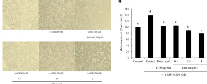
Effect of U. rhynchophylla on melanin content To analyze the melanogenesis effects of the U. rhyncho- phylla extracts, the levels of melanin content were measured in both α-MSH-untreated and α-MSH-treated B16F10 cells.
B16F10 cells are widely used to evaluate the anti-melano- genic effect of test materials because they share most of the melanogenic mechanisms of normal human melanocytes.
The cells were pretreated either with or without α-MSH for 12 hr, followed by treatment with extracts of U. rhynchophylla at concentrations of 0.1-1 mg/ml or with 100 μg/ml kojic acid for an additional 24 hr. As shown in Fig. 5A and Fig.
5B, the U. rhynchophylla extract at a concentration of 1 mg/ml was found to significantly decrease the cellular melanin content. Moreover, the α-MSH treatment alone enhanced melanin content compared to the control, whereas the U.
rhynchophylla extract pretreatment significantly inhibited melanin synthesis of the α-MSH induced pigmentation in a dose-dependent manner.
Effect of U. rhynchophylla on cellular tyrosinase activity
Since tyrosinase plays a key role in melanogenesis, we
examined the inhibitory effects of the U. rhynchophylla ex-
tracts on cellular tyrosinase in B16F10 cells. Kojic acid, well
known as an inhibitor of melanin synthesis, was used as
a positive standard in this study. The results indicate that
the tyrosinase activity in the B16F10 cells was inhibited by
various concentrations of U. rhynchophylla extracts (0.1-1
mg/ml). Compared to the kojic acid, which inhibited the
A B
Fig. 5. Effect of U. rhynchophylla on melanin content. (A) Cell morphology was observed under a phase contrast microscope. (B) Effect of U. rhynchophylla on melanin content in B16F10 cells. The cells were treated with 0.1-1 mg/ml of URE or kojic acid (positive control) for 24 hr after treatment with/without 200 nM α-MSH for 4 hr. URE, 70% ethanolic extract of U.
rhynchophylla. All values are expressed as mean ± S.D. (n=3). The letters a, b, c, and d above the bars stand for significantly different (p<0.05) groups by one-way ANOVA, followed by Duncan’s multiple test.
Fig. 6. Effect of U. rhynchophylla on cellular tyrosinase activity.
The cells were treated with 0.1-1 mg/ml of URE or kojic acid as positive control for 24 hr after treatment with/without 200 nM α-MSH for 4 hr. The supernatant lysed in RIPA buffer was incubated with 5 mM L-DOPA for 1 hr and each sample was normalized to total celluar protein. URE, 70% ethanolic extract of U. rhynchophylla.
All values are expressed as mean ± S.D. (n=3). The let- ters a, b, c, d, and e above the bars stand for significantly different (p<0.05) groups by one-way ANOVA, followed by Duncan’s multiple test.
tyrosinase activity as expected, U. rhynchophylla showed a similar tyrosinase inhibition activity at the 1 mg/ml concen- tration. Increasing the concentration of the extract in the re- action mixture increased the inhibitory activity of the en- zyme (Fig. 6).
Effect of U. rhynchophylla on tyrosinase zymography The effect of the U. rhynchophylla extracts on the amount of cellular tyrosinase protein was evaluated using a gel stain- ing of tyrosinase activity (zymography) analysis. The B16F10 cells were treated with α-MSH alone or with α-MSH plus treatments (U. rhynchophylla extracts or kojic acid), and the total proteins in the cells were separated by gel electro- phoresis and stained with L-DOPA. As shown in Fig. 6, the tyrosinase activity in the untreated cells was very light while the α-MSH-treated cells displayed a dark band with higher activity. The U. rhynchophylla extracts seemed to become more effective with increasing concentration, as the activity of the tyrosinase was reduced and lower bands were ob- served. This demonstrates that U. rhynchophylla extracts can significantly reduce the amount of tyrosinase protein in cells, which means that U. rhynchophylla extracts appear to inhibit melanogenesis of B16F10 cells by decreasing the cellular ty- rosinase content and, hence, its activity.
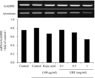
Effect of U. rhynchophylla on the mRNA expression of tyrosinase
To confirm the effect of the U. rhynchophylla extracts on
cellular melanin production, we assessed the mRNA levels
Fig. 7. Effect of U. rhynchophylla on the mRNA expression of tyrosinase. Total RNA from B16F10 cells treated with U. rhynchophylla after 200 nM α-MSH collected at the indicated time. URE, 70% ethanolic extract of U. rhyncho- phylla. Total mRNA levels were examined by RT-PCR, using glyceraldehyde 3-phosphate dehydrogenase (GADPH) as an internal control. The graph represents the fold val- ue of exposed band intensity.
of tyrosinase along with the control GAPDH in cells treated with α-MSH and U. rhynchophylla extracts. As expected, the cells that had the α-MSH and U. rhynchophylla treatments showed increased levels of tyrosinase (Fig. 7). While lower concentrations of U. rhynchophylla tended to decrease ty- rosinase mRNA levels, higher concentrations of U. rhyncho- phylla significantly inhibited mRNA levels. It appears that cells treated with U. rhynchophylla extracts show a dramatic reduction in the expression of tyrosinase mRNA levels.
Discussion
This research has identified a helpful and potential anti- melanogenic agent from a natural source. Although kojic acid is a common skin-whitening product, there is a need to find safer and more effective skin-lightening agents due to the skin irritation, low stability, and even carcinogenic possibility of kojic acid [11, 20]. This is the first report on the effect of U. rhynchophylla on melanin production.
In this study, it was determined that U. rhynchophylla ex- tracts have a high scavenging activity, which could be attrib- uted mainly to their levels of polyphenols and flavonoids.
Tyrosinase is a multifunctional copper-containing enzyme that plays a key role in the melanin pathway and is also
known as monophenol monooxygenase or polyphenol oxi- dase [3]. Tyrosinase catalyzes the oxidations of both mono- phenols and o-diphenols into reactive o-quinones. Tyrosinase contains two copper ions, which bind with histidine residues in the active site and are critical for the catalytic activity [3]. Mushroom tyrosinase is widely used as the target en- zyme for screening probable inhibitors of melanogenesis. As can be seen in Fig. 3, U. rhynchophylla extracts displayed a higher inhibitory effect on mushroom tyrosinase activity than kojic acid. To clarify, the specific inhibitory effect of the U. rhynchophylla extracts on melanogenesis, B16F10 mela- nin content, and intracellular tyrosinase activity were evaluated. Melanin is formed from L-tyrosine in the melano- somes and is responsible for skin color. Although melanin plays a protective role against ultraviolet light and stress, the overproduction and accumulation of melanin results in hyperpigmentation and various skin problems, including age spots, freckles, and melisma [25, 29], which makes mela- nogenesis inhibition a matter of interest. The U. rhynchophylla extracts had a stronger inhibitory effect on melanin for- mation in B16F10 cells than kojic acid, as shown in Fig. 5B.
The cell morphology was observed under a phase contrast microscope, which revealed that the U. rhynchophylla extracts decreased the melanin content (Fig. 5A). It has been reported that α-MSH can bind the melanocortin 1 receptor (MC1R) on the surface of melanocytes; this binding leads to the acti- vation of adenylate cyclase, which leads to an elevated level of intracellular cAMP (cyclic adenosine monophosphate) [19]. In the study, α-MSH was used as a cAMP inducer to stimulate melanin synthesis. Research into molecules that in- hibit tyrosinase has become increasingly important for cos- metic products that may be used as stronger skin-whitening agents for treating skin disorders. As shown in Fig. 6, U.
rhynchophylla extracts were able to repress melanogenesis in- duced by α-MSH mediated intracellular cAMP up-regulation.
To determine whether the inhibition of melanin synthesis by U. rhynchophylla was associated with melanogenesis-re- lated gene expression, a RT-PCR reaction was performed to observe the mRNA levels of tyrosinase. As shown in Fig.
7, the mRNA levels of the genes tended to be reduced by
the U. rhynchophylla extracts in a concentration-dependent
manner. It is notable that the inhibition levels of the U. rhyn-
chophylla extracts at higher concentrations were superior to
kojic acid, which is a renowned skin whitening factor in
functional cosmetics. Based on these results, U. rhynchophylla
may act as a good tyrosinase inhibitor.
In conclusion, U. rhynchophylla showed inhibitory effects on melanogenesis similar to kojic acid with no adverse skin reactions such as burning, pruritus, or erythema. These find- ings indicate that U. rhynchophylla may be a useful source of bioactive compounds for anti-hyperpigmentation and as an antioxidant agent in human skincare products. In future studies, we plan to investigate the effects of various protein kinase inhibitors on melanin production in B16F10 melano- ma cells after treatment with U. rhynchophylla and further analyze the potential active components in U. rhynchophylla to identify the possible skin-whitening mechanisms.
Acknowledgment
This study was supported by the Brain Busan 21+ project (BB21+).
The Conflict of Interest Statement
The authors declare that they have no conflicts of interest with the contents of this article.
References
1. Bradford, M. M. 1976. A rapid and sensitive method for the quantitation of microgram quantities of protein utilizing the principle of protein-dye binding. Anal. Biochem. 72, 248- 254.
2. Busca, R. and Ballotti, R. 2000. Cyclic AMP a key messenger in the regulation of skin pigmentation. Pigment Cell Res. 13, 60-69.
3. Chang, T. S. 2012. Natural melanogenesis inhibitors acting through the down-regulation of tyrosinase activity. Mater 5, 1661-1685.
4. Chang, T. S. 2009. An updated review on tyrosinase inhibi- tors. Int. J. Mol. Sci. 10, 2440-2475.
5. Delogu, G. L., Matos, M. J., Fanti, M., Era, B., Medda, R., Pieroni, E., Fais, A., Kumar, A. and Pintus, F. 2016. 2-Phe- nylbenzofuran derivatives as butyrylcholinesterase inhibi- tors: synthesis, biological activity and molecular modeling.
Bioorg. Med. Chem. Lett. 26, 2308-2313.
6. Draelos, Z. D. 2007. Skin lightening preparations and the hydroquinone controversy. Dermatol. Ther. 20, 308-313.
7. Galato, D., Ckless, K., Susin, M. F., Giacomelli, C., Ribeiro- do-Valle, R. M. and Spinelli, A. 2001. Antioxidant capacity of phenolic and related compounds: Correlation among electrochemical, visible spectroscopy methods and structure- antioxidant activity. Redox. Rep. 6, 243-250.
8. Gillbro, J. M. and Olsson, M. J. 2011. The melanogenesis and mechanisms of skin-lightening agents—existing and new approaches. Int. J. Cosmet. Sci. 33, 210-221.
9. Hou, W. C., Lin, R. D., Chen, C. T. and Lee, M. H. 2005.
Monoamine oxidase B (MAO-B) inhibition by active princi- ples from Uncaria rhynchophylla. J. Ethnopharmacol. 100, 216-220.
10. Hunt, G., Todd, C., Cresswell, J. E. and Thody, A. J. 1994.
Alpha-melanocyte stimulating hormone and its analogue Nle4DPhe7 alpha-MSH affect morphology, tyrosinase activ- ity and melanogenesis in cultured human melanocytes. J.
Cell Sci. 107, 205-211.
11. Juan, G. G., Daniel, G. V., Virginia, F. R. and Jaime, T. 2010.
Pigmented contact dermatitis due to kojic acid, a para- doxical side effect of a skin lightener. Contact Dermatitis 62, 63-64.
12. Kamakshi, R. 2012. Fairness via formulations: a review of cosmetic skin-lightening ingredients. J. Cosmet. Sci. 63, 43-54.
13. Kang, T. H., Murakami, Y., Matsumoto, K., Takayama, H., Kitajima, M., Aimi, N. and Watanabe, H. 2002. Rhyncho- phylline and isorhynchophylline inhibit NMDA receptors expressed in Xenopus oocytes. Eur. J. Pharmacol. 455, 27-34.
14. Karmanoli, K. 2002. Secondary metabolites as allelochem- icals in plant defence against microorganisms of the phyllo- sphere. Reigosa M. Pedrol. N. 2002, 277-288.
15. Kim, Y. S., Hwang, J. W., Kim, S. E., Kim, E. H., Jeon, Y.
J., Moon, S. H., Jeon, B. T. and Park, P. J. 2012. Antioxidant activity and protective effects of Uncaria rhynchophylla ex- tracts on t-BHP-induced oxidative stress in chang cells.
Biotechnol. Bioprocess Eng. 17, 1213-1222.
16. Kuramochi, T., Chu, J. and Suga, T. 1994. Gou-teng (from Uncaria rhynchophylla Miquel)-induced endothelium-de- pendent and -independent relaxations in the isolated rat aorta. Life Sci. 54, 2061-2069.
17. Leyden, J. J., Shergill, B., Micali, G., Downie, J. and Wallo, W. 2011. Natural options for the management of hyper- pigmentation. J. Eur. Acad. Dermatol. Venereol. 25, 1140-1145.
18. Mosmann, T. 1983. Rapid colorimetric assay for cellular growth and survival: application to proliferation and cyto- toxicity assays. J. Immunol. Methods 65, 55-63.
19. Mukhtar, H. and Elmets, C. A. 1996. Photocarcinogenesis:
mechanisms, models and human health implications:
introduction. Photochem. Photobiol. 63, 356-357.
20. Nakagawa, M. and Kawai, K. 1995. Contact allergy to kojic acid in skin care products. Contact Dermatitis 32, 9-13.
21. Pintus, F., Spanò, D., Corona, A. and Medda, R. 2015. Anti- tyrosinase activity of Euphorbia characias extracts. Peer J.
3, e1305.
22. Pintus, F., Spanò, D., Mascia, C., Macone, A., Floris, G. and Medda, R. 2013. Acetylcholinesterase inhibitory and anti- oxidant properties of Euphorbia characias latex. Rec. Nat. Prod.
7, 147-151.
23. Pisano, M. B., Cosentino, S., Viale, S., Spanò, D., Corona, A., Esposito, F., Tramontano, E., Montoro, P., Tuberoso, C.
I. G., Medda, R. and Pintus, F. 2016. Biological activities of aerial parts extracts of Euphorbia characias. Biomed. Res. Int.
2016, 1-11.
24. Rendon, M. I. and Gaviria, J. I. 2005. Review of skin-light- ening agents. Dermatol. Surg. 31, 886-890.
초록:B16F10세포에서 멜라닌 생성 억제제 및 항산화제로서 조구등의 억제 효과
동원원
1․우영민
2․차지현
1․차재영
1,3․려내유
1․백민우
1․박준성
4․이상현
1․하종명
1,3․김안드레
1,3*
(1신라대학교 의생명과학 제약공학과, 2신라대학교 자연과학연구소, 3한국 리포좀(주), 4신라대학교 웰빙체육학부)
최근 모든 연령대의 사람들은 미용적인 이유로 더 밝은 피부를 원하고 있으며, 천연 제품은 화학적으로 합성 된 화합물보다 더 많은 관심을 받고 있다. 조구등은 아시아에서 전통 한약재로 널리 사용되어 왔다. 새로운 피부 미백제를 찾기 위해 본 연구에서는 조구등의 항산화 활성과 잠재적인 tyrosinase 억제 작용을 확인하였다. 조구등 을 70% 에탄올로 추출하여 항산화 활성을 분석하고 tyrosinase 활성과 melanin 합성에 미치는 영향을 평가했다.
총 mRNA 발현은 RT-PCR을 사용하였다. 그 결과 조구등 추출물은 B16F10 세포에서 뛰어난 항산화 능력과 상당 한 수준의 폴리페놀 및 플라보노이드 화합물을 함유하였다. 또한, 세포 내 tyrosinase 활성을 억제하고 처리된 세 포에서 melanin의 양을 감소시켰다. Tyrosinase의 mRNA 발현은 1 mg/ml 농도에서 현저히 감소하였다. 이와 같은 결과는 조구등이 미백 효과가 있는 화장품의 천연 소재로 사용될 수 있는 높은 잠재력을 가지고 있음을 시사 한다.
25. Seiberg, M., Paine, C., Sharlow, E., Gordon, P. A., Costanzo, M., Eisinger, M. and Shapiro, S. S. 2000. Inhibition of mela- nosome transfer results in skin lightening. J. Invest. Dermatol.
115, 162-167.
26. Shim, E., Song, E., Choi, K. S., Choi, H. J. and Hwang, J.
2017. Inhibitory effect of Gastrodia elata Blume extract on alpha-melanocyte stimulating hormone-induced melano- genesis in murine B16F10 melanoma. Nutr. Res. Pract. 11, 173-179.
27. Solano, F., Briganti, S., Picardo, M. and Ghanem, G. 2006.
Hypopigmenting agents: an updated review on biological, chemical and clinical aspects. Pigment Cell Res. 19, 550-571.
28. Tada, H., Shiho, O., Kuroshima, K., Koyama, M. and Tsuka- moto, K. 1986. An improved colorimetric assay for inter- leukin 2. J. Immunol. Methods 93, 157-165.
29. Tobin, D. and Thody, A. 1994. The superoxide anion may mediate short-but not long-term effects of ultraviolet radia- tion on melanogenesis. J. Exp. Dermatol. 3, 99-105.
30. Tsuboi, T., Kondoh, H., Hiratsuka, J. and Mishima, Y. 1998.
Enhanced melanogenesis induced by tyrosinase gene-trans- fer increases boron-uptake and killing effect of boron neu- tron capture therapy for amelanotic melanoma. Pigment Cell Res. 11, 275-282.
31. Xie, L. P., Chen, Q. X., Huang, H., Wang, H. Z. and Zhang, R. Q. 2003. Inhibitory effects of some flavonoids on the ac- tivity of mushroom tyrosinase. Biochem. Moscow 68, 487-491.
32. Xie, S., Shi, Y., Wang, Y., Wu, C., Liu, W., Feng, F. and Xie, N. 2013. Systematic identification and quantification of tetra- cyclic monoterpenoid oxindole alkaloids in Uncaria rhyn- chophylla and their fragmentations in Q-TOF-MS spectra.
J. Pharm. Biomed. Anal. 81-82, 56-64.
33. Zhu, W. and Gao, J. 2008. The use of botanical extracts as topical skin-lightening agents for the improvement of skin pigmentation disorders. J. Investig. Dermatol. Symp. Proc. 13, 20-24.