저작자표시-비영리-변경금지 2.0 대한민국 이용자는 아래의 조건을 따르는 경우에 한하여 자유롭게 l 이 저작물을 복제, 배포, 전송, 전시, 공연 및 방송할 수 있습니다. 다음과 같은 조건을 따라야 합니다: l 귀하는, 이 저작물의 재이용이나 배포의 경우, 이 저작물에 적용된 이용허락조건 을 명확하게 나타내어야 합니다. l 저작권자로부터 별도의 허가를 받으면 이러한 조건들은 적용되지 않습니다. 저작권법에 따른 이용자의 권리는 위의 내용에 의하여 영향을 받지 않습니다. 이것은 이용허락규약(Legal Code)을 이해하기 쉽게 요약한 것입니다. Disclaimer 저작자표시. 귀하는 원저작자를 표시하여야 합니다. 비영리. 귀하는 이 저작물을 영리 목적으로 이용할 수 없습니다. 변경금지. 귀하는 이 저작물을 개작, 변형 또는 가공할 수 없습니다.
A Thesis
For the Degree of Master of Science
Induction of Melanogenesis by Fosfomycin in
B16F10 Cells Through the Upregulation of P-JNK
and P-p38 Signaling Pathways
Sana Ullah
Department of Chemistry and Cosmetics
Graduate School
Jeju National University
Dedicated to my parents and my
family for their love and support.
Acknowledgment
All praise to Allah Almighty for bestowing upon me his countless blessings. He became my strength when I felt weakened, He gave me courage when I felt defeated, and he blessed me with the patience to finally achieve success in this huge endeavor.
I would like to express my sincere gratitude to my supervisor Prof. Chung-Gu Hyun for his continuous support of my MS. Study and related research, for his patience, motivation, and encouraging attitude. He has not only been an excellent scientific mentor, but is a great human being. Besides my advisor, I would like to thank my thesis evaluation committee for their insightful comments and valuable suggestions during the thesis defense. Their input helped me in elevating the quality of this thesis. I really appreciate the support and encouragement given to me by my current lab mates Dr. You Chul Chung, Mr. Moon Seung Hyun and Mr.Kang Hyun Kyu and dear friends Dr. Faisal Jamil and Dr. Faisal Mehmood and specially my brother Dr. Shabir Ahmad.
I wish I could celebrate this huge achievement with my late parents whose endless prayers made me what I am today. I am deeply thankful to my siblings for their support, prayers and love towards my success. Finally, I would like to appreciate the undiminishing and unconditional support, love and care of my life mate Aneesa Hayat for being there for me each and every step of this journey. Without her support and encouragement, I would not have achieved my goals.
i
Table of Contents
Acknowledgment i
Abstract 1
Chapter 1: Introduction 2
1.1. Melanocytes (pigment producing cells) ... 3 1.2. Melanin (coloring pigment) ... 4 1.3. Melanogenesis and their enzymes ... 4
1.3.1. Tyrosinase 5
1.3.2. TRP-1 and TRP-2 5
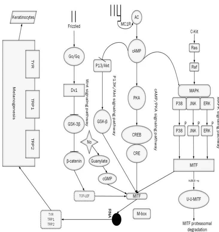
1.4. Melanogenesis (a complex reaction) ... 6 1.5. Regulation of melanogenesis and pathways ... 8 There are some basic steps involves in almost every melanogenesis reaction. There are some proteins and enzymes, which are involve in regulation of melanogenesis. 8
1.5.1. MC1R and αMSH 8
1.5.2. cAMP/PKA Pathway 9
1.5.3. MITF 11
1.5.4. MAPK/ERK 11
Chapter 2: Pigmentation Disorders 13
ii 2.1.1. Melasma 13 2.1.2. Solar Lentigo 14 2.2. Hypopigmentation ... 16 2.2.1. Vitiligo 16 Chapter 3: Antibiotics 18
3.1. Antibiotics and Melanogenesis ... 22
Chapter 4: Material and Methods 31
4.3. Cell viability 32
4.5. Measurement of cellular Tyrosinase contents ... 33 4.6. Western blot analysis ... 33 4.7. Statistical analysis ... 34
iii
List of Figures
Figure 1. ... 7
Figure 2.. ... 10
Figure 3. ... 14
Figure 4. Solar lentigo, a disorder of hyperpigmentation ... 15
Figure 5. Vitiligo, disorder of hypopigmentation ... 17
Figure 6. Structure of streptomycin ... 25
Figure 7. Structure of Netilmicin ... 26
Figure 8. Structure of amikacin ... 27
Figure 9. Structure of Kanamycin ... 28
Figure 10. Structure of gentamicin ... 29
Figure 11. Structure of Tobramycin ... 29
Figure 12. Structure of Fosfomycin disodium salt (FDS) ... 30
Figure 13. ... 36 Figure 14. ... 38 Figure 15. ... 40 Figure 16. ... 42 Figure 17. ... 44 Figure 18.. ... 46
iv
List of Tables
Table 1. Timeline and history of antibiotics ... 19 Table 2. List of Aminoglycosides and there genero and discovery year. ... 24
1
Abstract
With the discovery of penicillin, the first antibiotic, the development of antibiotics has started and many new antibiotics have been discovered. In the earlier days, antibiotics were used to treat bacterial infections. However, in recent, antibiotics have been used for many other diseases. Gradually the old antibiotics were getting reduced in usage or are no longer used due to antibiotic resistance, side effects, and the development of more effective antibiotics. Previously tobramycin and some other antibiotics were used to target melanogenesis and skin coloration. In this study, we investigated the effects of, Fosfomycin disodium salt (FDS) on melanogenesis in B16F10 melanoma cells. First, we investigate the effect of FDS on the viability of B16F10 cells was confirmed with MTT assay. The antibiotic FDS showed no cytotoxicity in B16F10 cells along with α-MSH.
We also confirmed the effect of FDS on Cellular tyrosinase and cellular melanin contents with and without α-MSH in B16F10 murine melanoma cells. The cellular tyrosinase (an enzyme important for melanin synthesis) and cellular melanin contents were increased by FDS with the increase in concentration compared with control.
Furthermore, from the results of western blot, we also confirmed that FDS up regulates the phosphorylation of p38, JNK and ERK, one of the MAPK proteins, which increasing the expression of MITF protein. The increased expression of MITF increased the expressions of tyrosinase, TRP-1, TRP-2, an enzyme important for the synthesis of melanin, in a concentration dependent manner. These results are clear evidence of the melanogenesis-inducing effect of FDS in B16F10 murine melanoma cells.
2
Chapter 1: Introduction
The skin is majorly divided into two layers: the epidermis and the dermis layer. The epidermis is further divided into several layers. These layers start with the stratum basale followed by stratum spinosum, stratum granulosum, and ends with the stratum corneum [1]. The uppermost layer comprised of dead cells. The other layers consist of special cells known as keratinocytes, which produce and store the keratin protein. Keratin is an intercellular fiber, which is responsible for the resistant properties in skin, nails and hairs. There are some other cells as well, which are present in different regions and layers of the epidermis.
Markel cells, which are known for sensory receptors, melanocytes, which function is to produce skin pigmentation and Langerhans, which act as a macrophage and engulf bacteria and damaged cells [2].
The dermis layer, which is known as the “core” of the integumentary system, composed of connective tissues and network of elastin and collagen fibers. The dermis layer contains blood vessels, hair follicles, proteins and sweat glands. These are helpful for skin elasticity, maintaining the structure of the skin, resistance and thermoregulation with environment [3].
The importance of the skin is not only important to looks handsome and perfect, but our skin performs so many functions for our body. The skin protects our internal bod from many small bacteria and viruses. It also protects our body from harmful radiations such as ultraviolet (UV) that can damage cells, DNA, proteins and other important organs [4]. The skin also produces vitamin D, when it exposed to light and helps to maintain the body temperature. Skin also maintains the homeostasis between body and external environment [5].
3
1.1. Melanocytes (pigment producing cells)
Melanin, known for skin or hair pigmentation is synthesized in melanocytes in the basal layer of the skin, from where it is distributed to neighboring cells. Melanocytes originate from the neural crest during embryo development, like neurons and glial cells [6].
Two essential proteins for the development of melanocyte are the proto-oncogene c-Kit and its ligand “steel factor” (also called stem cell factor (SCF) or mast cell growth factor (MGF)) [7]. Studies in mice have shown that loss-of function mutations in either gene lead to improper migration of melanoblasts outward from the dorsal neural tube, preventing melanocyte formation in the region’s most distant from the neural crest. The same is seen in humans, where mutations in c-Kit cause piebaldism, which is characterized by a lack of pigmentation on the forehead, chest, abdomen, and extremities [8]. Mutations in the microphthalmia transcription factor (MITF) have a similar effect on melanocyte development, but since MITF also plays an important role in melanin synthesis in mature melanocytes, this transcription factor will be discussed in more detail in a later section.
The pigmentation pattern in piebaldism is the result of a localized absence of melanocytes, but the majority of pigmentation regulation occurs within melanocytes. Studies of mouse models and human pigmentary disorders have elucidated more than a hundred genes involved in the regulation of skin pigmentation, and most of these regulate pigmentation at the subcellular level [9].
In addition to genetic studies, cell biological and biochemical analyses of human and mouse melanocytes or melanocyte-derived cell lines have provided a more detailed look at pathways of melanin synthesis and distribution. The next section will summarize some of these findings.
4
1.2. Melanin (coloring pigment)
Melanin, a coloring pigment which is found in almost all of the living organisms (from fungi and plants to mammals), and perform different functions in all of these organisms. For example, in invertebrates melanin is part of the immune system, and in the fungus C. neoformans it determines virulence [10].In mammals melanin is the main determinant of skin-, hair-, and eye color, but it is also found in the substantia nigra area of the brain in the form of neuromelanin [11].This work focuses on the study of mechanisms of skin pigmentation in mouse and human cells. Skin pigment consists of two forms of melanin: red/yellow pheomelanin and the more abundant brown/black eumelanin. In humans, pigmentation by pheomelanin is noticeable in the (near) absence of eumelanin, e.g. in red-haired, fair-skinned individuals, but levels of eumelanin more closely correlate with phenotypical pigmentation differences [12].
1.3. Melanogenesis and their enzymes
Skin consists of three different types of cells keratinocytes, Fibroblasts and Melanocytes. Among these Melanocytes are the color producing cells arises from the follicular and interfollicular epidermis, which produces a Specific type of lysosomal related organelle known as the Melanosomes. Melanins are produced their within the melansomes, which are responsible to give color to hair, skin and some other tissues as well. These melansomes fused with some structural proteins which is departed from endoplasmic reticulum and carried through Golgi vesicles [13, 14].
Melanosomes are organelles which produce, store and event transport melanin and these involves some enzymatic complex reaction known as melanogenesis. Melanin is the end product of Melanogenesis. It involves the catalysis of tyrosine and other proteins and biomolecules. Tyrosinase and tyrosinase related proteins TRP-1 and TRP-2 help in catalysis. These enzymes are
5
very important in research point of view and this pathway studied a lot while analyzing cosmetics products [15-18]. There are some basic steps involves in almost every melanogenesis reaction. Let’s discuss one by one
1.3.1. Tyrosinase
Tyrosinase is a type-3 copper protein monooxygenase also known as polyphenol oxidase, which is multifunctional and situated in the membrane of melanosomes and produced by melanocytes [19, 20]. It is mostly found in fungi, higher plants and animals as well [21]. Its synthesis and other processes occur in Endoplasmic reticulum and Golgi bodies and transferred to melanosomes. Tyrosinase has two domains the cytoplasmic domain and the internal domain. The cytoplasmic domain helps in the transport of the enzyme from the nucleus to melanosomes while the internal domain has the catalytic regions [22]. From structural point of view, the internal domain each copper surrounded by three histidine proteins. If copper get oxidized it has been inactivated and it can be activated by some electron donors such as ascorbic acid, L-DOPA etc. [23, 24]. It also get activated when two serine residues of the cytoplasmic domain phosphorylated by protein kinas C- β (PKC-β) [25]. The main function of tyrosinase is hydroxylation. At the active sites it converts monophenols to diphenols and also do oxidation of o-phenols to o-quinones. Furthermore it also converts dopamine to dopaquinone and dopachrom, and all of them lead to enhance melanogenesis [26-28].
1.3.2. TRP-1 and TRP-2
Melanogenesis complex pathway for production of melanin involves specific enzymes like Tyrosinase, tyrosinase related protein 1 and tyrosinase related protein 2. Both TRP-1 and TRP-2 are involved in downstream of the modulation of melanin. The discussion for molecular structures of TRPs began in 1980. In 1986, Shibahara et al. find tyrosinase related gene and call it isoenzyme
6
later on it called TRP-1. Later on a second gene homology of tyrosinase also identified and called it TRP-2[29, 30].
In human melanocytes the work of these TRPS are still unclear, while in mouse these TRPs act as oxidase enzyme and oxidase dihydroxyindole carboxylic acid (DHICA). Both are responsible for the production of Eumelanin. TRP-1 is also involved in some other functions such as maintaining stability of tyrosinase, effect proliferation and cell death of melanocytes and maintenance the ultrastructure of melanocytes. If there is any sort of gene mutation occurs, it leads to Albinisms type-3 [31].
1.4. Melanogenesis (a complex reaction)
Melanin is the major determining factor for skin color and also provides defense against harmful radiation. It is present in almost all type of organisms. Melanins are the heterogeneous biopolymers of phenolic compounds which are produced, synthesize and store in special organelles (Melanosomes) via enzymatic complex reactions called melanogenesis [32, 33].
The amount of melanin produced in melanocytes is predominantly determined by the direct or indirect regulation of levels or activity of the enzymes responsible for various steps of the melanin synthesis pathway, shown in Figure 1.1. The first, and rate-limiting, step of melanin synthesis is the conversion of tyrosine to DOPA and dopaquinone by the oxidating enzyme tyrosinase. This step is common to both eumelanin and pheomelanin synthesis [34].
From there, synthesis of pheomelanin requires the addition of cysteine to dopaquinone, forming 5-S-cysteinyl-DOPA or 2-S-cysteinyl-DOPA. Alternatively, dopaquinone can enter the eumelanin-specific pathway by oxidation to dopachrome, which is further processed to form the two building blocks of eumelanin: DHI (5,6-dihydroxyindole) and DHICA (6-dihydroxyindole- 2-carbolic acid).
7
Figure 1. Melanogenesis basic pathway for synthesis of Eumelanin and pheomelanin, Tyrosinase and their related proteins TRP-1 and TRP-2 play a key role in degradation and hydroxylation and leads to formation of melanin.The formation of eumelanin requires additional enzymes beyond tyrosinase: TYRP1 (tyrosinase-related protein 1) and TYRP2 (tyrosinase-related
8
1.5. Regulation of melanogenesis and pathways
There are some basic steps involves in almost every melanogenesis reaction. There are some proteins and enzymes, which are involve in regulation of melanogenesis.
1.5.1. MC1R and αMSH
The Melanocortin receptors (MC1R) belong to one of the members of G-protein-coupled receptors. (MC1R) is a seven trans membrane domain G protein-coupled receptor. It is expressed predominantly in melanocytes, but is also found in other skin cells [36]. Other tissues contain melanocortin receptors as well: MC2R in the adrenal cortex, MC3R and MC4R in the brain, and MC5R in peripheral tissues (Exocrine glands). The different melanocortin receptors have unique functions (for example, MC4R plays a role in the hypothalamic regulation of food intake [37]) but they are all activated by ligands derived from the precursor protein proopiomelanocortin (POMC) [38].The predominant POMC derived ligand produced in skin cells is α-melanocyte stimulating hormone (αMSH), produced by melanocytes, keratinocytes, fibroblasts, endothelial cells and inflammatory cells [39].Stimulation of the MC1R by αMSH leads to intracellular release of the GTPbound Gsα subunit, which then activates adenylate cyclase to produce cAMP. cAMP interacts with several downstream pathways to regulate melanin synthesis, as well as dendrite extension and melanosome transport. These pathways will be outlined in the next section.
Regulation of melanin synthesis by the MC1R only affects eumelanin, not pheomelanin. Loss-of function mutations in the MC1R therefore lead to a phenotype in which synthesis of the brown/black pigment eumelanin is impaired, but the red/yellow pheomelanin is still produced in normal levels. This is a relatively common phenotype: most individuals with red hair and pale skin carry MC1R mutations [40].These individuals often show an impaired tanning response: UV radiation up regulates melanin synthesis through several mechanisms, including the increase of
9
αMSH secretion from melanocytes and keratinocytes [41].Since αMSH needs the MC1R to induce pigmentation, a non-functional MC1R does not respond to the UV-induced increase in αMSH.
The endogenous antagonist of several melanocortin receptors (including MC1R) is the Agouti signaling peptide (ASP), which may even act as an inverse agonist [42].Mice with a dominant mutation in Agouti produce excessive amounts of ASP, leading to a yellow coat color through the MC1R and to obesity through the MC4R [43].
The following section will outline several pathways related to melanin synthesis that are affected by MC1R activation of adenylate cyclase and subsequent elevation of intracellular cAMP. While the overall effect of cAMP stimulation is an increase in pigmentation, cAMP is the starting point of several interacting signaling cascades that together fine-tune melanin synthesis.
1.5.2. cAMP/PKA Pathway
Cyclic adenosine monophosphate/protein kinase A (cAMP/PKA) is a well-known signaling pathway for melanogenesis regulation. Apart from melanogenesis it also regulates aging, ion channel conductivity, metabolism, cell growth and division, sperm motility, cell differentiation and gene regulation [44-49].
Protein kinase A (PKA) is a well-known and well-studied downstream effector of cAMP. The cAMP bind with the regulatory subunit of PKA activates the catalytic domain via conformational change, allowing PKA to phosphorylate several intracellular targets, including the transcription factor CREB (cAMP responsive element binding protein) and CREB-binding protein (CBP) [50]. Phosphorylated CREB then activates the expression of genes with CRE consensus sequences in their promoter [51]. One protein that is expressed in response to CREB activation is MITF [52], which in turn activates the transcription of tyrosinase and other melanin synthesizing genes, as described above.
10
Figure 2. Melanogenesis signaling pathways involves in regulation of melanin. All of them end with melanin production. The up-regulation or down-regulation of these molecules is the possible answer for the rise and fall of
11
To summarize this pathway: cAMP up regulates melanin synthesis through PKA activation, CREB phosphorylation, MITF expression and increased transcription of tyrosinase and related genes. However, PKA is not the only target of cAMP in melanocytes.
1.5.3. MITF
MITF is the only member of microphthalmia family which is essential for development of melanocytes and acts as a master regulator for production of melanin. The gene of MITF contains several promoters for its expression. Among these promoters M promoter is known for melanocytes development it regulates by itself or some other transcription factors such as cAMP, CREB, SOX9, SOX10, PAX3 and LEF-1/TCF [53]. MITF also regulated by some known pathways like MAPK, ERK2, STATE, JNK, AKT and P90SRK in melanocytes [54].
A highly conserved 10 base pair motif (GTCATGTGCT) which is known as M-Box, with the help of this M-Box, tyrosinase and tyrosinase related protein 1 and 2(TRP-1 , TRP-2) genes are transcribed by MITF [55,56]. The transcription of tyrosinase, TRP-1, and TRP-2 is regulated by the microphthalmia transcription factor (MITF), which binds a region in the promoters of these genes called M-Box (AGTCATGTGCT)[57. MITF is also involved in melanocyte development, and various mutations in MITF in mice lead to a deficiency in melanocytes in skin and ear, and decreased pigmentation and size of the eyes [58]. These enzymes had studied for a number of occasions while triggered melanogenesis and curing dermatological diseases.
1.5.4. MAPK/ERK
Mitogen-activated protein kinases (MAPK)/ extracellular signal-regulated kinases (ERK) is another important pathway in melanogenesis. This pathway is not only studied for
12
melanogenesis but for apoptosis, cell proliferation and migration, skeletal muscles cell activation and senescence [59-62].
The pathway initiates when the extracellular ligand attach to the receptor (Trans membrane protein receptor or receptor tyrosine kinase RKT). As shown in figure 2, the binding of the ligand dimerise the two sub-units of RTK. Further after it activates a complex mechanism (RAS-RAF-MEK-ERK).
The small protein RAS activated by guanine-nucleotide exchange factor (GEFs) which activate kinase cascade. RAS first activate MAP-KKK (with three kinases) also known as BRAF activates MAP-KK (with two kinases) also known as MEK. At the end MEK or MAP-KK activate MAP-K (with one kinase) or ERK. ERK activates MITF for gene expression like TYR, TRP-1 and TRP-2. Hence the up-regulation or down-regulation of ERK leads to rise and fall of melanin [63-67]. The effect of the Ras/ERK pathway on melanin synthesis appears to be a negative feedback mechanism for the cAMP and PKA mediated increase in tyrosinase expression. When B16 cells are treated with PD98059, a MEK-1 specific inhibitor, tyrosinase levels and activity are increased, indicating that the ERK pathway in these cells has a negative effect on melanin synthesis. Indeed, overexpression of dominant negative Ras or MEK-1 also increases tyrosinase expression, while constitutively active mutants of the same proteins inhibit tyrosinase expression [68].
13
Chapter 2: Pigmentation Disorders
Human skin has a variety of colors from the darkest brown to lightest hues. Melanin, a heterogeneous biopolymer is responsible for skin coloration as well as protection of internal body from external assaults. Thus, the required amount of melanin must be available for skin. When the required amount of melanin is not available for skin it results hyperpigmentation (excess of melanin) or Hypopigmentation (lesser amount of melanin).
2.1. Hyperpigmentation
The natural skin color for human is balance by three chromophores: Hemoglobin (red color), carotenoid (yellow/orange in color) and melanin (blow/brown) in color [69]. Melanin is the most important among these. Hyperpigmentation occurs because of the production of excess of melanin. In hyperpigmentation, the skin appears darker than surroundings and some dark spots occur on skin [70]. Melasma, solar lentigo, ephelides, Café-au-lait macules, Nevi and melanoma are the most common diseases of hyperpigmentation [71]. Melasma, solar lentigo and melanoma are mostly studied by researchers.
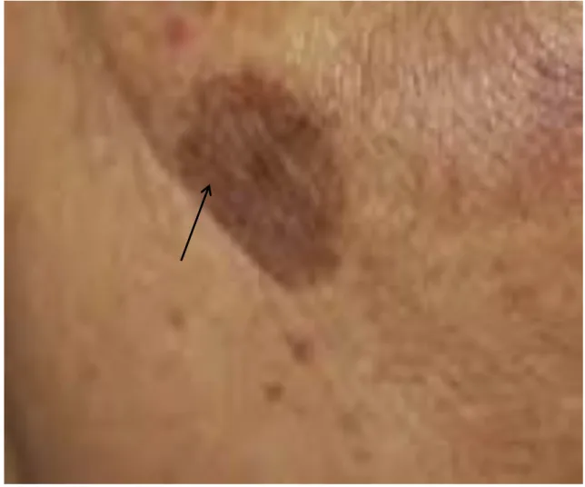
2.1.1. Melasma
Melasma is a Greek word derived from melas means black, is a common acquired, non-scalar, progressive pigmentation disorder which generally appears on dorsal forearms, cheeks and mandibles. There is a lot of factors which causes melasma such as exposure to UV light, birth control pills, pregnancy, oral contraceptives and hormonal therapy [72-75]. In melasma, the number of melanocytes and the production of melanin increases. Women are about 9 times more affected by melasma compared to men [76].
14
Figure 3. Melasma, the most common hyperpigmentation disorder affects about 80% of teenagers.
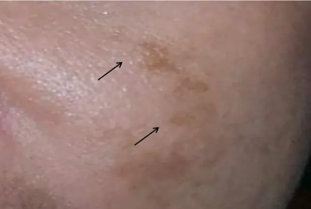
2.1.2. Solar Lentigo
Solar lentigo is a common macule hyperpigmentation disease which appears as aging spots on skin exposure. The color of spots varies from light yellow to dark brown. It is most commonly appearing on face, chest, back, hands and forearms. It is due to these organs are exposed to sun light [77]. The persons with white skin or Asian persons are most likely suffered from solar lentigo [78].
Histologically confirmed that solar lentigo is due to large melanosomal complexes, extra deposition of melanin in the basal layer and elongation of epidermal ridges. Solar lentigo suffered persons having activated melanocytes in their tissues [79].
15
Solar lentigo is commonly targeted in those people whose age is above 20years. The sun exposure gradually activates melanocytes, melanogenic pathways related enzymes and it leads to enhancement of melanin. Less exposure to sun light, using sun screen regularly, and protection from sun burn are the most common prevention from solar lentigo. Hydroquinone has been used for many years for curing solar lentigo but it has also some adverse effects and the seriousness of the diseases in specially adults increasing so ultimately it required more attention of the dermatologists and scientists [80-86].
16
2.2. Hypopigmentation
Hypopigmentation is characterized as when the skin generates lesser amount of melanin than normal skin. The result could be patches of skin and loss of pigmentation. If skin melanocytes do not produce balance melanin, the skin coloration damaged badly, results some light spots appearance [87]. The color of skin depends on location of skin. Inflammation is also a cause of hypopigmentation. Several factors include sun light exposure, medicines, genes mutation and some chemical compounds as well can enhance the hypopigmentation. Vitiligo, Albinism, Pityriasis alba, Tuberous sclerosis, Tinea versicolor, Piebaldism, Hypo-melanosis of Ito are the common disorders of hypopigmentation [88].
2.2.1.
Vitiligo
Vitiligo is a hypopigmentation disorder caused by deficiency of melanin which results disfiguring of pigmentation. Some scientist believes that is hereditary disease but the exact etiology of this disease is still unknown. It affects almost all type of skins but for black patients it is a massive burden and stress. Although the number of patients is less but it is increases now because of increase of pollution and other environmental and food factors [89, 90]. Most researchers relate the vitiligo with abnormality in keratinocytes apart from melanocytes. Vitiligo may also be due to over expression of tyrosinase enzyme and tyrosinase related proteins TRP-1 and TRP-2, MITF, p38, JNK or low expression or down-regulation of ERK/AKT, WNT/β-catenin signaling [91, 92]. Based on area of appearance of vitiligo, it has four types: localized, generalized, acral/acrofacial, and segmental. Treatment of disease varies based on their type.
17
18
Chapter 3: Antibiotics
The word antibiotic means “against life” used to refer any substances that are used against bacterial infections. The history of the antibiotics almost all of people know even without the background knowledge of life sciences. Everyone has heard about the accidental discovery of penicillin by Alexander Fleming. He coincidently contaminated his agar plate with mould back in 1929 [93].
Before penicillin, with lesser common knowledge, another pioneering work done by Alexander Fleming and Sahachiro Hata, led to the discovery of salvarsan in 1909. Salvarsan was a novel drug, used to treat sexual transmitted disease, which is caused by the spirochete Treponema
palladium [94].
Ehrlich and Hata used the large-scale screening method for the discovery of salvarsan, became the well-known standard for searching novel drugs and led to the discovery of the first sulfa drug in 1934. Sulfonamidochrysoidine, a precursor of the active compound sulfanilamide, inhibits folic acid synthesis in bacteria [95].
The golden age of antibiotics begins with the discovery of sulfa drugs and the release of penicillin for clinical research. From 1940 to 1970, almost all of the antibiotic drugs classes were discovered and most of them were isolated from natural resources such as plant extracts and microorganisms [96]. Before the earlier 20th century, cholera, typhoid fever, syphilis, tuberculosis
and smallpox etc. were the most dangerous diseases, spread all over the world. At that time, people use natural products, herbs, plant extracts and some known antibiotics for its treatment [97, 98]. Bacteremia caused by staphylococcus aureus was the main headache at that time, and it reached to 82% mortality, in which only 2 % of the patient’s age was above 50 years. The interesting fact
19
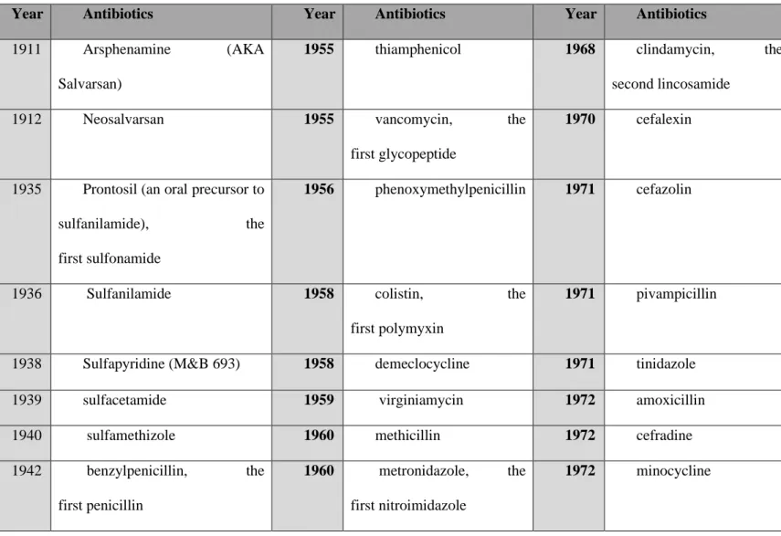
was that, the older patients were able to survive. Many people died due to this infectious disease before antibiotics were used [99]. A report commissioned by the government of the United Kingdom in 2014, estimated that the annual number of deaths attributable to antimicrobial resistance would be 10 million by 2050 and that it will generate a loss of 100 trillion dollars globally [100]. Therefore, researchers develop new antibiotics with high resistance. Table 1 shows the timeline of the developed antibiotics till date.
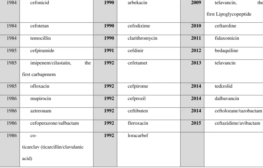
Table 1. Timeline and history of antibiotics
Year Antibiotics Year Antibiotics Year Antibiotics
1911 Arsphenamine (AKA Salvarsan)
1955 thiamphenicol 1968 clindamycin, the second lincosamide
1912 Neosalvarsan 1955 vancomycin, the first glycopeptide
1970 cefalexin
1935 Prontosil (an oral precursor to sulfanilamide), the first sulfonamide
1956 phenoxymethylpenicillin 1971 cefazolin
1936 Sulfanilamide 1958 colistin, the first polymyxin
1971 pivampicillin
1938 Sulfapyridine (M&B 693) 1958 demeclocycline 1971 tinidazole 1939 sulfacetamide 1959 virginiamycin 1972 amoxicillin 1940 sulfamethizole 1960 methicillin 1972 cefradine 1942 benzylpenicillin, the
first penicillin
1960 metronidazole, the first nitroimidazole
20 1942 gramicidin S, the
first peptide antibiotic
1961 ampicillin 1972 pristinamycin
1942 sulfadimidine 1961 spectinomycin 1973 Fosfomycin 1943 sulfamerazine 1961 sulfamethoxazole 1974 talampicillin 1944 streptomycin, the
first aminoglycoside
1961 trimethoprim, the first dihydrofolate reductase inhibitor
1975 talampicillin
1947 sulfadiazine 1962 cloxacillin 1975 bacampicillin 1948 chlortetracycline, the
first tetracycline
1962 fusidic acid 1975 ticarcillin
1949 chloramphenicol, the first amphenicol
1963 fusafungine 1976 amikacin
1949 neomycin 1963 lymecycline 1977 azlocillin 1950 oxytetracycline 1964 gentamicin 1977 cefadroxil 1950 penicillin G procaine 1964 cefalotin, the
first cephalosporin
1977 cefamandole
1952 erythromycin, the first macrolide
1966 doxycycline 1977 cefoxitin
1954 benzathine penicillin 1967 carbenicillin 1977 cefuroxime 1955 spiramycin 1967 rifampicin 1977 mezlocillin 1955 tetracycline 1967 nalidixic acid, the
first quinolone
1977 pivmecillinam
21
1980 cefmetazole 1987 cefixime 1992 rufloxacin 1980 cefotaxime 1987 roxithromycin 1993 brodimoprim 1980 piperacillin 1987 sultamicillin 1993 dirithromycin 1981
co-amoxiclav (amoxicillin/clavulanic acid)
1987 ciprofloxacin, the first 2nd-gen fluoroquinolone
1993 levofloxacin
1981 cefoperazone 1987 rifaximin, the first ansamycin
1993 nadifloxacin
1981 cefotiam 1988 azithromycin 1993 panipenem/betamipron 1981 cefsulodin 1988 flomoxef 1993 sparfloxacin
1981 latamoxef 1988 isepamycin 1994 cefepime 1981 netilmicin 1988 midecamycin 1996 meropenem
1982 ceftriaxone 1988 rifapentine 1999 quinupristin/dalfopristin 1982 micronomicin 1988 teicoplanin 2000 linezolid, the
first oxazolidinone
1983 cefmenoxime 1989 cefpodoxime 2001 telithromycin, the first ketolide
1983 ceftazidime 1989 enrofloxacin 2003 daptomycin
1983 ceftizoxime 1989 lomefloxacin 2005 tigecycline, the first glycylcycline
22
1984 cefonicid 1990 arbekacin 2009 telavancin, the first Lipoglycopeptide 1984 cefotetan 1990 cefodizime 2010 ceftaroline 1984 temocillin 1990 clarithromycin 2011 fidaxomicin 1985 cefpiramide 1991 cefdinir 2012 bedaquiline 1985 imipenem/cilastatin, the
first carbapenem
1992 cefetamet 2013 telavancin
1985 ofloxacin 1992 cefpirome 2014 tedizolid 1986 mupirocin 1992 cefprozil 2014 dalbavancin
1986 aztreonam 1992 ceftibuten 2014 ceftolozane/tazobactam 1986 cefoperazone/sulbactam 1992 fleroxacin 2015 ceftazidime/avibactam 1986
co-ticarclav (ticarcillin/clavulanic acid)
1992 loracarbef
Table 1. Timeline and history of antibiotics
3.1. Antibiotics and Melanogenesis
From the last twenty to thirty years, researchers use melanocytes cell cultures in order to investigate new drugs to regulate melanogenesis and their enzymes and pathways. For this purpose, many plant extracts, natural products and compounds were used investigate novel compound to help in melanin synthesis. From the last decades, researchers and biochemists are interested in antibiotics to find the new antibiotic to cure melanogenic diseases. Penicillin and streptomycin are of the most widely used antibiotics used in mammalian cell culture [101]. There
23
are some other antibiotics which regulates the melanogenesis pathways and their enzymes. Here we will discuss few of them.
3.2.
Aminoglycosides related antibiotics
Aminoglycosides, derived from actinomycetes are the broad range of natural or semisynthetic antibiotics. Most of these aminoglycoside antibiotics end with “-mycin”, “-micin” or simply “-sin” and aminoglycosides with suffix “-thromycin” are the macrolides like erythromycin and erythromycin. The main bacterial target for the aminoglycosides is gram-negative bacteria or gram-gram-negative amino bacilli. They are no used for gram positive bacteria. The aminoglycosides are also active against mycobacteria like mycobacterium tuberculosis.
The aminoglycosides are used previously for some complicated infections, such as complicated UTIs, complicated skin or skin soft tissues, septicemia, osteomyelitis, plague, tularemia and pneumonia [102-104]. The structure of aminoglycosides comprised of two or amino sugars joined by glycoside linkage to a hexose nucleus. Different aminoglycosides are differentiated by the amino sugars attached to the aminocyclitol ring by glycosidic bond. This antibiotic binds to 30S ribosomes and inhibits translations, thereby synthesizing proteins which act as an inhibitor [105,106].
24
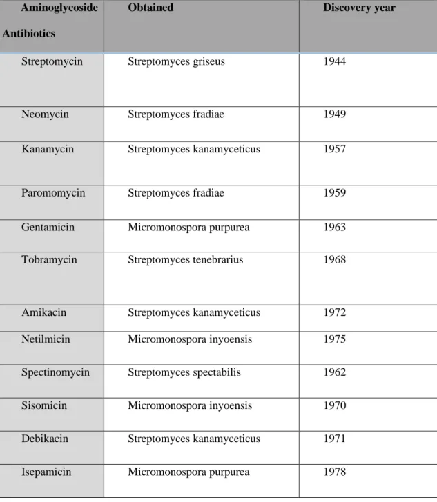
Table 2. List of Aminoglycosides and there genero and discovery year.
Aminoglycoside Antibiotics
Obtained Discovery year
Streptomycin Streptomyces griseus 1944
Neomycin Streptomyces fradiae 1949
Kanamycin Streptomyces kanamyceticus 1957
Paromomycin Streptomyces fradiae 1959
Gentamicin Micromonospora purpurea 1963
Tobramycin Streptomyces tenebrarius 1968
Amikacin Streptomyces kanamyceticus 1972 Netilmicin Micromonospora inyoensis 1975
Spectinomycin Streptomyces spectabilis 1962
Sisomicin Micromonospora inyoensis 1970
Debikacin Streptomyces kanamyceticus 1971
Isepamicin Micromonospora purpurea 1978
25
Streptomycin is the first aminoglycoside antibiotic discovered by Albert Schatz from Streptomyces griseus in 1944. It is used to treat against numerous gram-negative bacterial infections include septicemia, osteomyelitis, plague, tularemia and pneumonia. It exerts a strong action against gram-negative bacteria that are not affected by penicillin [107].
It is reported that Streptomycin inhibits the activity of tyrosinase and other melanogenic enzymes to reduce melanin contents and it also act as an antioxidant [108].
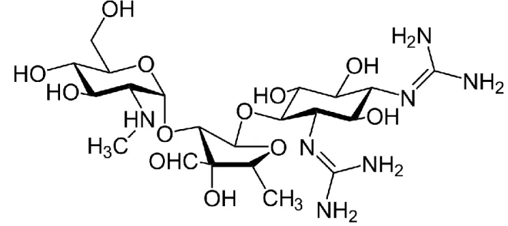
Figure 6. Structure of streptomycin
3.2.2. Netilmicin
Netilmicin is another member of aminoglycoside antibiotic. It is discovered in Micromonospora inyoensis in 1975. It also has the potential to demolish various bacterial infections. It is used only for treatment of serious infections, which are resistant to gentamicin [109].
26
The Netilmicin inhibit the melanin biosynthesis by inhibiting the cellular tyrosinase activity in human melanocytes [110].
Figure 7. Structure of Netilmicin
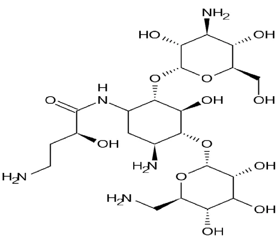
3.2.3. Amikacin
Amikacin is an aminoglycoside antibiotic used for a wide range of bacterial infections such as meningitis, sepsis, pneumonia, joint infections and intra-abdominal infections. It is discovered in Streptomyces kanamyceticus in 1972. Amikacin can cause hair loss just like other aminoglycoside antibiotics [111].
Just like Netilmicin it also inhibits the synthesis of melanin by inhibiting the cellular tyrosinase activity in human melanocytes [112].
27
Figure 8. Structure of amikacin
3.2.4. Kanamycin
Kanamycin was discovered from Streptomyces kanamyceticus by Japanese microbiologist Hamao Umezawa in 1957. It is also used for several bacterial infections and tuberculosis just like other aminoglycoside antibiotic. It is also called kanamycin A, as there are several others like kanamycin B, kanamycin C and kanamycin D [113].
Kanamycin suppresses the synthesis of melanin by decreasing the cellular tyrosinase activity in human melanocytes. Kanamycin also acts as an antioxidant [114].
28
Figure 9. Structure of Kanamycin
3.2.5. Gentamicin
Just like other aminoglycoside antibiotics, gentamicin is also an important antibiotic which is used for number of bacterial infections. It was found in 1963 by Weinstein, Wagman et al. in Micromonospora purpurea in fermentation. Like others, it is also active against gram-negative bacteria such as Pseudomonas, Proteus, Escherichia coli, Klebsiella pneumoniae, Enterobacter aerogenes, Serratia [115].
Like kanamycin, streptomycin and Netilmicin gentamicin also reduce the biosynthesis of melanin by suppressing the cellular tyrosinase activity in human normal melanocytes [116].
29
Figure 10. Structure of gentamicin
3.2.6. Tobramycin
Tobramycin is also an aminoglycoside antibiotic used for wide verity of bacterial infections. Tobramycin was founded in1967 by Eli Lilly and his company workers in Streptomyces tenebrarius. It is active against most of gram-negative bacteria specially pseudomonas [117].
Tobramycin induces melanogenesis by induces the cellular tyrosinase activity, TRP-1, TRP-2 and by up-regulation of p38 MAPK protein phosphorylation in B16F10 cells [118].
30
3.3.
Fosfomycin
Fosfomycin is a novel class of antibiotic and belongs to phosphonic acid antibiotics. Fosfomycin was discovered by Merck and his company in 1969 and approved for use in1996. It is isolated from Streptomyces fradiae. It is active against both gram-negative such as Citrobacter and Proteus and gram-positive E. faecalis, E. coli [119].
Fosfomycin is generally used to treat against bladder infections. Fosfomycin disodium salt (FDS) is a bactericidal drug, which is generally used for inhibiting the cell synthesis. It is also used for the treatment of urinary tract infections [120].
31
Chapter 4: Material and Methods
4.1.
Sample and reagents
Fosfomycin purchased from Tokyo Chemical Industry (Chuo-ku, Tokyo, Japan). Dulbecco modified eagle medium (DMEM), fetal bovine serum (FBS), penicillin/streptomycin, trypsin-ethylenediaminetetraacetic acid, PD98059, BCA protein assay kit purchased from Thermo Fisher Scientific (Waltham, MA, USA).
α-MSH, NaOH, L-DOPA, Griess reagent, proteasome inhibitor cocktail, H89 (PKA inhibitor) purchased from Sigma-Aldrich (St. Louis, MO, USA). 3-(4,5-dimethylthiazol-2-yl)-2,5-diphenyltetrazolium bromide (MTT) purchased from VWR (Radnor, Pennsylvania, U.S.A). Tyrosinase, TRP-1, TRP-2, and MITF purchased from Santa Cruz Biotechnology (Dallas, TX, USA).
p-p38, p38, p-JNK, JNK, p-ERK, ERK, p-AKT, AKT, β-actin primary anti-rabbit lgG HRP-linked Antibody, anti-mouse lgG HRP-linked Antibody, LY294002 (AKT inhibitor) purchased from Cell Signaling Technology (Danvers, MA,
USA).
Radioimmunoprecipitation assay (RIPA) buffer, Dimethyl sulfoxide (DMSO),
Enhanced chemiluminescence (ECL) kit, 2× Laemmli sample buffer purchased from Bio World (Sungnam, Gyeonggi-do, Korea). SP600125 (JNK inhibitor) purchased from Cayman Chemical (Ann Arbor, MI, USA). SB203580 (p38 inhibitor) purchased from Calbiochem (San Diego, CA, USA).
32
4.2.
Cell Culture
B16F10 Murine melanoma mice cells were ordered from Korean cell line bank (Seoul, Korea), and cultured in (DMEM) Dulbecco's Modified Eagle's Medium with 10% (FBS) fetal bovine serum and 1% penicillin streptomycin. Cells were incubated in 95% air humidified atmosphere containing 5%CO2.
4.3.
Cell viability
To check the cell viability of FDS we use [3-(4,5-dimethylthiazol-2-yl)-2,5 diphenyltetrazolium bromide] (MTT) a dye. 0. 3× 10⁴ B16F10 mice cells were mantained in
24-well plates and incubated in 95% air humidified atmosphere containing 37℃ and 5%CO2 for 24
hours. Different concentration of FDS were treated followed by 25µL MTT 0.1% reagent to each well and incubated for two hours, Remove medium carefully and add 500 µL (DMSO) dimethyl sulfoxide to each well dissolve the dye formazan. Plates were placed in shaker to shake well and absorbance was measured at 540nm. The percentage of cell viability was determined according to previously described methods.
4.4.
Measurement of cellular melanin contents
Cellular melanin contents of B16F10 murine melanoma cells was measured . 1× 10⁵
B16F10 cells were seeded in 60mm π dishes and incubated for 24 hours. Add new medium with 200nM αMSH along with FDS with different concentrations(125-500)µg/mL, and incubated for 72 hours, Remove the medium and wash with (PBS) cold phosphate-buffered saline, the cell pellets were dissolved with 10%DMSO contain 1N NaOH and kept in UV sterilizer with 70 ℃ for 1 hour and measure absorbance at 405nm. Each experiment was performed in triplet
.
33
4.5.
Measurement of cellular Tyrosinase contents
Intercellular tyrosinase activity assay was measured. 1× 10⁵ murine melanoma cells were
seeded in 60mm π dishes and incubated for 24 hours. Different concentrations of FDS were loaded and incubated for 72 hours. Add 200µl Radioimmunoprecipitation assay buffer (RIPA buffer) containing 1% enzyme inhibition cocktail to each plate. The cell lysates were clarified by centrifugation for 20 minutes at 15000 rpm. Remove 150 µL protein concentrations carefully in a separate tube and diluted with sodium phosphate buffer. Different concentrations were loaded in 96-well plates and followed by 15mM of L-DOPA to each well and incubated for 30 min at 300 rpm and 37℃. The absorbance was measured at 475nm with ELISA reader. Experiment was performed in triplicate.
4.6.
Western blot analysis
1× 10⁵ Cells/well of B16F10 murine melanoma mice cells were seeded in 100mm dishes
for 24 hours. Various concentrations of FDS ranges from (125-500) µg/mL were added and incubated for 72hours. The cells pellets were lysed with (RIPA) Radioimmunoprecipitation assay buffer with 1% protease inhibition cocktail. Kept the lysate in refrigerator under -4℃ for 20min. the lysate were scraped. The cell lysates were collected and centrifuged at 15,000rpm under -10℃ for 30 minutes. The supernatants were collected from the surface carefully and normalized using the Bradford standard assay (BSA) protein assay. 2x Laemmli sample and the supernatants were mixed 1:1 using BSA table in an e-tube and make sample for western blot. FEGT sample was regulated to make an equal concentration of 20µg of proteins. Heat the sample at 100℃ for 5min and from each sample 20µL/well in 10 rows lane of 10% SDS-Polyacrylamide gel electrophoresis were loaded. Isolated proteins were transferred into a (PVDF) polyvinylidene
34
difluoride membrane, and the membrane blocked with (5% non-fate) skim milk in TBST buffer. Shake for 2hrs, and wash thrice with Tris-Buffered Saline with Tween (TBST) buffer thrice each 10 minutes. The membrane was incubated with rabbit primary antibody (1:1000) diluted with TBST buffer overnight, Wash with TBST three times and add secondary anti-rabbit antibody (1:3000) times diluted and incubated for 2hrs. The bands of protein were visualized with ECL kit.
4.7.
Statistical analysis
All the data were shown as the mean of ± standard deviation of at least three replicates. The results were analyzed with a Student’s t-test. The Statistical significance was considered at * p < 0.05, ** p < 0.01, or *** p < 0.001.
35
5. Results
5.1.
Effect of FDS on cell viability
The cell toxicity of FDS was performed on B16F10 melanoma cells with various concentration ranges from (0.1mg to 1mg)/mL in 24-well plates and maintained in incubated atmosphere. (3 × 10⁴) B16F10 cells were treated with FDS and the
3-(4,5-dimethylthiazol-2-yl)-2,5-diphenyl tetrazolium bromide (MTT) solution were incubated at 37 °C and 5% CO2 for 2
hours. MTT is water soluble salt containing yellow color. MTT converts into another water-insoluble dye (Formazan) by the active cells or the viable cells. At molecular level this is happened due to reductive cleavage of tetrazolium ring. Formazan is DMSO soluble dye and absorb UV radiation at 450±100nm. The precentage of viable cells can be known from how much of the MTT converted to Formazan by active cells. More the formation of formazan more will be the active cells and vice versa. In the present study, the FDS shows no cellular toxicity. As shown in figure 13, the viability of cells is almost 90-100% with concentrations ranging from 0.0625 to 1 mg/mL. Therefore, we used FDS at concentrations of 0.125, 0.25, and 0.5 mg/mL for further study.
36
Figure 13. Cytotoxicity of Fosfomycin disodium salt (FDS) on B16F10 cells were analyzed. Murine melanoma cells were treated with different concentrations of FDS and MTT assay was performed. Cell viability is expressed as percentages compared to the respective values obtained for
untreated control cells. The absorbance was measured at 540nm. The data represents the mean± standard deviation of the triplicate experiment. SD: standard deviation.
37
5.2.
Effect of FDS on cellular Melanin production
To examine melanin content in B16F16, 1 × 10⁵ B16F10 murine melanoma cells were
treated with FDS, incubated for 24 h, and further incubated for 72 hours with different concentrations of FDS (0.125–0.5 mg/mL). α-melanocyte-stimulating hormones (α-MSH) were used as a positive control. Melanin content was then analyzed. Melanin concentration percentage increased as the concentration of FDS increased. We also found that FDS with this range of concentration showed no cytotoxicity. α-MSH-treated cells markedly increase the melanin content up to 249% compared to the control (100%) as shown in Figure 13. This means more than 50% of enhancement in melanin content. Fosfomycin also enhance melanin content compared to the control at different level of concentrations. At 0.125 mg/mL, FDS slightly increased the melanin content up to 118%, FDS treated cells with 0.25 mg/mL enhance melanin content up to 159%., while at 0.5 mg/mL FDS had melanin content of 210%.
38
Figure 14. Effects of FDS on melanin content in B16F10 murine melanoma cells. The cells were treated with various concentration of FDS (0.125, 0.25, and 0.5 mg/mL). α-melanocyte-stimulating hormones (α-MSH) (200 nM) were used as a positive control. The contents of melanin in FDS treated cells were exhibited as percentages compared to the respective values which is obtained for the FDS untreated cells. The data are presented as mean ± standard deviation (SD) of at least three independent experiments. ** p < 0.01, *** p < 0.001 vs. untreated cell. SD: standard deviation.
5.3.
Effect of FDS on Cellular Tyrosinase Activity
Tyrosinase is a major enzyme in melanogenesis, plays a key role melanin synthesis, which is the skin-coloring pigment. Tyrosinase is involved in the first two steps of melanogenesis complex reaction. Increasing the tyrosinase activity will ultimately increase the production of melanin. For the analysis of cellular tyrosinase activity, B16F10 (1 × 10⁵) murine melanoma cells
39
used as a positive control. Proteins, extracted from these cells were treated with FDS or α-MSH were then mixed with 15 mM L-DOPA. The results showed that the tyrosinase activity increased in a dose-dependent manner. α-MSH increased the activity of tyrosinase up to 235% compared with that in the control cells (100%), which contained no sample and no FDS as shown if Figure14. At 0.125 mg/mL FDS slightly decreased the activity of tyrosinase compared with the control. However, at 0.25 mg/mL FDS increased the activity of tyrosinase up to 149% and at 0.5 mg/mL FDS increased the activity up to 240%, when compared to the control.
40
Figure 15. Effect of FDS on cellular tyrosinase activity in B16F10 melanoma cells. The B16F10 melanoma cells were treated with different concentrations of FDS for 72 hours, and α-MSH was used as a positive control. Data are represented as mean ± standard deviation (SD) of at least three
independent experiments. ** p < 0.01, *** p < 0.001 vs. untreated cell. SD: standard deviation.
5.4.
Results of Western Blotting
To evaluate the effect of FDS whether it increases the expression of melanogenic proteins and enzymes, western blot analysis was performed. 1 × 10⁵B16F10 cells were treated
for 72 hours at different concentrations ranges of FDS (0.125 - 0.5 mg/mL). 25 µg of proteins were separated by gel electrophoresis and transferred to a polyvinylidene fluoride (PVDF) membrane, blocked with 5% skim milk and primary antibody was added and incubated to detect the expression of proteins. Tyrosinase is an enzyme, play one of the important roles in the melanogenesis pathway. Tyrosinase converts L-tyrosine and L-DOPA into DOPAquinone. That’s because, the expression of this enzyme was analyzed. The FDS enhanced the expression of the tyrosinase in a concentration dependent manner as shown in
41
figure15. Tyrosinase related proteins TRP-1 and TRP-2 are also play key role in the synthesis of melanin. Therefore, to examine the effect of FDS on these two enzymes the B16F10cells were treated with various concentrations for 48hrs. The results also clarified that these melanogenic enzymes expressions were increased by FDS as the concentration were grown up. we also clarify the effect of MITF (a transcription factor) which play a vital role in the synthesis of melanin. MITF is a well-known enzyme, play an important role in various pathways by binding with the M-box inside the tyrosinase promoter and enhance tyrosinase expression. Up-regulation of MITF enzyme leads to increase in melanogenesis. The result gives a strongclarification that FDS treated cells induce the expression of MITF as compared to FDS untreated cells.
42
Figure 16. Effect of FDS on tyrosinase and tyrosinase related proteins TRP-1, TRP-2 and MITF expressions in B16F10 cells. Cells were treated with different concentrations of FDS (0.125- 0.5 mg/mL). The proteins level was inspected by Western blotting. (a) Result of Western blotting, and protein levels of (b) MITF, (c) TRP-1, (d) TRP-2, and (e) tyrosinase. Results are expressed as a percentage of the control. The data are presented as mean ± SD of at least three independent experiments. * p < 0.05, ** p < 0.01, *** p < 0.001 vs. untreated cell. SD: standard deviation. TRP: tyrosinase-related protein. MITF: Microphthalmia-associated transcription factor.
5.5.
Fosfomycin effect on Mapk Signaling Pathways
JNK, Protein kinase B (AKT) and p38 are one of the basic pathways to be involved in melanin synthesis. Enhancement in the phosphorylation of p38 and JNK leads to promote melanogenesis whereas, decrease in the phosphorylation of AKT leads to induction of melanin production. Therefore, to inspect the Fosfomycin effect on these signaling pathways, cells were treated with selected range of concentrations of FDS (0.125 to 0.5 mg/mL), and α-MSH was used
43
as a positive control. FDS up-regulates the expression of p38 and JNK, which turns to increase melanogenesis with the increase in concentration of fosfomycin. Both phosphorylated JNK and phosphorylated p38 increased with increase in the concentration of FDS. AKT expression remains almost same in a dose-dependent manner. These results give us a clear evidence that FDS turns to increase in the phosphorylation of JNK and p38 signaling pathway in induction of melanogenesis in B16F10 cells.
44
Figure 17. Effect of Fosfomycin on phosphorylation of P-JNK, P-AKT and P-p38. The murine melanoma cells were treated with FDS with different concentrations. (a) Result of Western blotting and protein levels of (b) P-JNK, (c) P-p38, and (d) P-AKT. The data are presented as mean ± SD of at least three independent experiments. *** p < 0.001 vs. untreated cell. AKT: protein kinase B. JNK: c-Jun N-terminal kinase. P: phosphorylated. T: total. S: standard deviation.
45
6.
Discussion
The color of skin is a major problem around the globe because the skin protects all of the important organs inside the body from harmful invaders and some toxic radiations. Melanin, a heterogeneous biopolymer plays a key role by providing coloration to the skin and blocking of UV radiation from entering the body.
Hair is an important and major issue around the world. Hair coloring is one of the signs of attractiveness and beauty. Whitening of hair is now become a major problem, and many people are suffering from such condition. Researcher and biochemists suggest different types of dyes, some natural compounds and anti-hair whitening agents for coloring of hair. Thus, the demand of these pigmenting compounds is increasing day by day.
Antibiotics are also used to develop and induce melanogenesis now days. Thus, to develop a new antibiotic to induce melanogenesis, we used various antibiotics. We found FDS antibiotic effectively. To examine the cytotoxicity of FDS, we performed MTT assay with different ranges of concentration of 0.125–0.5 mg/mL, and the results at these concentrations showed that FDS exerted no cellular toxicity. The results are showing the cell viability of almost 100%. Therefore, FDS at this range of concentration from 0.125–0.5 mg/mL selected for other experiments as well.
As Melanocytes can be stimulated by many factors and α-MSH is one of the factors; in this paper, we used (200 nm) α-MSH to stimulate the melanogenesis pathway in B16F10 cells. To examine the effect of Fosfomycin on cellular melanin content and cellular tyrosinase activity, murine melanoma cells were treated with selected ranges of concentrations of FDS for 72 hours. FDS clearly increased both cellular melanin and cellular tyrosinase activity content in a concentration-dependent manner.
Tyrosinase, TRP-1, and TRP-2 are the key enzymes in melanogenesis; therefore, up-regulation of these enzymes leads to excess formation of melanin, whereas down-regulation of these enzymes
46
is a major strategy and helpful for developing of new cosmetics products. The effect of Fosfomycin on the activity of intercellular tyrosinase was performed in B1610 cells. FDS enhanced the activity of tyrosinase in a dose-dependent manner. Western blotting result also provided clear evidence that FDS enhance the production of melanin by increasing the expression of the melanogenic enzymes and having no cytotoxicity.
Figure 18. Melanogenesis signaling pathway in cells. FDS increased both melanin and tyrosinase levels. FDS: Fosfomycin disodium salt, TYR: tyrosinase, CREB: cAMP-Responsive Element Binding, α-MSH: α-melanocyte-stimulating hormone, MC1R: melanocortin 1 receptor, AC: adenylate cyclase, cAMP: cyclic adenosine monophosphate, MITF: Microphthalmia-Associated Transcription Factor.
Furthermore, western blotting analysis was also used to study the effect of fosfomycin on the phosphorylation of MAPK pathways. Previous studies showed that decreasing the phosphorylation of AKT leads to induction of melanogenesis [121]. Hence, the phosphorylation
47
of AKT was examined. However, FDS did not change the expression of AKT in a dose-dependent manner. Recent studies also suggested that enhancement in the phosphorylation of p38 and JNK ultimately leads to enhancement of melanogenesis. In the paper the Fosfomycin also enhanced the phosphorylated p38 and JNK expressions. All of our results come up with a clear evidence that FDS can be used the enhancement of melanogenesis, as FDS increased the activity of melanogenesis-related enzymes through the JNK and p38 signaling pathways.
48
7. References
1. Da Silva, D. L. P., Thiago, S. B., Pessôa, F. A., Mrestani, Y., Rüttinger, H. H., Wohlrab, J., & Neubert, R. H. H. (2008). Penetration profile of taurine in the human skin and its distribution in skin layers. Pharmaceutical research, 25(8), 1846.
2. Rawles, Mary E. "The integumentary system." Biology and comparative physiology of
birds 1 (1960): 189-240.
3. Yu, L., Yuan, W., & Liu, J. (2018). Effects of autologous adipose stem cells on dermis layer thickness and fibroblast cells number in photoaging nude mice model.
4. Song, Hongdong, et al. "The effect of collagen hydrolysates from silver carp (Hypophthalmichthys molitrix) skin on UV-induced photoaging in mice: molecular weight affects skin repair." Food & function 8.4 (2017): 1538-1546.
5. Holick, M. F. (2018). Photobiology of vitamin D. In vitamin D (pp. 45-55). Academic Press. 6. Henion, Paul D., and James A. Weston. "Timing and pattern of cell fate restrictions in the
neural crest lineage." Development 124.21 (1997): 4351-4359.
7. Zsebo, K. M., Williams, D. A., Geissler, E. N., Broudy, V. C., Martin, F. H., Atkins, H. L., ... & Jacobsen, F. W. (1990). Stem cell factor is encoded at the SI locus of the mouse and is the ligand for the c-kit tyrosine kinase receptor. Cell, 63(1), 213-224.
8. Fleischman, Roger A., et al. "Deletion of the c-kit protooncogene in the human developmental defect piebald trait." Proceedings of the National Academy of Sciences 88.23 (1991): 10885-10889.
9. Suzuki, Tamio, and Yasushi Tomita. "Recent advances in genetic analyses of oculocutaneous albinism types 2 and 4." Journal of dermatological science 51.1 (2008): 1-9.
49
10. García-Rodas, Rocío, et al. "Cryptococcus neoformans capsular enlargement and cellular gigantism during Galleria mellonella infection." PloS one 6.9 (2011).
11. Berg, D., Gerlach, M., Youdim, M. B. H., Double, K. L., Zecca, L., Riederer, P., & Becker, G. (2001). Brain iron pathways and their relevance to Parkinson's disease. Journal of
neurochemistry, 79(2), 225-236.
12. D'Orazio, J. A., Nobuhisa, T., Cui, R., Arya, M., Spry, M., Wakamatsu, K., ... & Ito, S. (2006). Topical drug rescue strategy and skin protection based on the role of Mc1r in UV-induced tanning. Nature, 443(7109), 340-344.
13. Ebanks, Jody, Randall Wickett, and Raymond Boissy. "Mechanisms regulating skin pigmentation: the rise and fall of complexion coloration." International Journal of Molecular Sciences 10.9 (2009): 4066-4087.
14. Nordlund, James J., Raymond E. Boissy, Vincent J. Hearing, Richard A. King, William S. Oetting, and Jean-Paul Ortonne, eds. The pigmentary system: physiology and pathophysiology. New York: Oxford University Press, 1998.
15. Wang, Ning, and Daniel N. Hebert. "Tyrosinase maturation through the mammalian secretory pathway: bringing color to life." Pigment cell research 19.1 (2006): 3-18.
16. Sanchez-Amat, Antonio, Francisco Solano, and Patricia Lucas-Elío. "Finding new enzymes from bacterial physiology: a successful approach illustrated by the detection of novel oxidases in Marinomonas mediterranea." Marine drugs 8.3 (2010): 519-541.
17. Stepien, K., Dzierżega-Lecznar, A., Tam, I., & Kurkiewicz, S. 9 Structure and Biological Activity of Natural Melanin Pigments. COMPOUNDS, 211.
18. Riley, P. A., et al. "Melanogenesis-targeted anti-melanoma pro-drug development: effect of side-chain variations on the cytotoxicity of tyrosinase-generated ortho-quinones in a model screening system." European journal of cancer 33.1 (1997): 135-143.
50
19. Kim, Y-J., and H. Uyama. "Tyrosinase inhibitors from natural and synthetic sources: structure, inhibition mechanism and perspective for the future." Cellular and molecular life sciences CMLS 62.15 (2005): 1707-1723.
20. Parveen, Ifat, et al. "Oxidative phenols in forage crops containing polyphenol oxidase enzymes." Journal of agricultural and food chemistry 58.3 (2010): 1371-1382.
21. Matoba, Yasuyuki, et al. "Crystallographic evidence that the dinuclear copper center of tyrosinase is flexible during catalysis." Journal of Biological Chemistry 281.13 (2006): 8981-8990.
22. Van Gelder, Celia WG, William H. Flurkey, and Harry J. Wichers. "Sequence and structural features of plant and fungal tyrosinases." Phytochemistry 45.7 (1997): 1309-1323.
23. Park, H. Y., et al. "Cellular mechanisms regulating human melanogenesis." Cellular and molecular life sciences 66.9 (2009): 1493-1506.
24. Schallreuter, Karin U., et al. "Regulation of melanogenesis–controversies and new concepts." Experimental dermatology17.5 (2008): 395-404.
25. Slominski, Andrzej, et al. "Melanin pigmentation in mammalian skin and its hormonal regulation." Physiological reviews 84.4 (2004): 1155-1228.
26. Busca, R., & Ballotti, R. (2000). Cyclic AMP a key messenger in the regulation of skin pigmentation. Pigment Cell Research, 13(2), 60-69.
27. Jiménez-Atiénzar, M., Escribano, J., Cabanes, J., Gandía-Herrero, F., & García-Carmona, F. (2005). Oxidation of the flavonoid eriodictyol by tyrosinase. Plant Physiology and Biochemistry, 43(9), 866-873.
28. Pillaiyar, T., Manickam, M., & Namasivayam, V. (2017). Skin whitening agents: Medicinal chemistry perspective of tyrosinase inhibitors. Journal of enzyme inhibition and medicinal chemistry, 32(1), 403-425.
51
29. Decker, H., & Tuczek, F. (2000). Tyrosinase/catecholoxidase activity of hemocyanins: structural basis and molecular mechanism. Trends in biochemical sciences, 25(8), 392-397. 30. Tsukamoto, Katsuhiko, et al. "A second tyrosinase‐related protein, TRP‐2, is a melanogenic
enzyme termed DOPAchrome tautomerase." The EMBO journal 11.2 (1992): 519-526. 31. Shibahara, S., Torruta, Y., Sakakura, T., Nager, C., Chaudhuri, B., & Müller, R. (1986).
Cloning and expression of cDNA encoding mouse tyrosinase. Nucleic acids research, 14(6), 2413-2427.
32. Quevedo WC, Fitzpatrick TB, Pathak MA, JimbowK: Light and skin color, Sunlight and Man.Edited by MA Pathak, LC Harber, M Seiji,A Kukita; TB Fitzpatrick, consulting editor.Tokyo, University of Tokyo Press, 1974, pp 165-194
33. Land, Edward J., Christopher A. Ramsden, and Patrick A. Riley. "Quinone chemistry and melanogenesis." Methods in enzymology. Vol. 378. Academic Press, 2004. 88-109.
34. Prota, Giuseppe. "Recent advances in the chemistry of melanogenesis in mammals." Journal
of Investigative Dermatology 75.1 (1980): 122-127.
35. Ito, Shosuke, and Kazumasa Wakamatsu. "Chemistry of mixed melanogenesis—pivotal roles of dopaquinone." Photochemistry and photobiology 84.3 (2008): 582-592.
36. Schaffer, Julie V., and Jean L. Bolognia. "The melanocortin-1 receptor: red hair and beyond." Archives of dermatology 137.11 (2001): 1477-1485.
37. Huszar, D., Lynch, C. A., Fairchild-Huntress, V., Dunmore, J. H., Fang, Q., Berkemeier, L. R., ... & Smith, F. J. (1997). Targeted disruption of the melanocortin-4 receptor results in obesity in mice. Cell, 88(1), 131-141.
38. Eves, Paula C., Sheila MacNeil, and John W. Haycock. "α-Melanocyte stimulating hormone, inflammation and human melanoma." Peptides 27.2 (2006): 444-452.