Received April 7, 2014, Revised August 7, 2014, Accepted for publication August 14, 2014
Corresponding author: Sung Eun Chang, Department of Dermatology and Research Institute of Dermatology, Asan Medical Center, University of Ulsan College of Medicine, 88 Olympic-ro, 43-gil, Songpa-gu, Seoul 138-736, Korea. Tel: 82-2-3010-3460, Fax: 82-2- 486-7831, E-mail: [email protected]
This is an Open Access article distributed under the terms of the Creative Commons Attribution Non-Commercial License (http://
creativecommons.org/licenses/by-nc/4.0) which permits unrestricted non-commercial use, distribution, and reproduction in any medium, provided the original work is properly cited.
ORIGINAL ARTICLE
Tranexamic Acid Diminishes Laser-Induced Melanogenesis
Myoung Shin Kim, Seung Hyun Bang1, Jeong-Hwan Kim2, Hong-Ju Shin2, Jee-Ho Choi1, Sung Eun Chang1
Department of Dermatology, Inje University, Sanggye Paik Hospital, 1Department of Dermatology, University of Ulsan College of Medicine, Asan Medical Center, Seoul, 2Aesthetic Research Team, Amore Pacific Corporation Research and Development Center, Yongin, Korea
Background: The treatment of post-inflammatory hyper- pigmentation (PIH) remains challenging. Tranexamic acid, a well-known anti-fibrinolytic drug, has recently demon- strated a curative effect towards melasma and ultra- violet-induced PIH in Asian countries. However, the precise mechanism of its inhibitory effect on melanogenesis is not fully understood. Objective: In order to clarify the inhibitory effect of tranexamic acid on PIH, we investigated its effects on mouse melanocytes (i.e., melan-a cells) and human melanocytes. Methods: Melan-a cells and human melano- cytes were cultured with fractional CO2 laser-treated kerati- nocyte-conditioned media. Melanin content and tyrosinase activity were evaluated in cells treated with or without tra- nexamic acid. Protein levels of tyrosinase, tyrosinase-related protein (TRP)-1, and TRP-2 were evaluated in melan-a cells.
Signaling pathway molecules involved in melanogenesis in melanoma cells were also investigated. Results: Tranexamic acid-treated melanocytes exhibited reduced melanin con- tent and tyrosinase activity. Tranexamic acid also decreased tyrosinase, TRP-1, and TRP-2 protein levels. This inhibitory effect on melanogenesis was considered to be involved in ex- tracellular signal-regulated kinase signaling pathways and subsequently microphthalmia-associated transcription fac-
tor degradation. Conclusion: Tranexamic acid may be an at- tractive candidate for the treatment of PIH. (Ann Dermatol 27(3) 250∼256, 2015)
-Keywords-
Hyperpigmentation, Melanocytes, Tranexamic acid
INTRODUCTION
Pigmentation disorders are common skin diseases whose dermatologic treatment remains challenging. Although pigmentation disorders can occur in both sexes regardless of skin type, they are more problematic in people with darker complexions, such as Asians1. In this context, post- inflammatory hyperpigmentation (PIH) after cosmetic pro- cedures including laser treatment and peeling is an im- portant issue that decreases treatment satisfaction in Asian women.
Tyrosinase is the key enzyme involved in this process and regulates the rate-limiting steps of melanogenesis2. There- fore, many studies have focused on finding tyrosinase in- hibitors3. Accordingly, tyrosinase inhibitors including hy- droquinone, ascorbic acid, kojic acid, and arbutin have been applied for the treatment of hyperpigmentation1. However, studies aiming to find more effective and safe treatments for hyperpigmentation are ongoing.
Tranexamic acid (trans-4-amino-methylcyclohexanecarbox- ylic acid, TA) has recently been used to reduce pigmenta- tion in melasma and ultraviolet-induced hyperpigmenta- tion4-7. TA is a well-known anti-fibrinolytic agent that in- hibits the plasmin/plasminogen system; thus, it has been used to prevent blood loss during surgery8. In addition to its hemostatic effects, TA exhibits anti-allergic and anti-in- flammatory effects on various skin diseases such as an-
gioedema7. Its anti-inflammatory mechanism appears to be related to its inhibitory effect on melanogenesis6,7. However, the exact action mechanisms of TA on melano- genesis and its related pathways have not been elucidated.
Moreover, most previous reports of its inhibitory effects on melanogenesis in vitro involved the condition of ultra- violet-induced hyperpigmentation using animal skin or melanocytes5,6. Therefore, it is necessary to determine the effects and mechanism of TA on laser-induced hyper- pigmentation as well as on human melanocytes.
Therefore, this study investigated the effects of TA on mur- ine and human melanocytes under laser-induced melanin production. In addition, we studied the effects of TA on the expressions of enzymes responsible for melanogenesis as well as melanogenesis-related signal transduction pathways.
MATERIALS AND METHODS
Cell culture
HaCaT keratinocytes, a spontaneously immortalized hu- man keratinocyte cell line, were cultured in Dulbecco’s modified Eagle medium supplemented with 10% heat-in- activated fetal bovine serum (FBS), 100 units/ml penicillin and 100 mg/ml streptomycin at 37oC in a 10% CO2 at- mosphere in a humidified incubator.
Murine melanocytes (melan-a cells) originally derived from C57BL/6 J (black, a/a) mice were a generous gift from Prof. Dorothy C. Bennett (St. George’s Hospital, London, UK). Melan-a cells were cultured in RPMI 1640 medium (Sigma-Aldrich Co., St. Louis, MO, USA) containing 10%
heat-inactivated FBS, 100 units/ml penicillin, 100 mg/ml streptomycin, and 200 nM phorbol 12-myristate 13-acetate at 37oC in 10% CO2. The culture medium was changed twice weekly, and the cells were subcultured once weekly.
B16F10 mouse melanoma cells were obtained from the American Type Culture Collection (Manassas, VA, USA).
B16F10 cells were cultured in Dulbecco’s modified Eagle medium supplemented with 100 units/ml penicillin, 100 mg/ml streptomycin, and 10% FBS. The cells were then incubated in 5% CO2 at 37oC and subcultured every 3 days.
Laser-treated keratinocyte-conditioned medium
HaCaT keratinocytes (6×10⁴ cells/ml) were seeded in 24-well plates and treated in serum-free conditions with a fractional CO2 laser (eCO2; Lutronic, Seoul, Korea). Each treatment session used pulse energy of 100 mJ per fixed 8-mm-diameter microbeam at a density of 100 spots/cm2. After 24 hours, the supernatant medium was harvested, mixed 1:1 with RPMI 1640 medium, and supplemented
with 10% heat-inactivated FBS, 100 units/ml penicillin, 100 mg/ml streptomycin, and 200 nM phorbol 12-myristate 13-acetate to make laser-treated keratinocyte-conditioned medium (LT-KCM).
Similarly, for human melanocytes (Invitrogen Co., Eugene, OR, USA), supernatant medium was harvested and mixed 1:1 with Medium 254 (Invitrogen Co.) supplemented with Human Melanocyte Growth Supplement (Invitrogen Co.) to make LT-KCM.
Measurement of inflammatory mediators in conditioned media
The concentrations of inflammatory mediators in KCM and LT-KCM, including interleukin (IL)-1α, IL-8, and pros- taglandin E2 (PGE2), were measured using commercial enzyme-linked immunosorbent assay (ELISA) kits (R&D Systems Inc., Minneapolis, MN, USA for IL-1α and IL-8;
Cayman Chemical, Ann Arbor, MI, USA for PGE2). For these assays, KCM and LT-KCM media samples were di- luted in ELISA buffer (supplied by the manufacturers) to a final volume of 200 μl. ELISAs were performed according to the manufacturers’ instructions.
Measurement of melanin content
The melanin contents of melan-a cells and human mela- nocytes grown in KCM and LT-KCM were determined.
Melan-a cells (2×10⁴ cells/ml) were seeded on 48-well plates with 10, 50, and or 100 mg/L TA (Hangzhou Huajin Pharma Co. Ltd., Hangzhou, China) in LT-KCM in triplicate. Similarly, human melanocytes (5×10⁴ cells/ml) were seeded on 24-well plates with 10, 50, and or 100 mg/l TA in LT- KCM in triplicate.
The medium was changed daily. After 5 days of culture, the cells were lysed with 1 ml 1 N NaOH. Then, 200 ml of each crude cell extract was transferred to 96-well plates. Melanin content was measured by a microplate reader (Molecular Devices Inc., Sunnyvale, CA, USA) at 405 nm and adjusted by total protein content in consid- eration of cell viability. Protein content was determined by the Bradford method using Bio-Rad protein assay kit (Bio-Rad Laboratories Inc., Hercules, CA, USA). Arbutin (Sigma-Aldrich Co.), a well-known melanogenesis in- hibitor, was used as a positive control.
Measurement of tyrosinase activity
The tyrosinase activities of melan-a cells and human mela- nocytes grown in KCM as well as that of melan-a cells grown in LT-KCM were determined. Melan-a cells were pretreated with arbutin and 10, 50, and or 100 mg/L TA for 3 days, washed twice with ice-cold phosphate-buffered saline, and lysed in lysis buffer (PO4 buffer 80 mM+1%
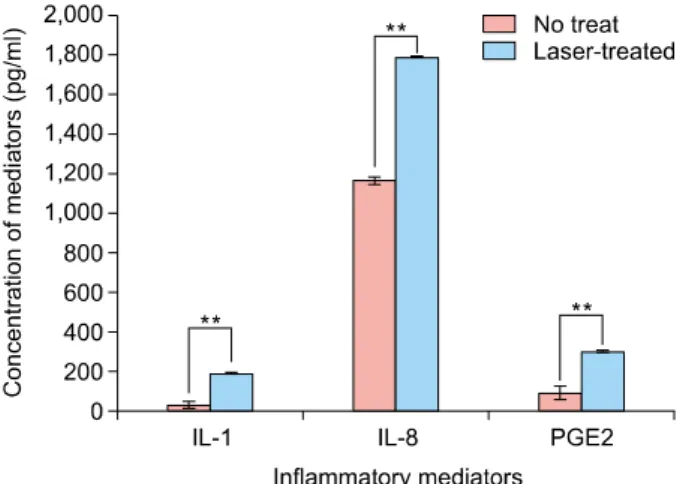
Fig. 1. Laser-treated keratinocyte-conditioned medium induced significant increases of inflammatory mediators. Inflammatory mediators including interleukin (IL)-1, IL-8, and prostaglandin E2 (PGE2) were significantly higher in laser-treated keratinocyte- conditioned medium (blue) than non-treated keratinocyte- conditioned medium (red). Data are mean±standard deviation.
**p<0.01; Student’s t-test.
Triton X-100+PMSF 100 mg/ml) for 2 hours in a deep freezer. Lysates were centrifuged at 12,000g for 15 mi- nutes to remove insoluble material. Protein content was determined by the Bradford method using Bio-Rad protein assay kit. A 50-μl aliquot of the samples was mixed with 100 ml sodium phosphate 100 mM (pH 7.0) at 37oC for 5 minutes, followed by the addition of 50 ml l-DOPA 100 mM. After incubation at 37oC, absorbance was measured at 475 nm using a microplate reader.
Western blot analysis
Melan-a cells (5×10⁵ cells/ml) were seeded on 60-mm culture dishes and treated with TA (10, 50, and or 100 mg/L). After 3 days of incubation, the cells were lysed with radioimmune precipitation assay buffer (Sigma- Aldrich Co.), and protein concentrations were determined using a Bio-Rad protein assay kit. Equal amounts of pro- teins were boiled for 3 minutes, chilled on ice, subjected to 8% to 10% sodiumdodecylsulfate-polyacrylamide gel electrophoresis (SDS-PAGE), and transferred to a nitro- cellulose membrane (Amersham International, Little Chalfont, UK). After the membranes were blocked using 5% skim milk in Tris-buffered saline containing 0.1%
Tween 20 for 1 hour, they were incubated with primary polyclonal anti-tyrosinase antibody, anti-tyrosinase-related protein (TRP)-1 antibody, and anti-TRP-2 antibody (Santa Cruz Biotechnology Inc., Santa Cruz, CA, USA). The membranes were subsequently incubated with the secon- dary antibody horseradish peroxidase-conjugated rabbit anti-goat immunoglobulin G antibody (Santa Cruz Biotechnology Inc.). Blotted antibodies were visualized by chemiluminescence (Amersham International). Anti-β-actin (Sigma-Aldrich Co.) was used as a loading control.
B16F10 cells (5×10⁵ cells/ml) were seeded on 60-mm culture dishes, serum starved for 24 hours, washed with phosphate-buffered saline, and treated with 10 mg/ml TA for 5 minutes, 30 minutes, 3 hours, 6 hours, 24 hours, 48 hours, or 72 hours. Cells were lysed with pro-prep protein lysis buffer (Intron, Seongnam, Korea) and centrifuged at 13,000g for 10 minutes. Protein concentrations were de- termined using a bicinchoninic acid protein assay kit (Sigma-Aldrich Co.). Protein (15 μg) was separated by SDS-PAGE and transferred to nitrocellulose membranes, which were subsequently blocked using 5% skim milk in Tris-buffered saline containing 0.1% Tween 20. The mem- branes were incubated with appropriate primary antibodies including phospho-extracellular kinase and extracellular kinase (ERK) (1:1,000; Cell Signaling Technology, Danvers, MA, USA) and microphthalmia-associated tran- scription factor (MITF) (Abcam, Cambridge, UK) for 4 hours and then with anti-rabbit or anti-mouse horseradish
peroxidase-conjugated antibody (1:2,000, Santa Cruz Biotechnology Inc.). Blotted antibodies were visualized by chemiluminescence (Pierce, Rockford, IL, USA). The membranes were re-probed with anti-β-actin antibody (1:10,000).
Image analysis was performed using ImageJ (http://rsb.
info.nih.gov/ij/) to quantify the relative band density.
Statistical analysis
Statistical analyses were performed by SPSS ver. 12.0 (SPSS Inc., Chicago, IL, USA). Each experiment was per- formed at least 3 times, and the results are expressed as mean±standard deviation. The statistical significance of the differences between groups was assessed by Student’s t-test. The level of significance was set at p<0.05.
RESULTS
Inflammatory mediators in conditioned media
As KCM itself can induce melanogenesis7, we measured the concentrations of inflammatory mediators in both KCM and LT-KCM. IL-1α, IL-8, and PGE2, which contrib- ute to the activation of melanocytes during inflammation9, were detected. The release of these mediators was sig- nificantly higher in LT-KCM (p<0.01), indicating laser treatment induced more inflammatory mediators (Fig. 1).
Effects of laser-treated keratinocyte-conditioned medium on melanin synthesis and tyrosinase activity
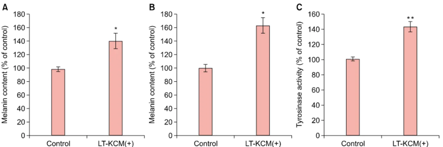
Both melan-a cells and human melanocytes grown in the
Fig. 2. Laser-treated keratinocyte-conditioned medium (LT-KCM) induced increased melanin content and tyrosinase activity. Melan-a cells and human melanocytes were cultured in the presence of LT-KCM. The melanin contents (% of control) in both melan-a cells (A) and human melanocytes (B) are shown. Tyrosinase activity (% of control) in cultured melan-a cells (C) is shown. Data are mean±standard deviation. *p<0.05, **p<0.01; Student’s t-test.
Fig. 3. Tranexamic acid (TA) decreased melanin content in melan-a cells. Melan-a cells were cultured in the presence of laser-treated keratinocyte-conditioned medium (LT-KCM). Arbu- tin and tranexamic acid inhibited melanin content in cultured melan-a cells. Data are mean±standard deviation. *p<0.05,
**p<0.01; Student’s t-test.
Fig. 4. Tranexamic acid (TA) decreased melanin content in human melanocytes. Human melanocytes were cultured in the presence of laser-treated keratinocyte-conditioned medium (LT-KCM). Data are mean±standard deviation.
presence of LT-KCM showed increased melanin content (adjusted for protein content) (Fig. 2A, B). Tyrosinase activ- ity was significantly higher (p<0.01) in melan-a cells cul- tured with LT-KCM than without (Fig. 2C).
Effects of tranexamic acid on melanin synthesis and tyrosinase activity
TA inhibited melanin synthesis in LT-KCM. TA-treated melan-a cells exhibited a dose-dependent reduction in melanin content adjusted by protein content (Fig. 3). In human melanocytes grown in LT-KCM, 10, 50, and 100
mg/L. TA seemed to inhibit melanin content; however, the inhibition was not significant (p=0.296, 0.238, and 0.584, respectively; Fig. 4). Although arbutin and 100 mg/L TA markedly reduced melanin content without adjustment (data not shown), the decreased protein content affected the adjusted melanin content.
Furthermore, TA significantly reduced tyrosinase activity in melan-a cells in a dose-dependent manner (Fig. 5). This finding corresponded with the reduction in melanin con- tent in melan-a cells (Fig. 3).
Fig. 5. Tranexamic acid (TA) reduced tyrosinase activity in melan-a cells. Melan-a cells were cultured in the presence of laser-treated keratinocyte-conditioned medium (LT-KCM).
Arbutin and tranexamic acid reduced tyrosinase activity (% of control) in cultured melan-a cells. Data are mean±standard deviation. *p<0.05, **p<0.01; Student’s t-test.
Fig. 7. Tranexamic acid (TA) stimulated the extracellular signal-regulated kinase (ERK) signaling pathway and decreased protein levels of microphthalmia-associated transcription factor (MITF). Protein levels of ERK, phosphorylated ERK (p-ERK), and MITF after TA treatment were evaluated by western blot analysis using B16F10 cells. Sustained activation of ERK was observed;
in particular, p-ERK was elevated until 24 hours after TA treatment. MITF protein level decreased in a time-dependent manner.
Fig. 6. Tranexamic acid (TA) decreased protein levels of melanogenesis-related enzymes. Protein levels of tyrosinase, tyrosinase-related protein (TRP)-1, and TRP-2 were higher in melan-a cells cultured with laser-treated keratinocyte-condi- tioned medium (LT-KCM) than keratinocyte conditioned medium (KCM) (CTL1 vs. CTL2) in western blot analysis. However, after TA treatment, their protein levels decreased in a dose-dependent manner. Arbutin (A100) was used as a positive control, and equal protein loading was confirmed by using β-actin. CTL1: control 1 (KCM without laser treatment), CTL2: control 2 (LT-KCM), A100: arbutin 100 mg/L, T10: 10 mg/L TA, T50: 50 mg/L TA, T100: 100 mg/L TA.
Effects of tranexamic acid on expressions of melano- genesis-related enzymes
In order to clarify the inhibitory effect of TA on melano- genesis, the protein levels of tyrosinase, TRP-1, and TRP-2 in melan-a cells were investigated by western blot analysis.
Tyrosinase, TRP-1, and TRP-2 protein levels were elevated in melan-a cells cultured with laser-treated KCM (Fig. 6).
However, after TA treatment, their protein levels de- creased in a dose-dependent manner, which was con- firmed by image analysis quantifying the densities of each band (data not shown).
Effects of tranexamic acid on expressions of signaling pathway molecules involved in melanogenesis
On the basis of the above results, we hypothesized TA af- fects the expressions of MITF, which plays a key role in melanogenesis. Therefore, we evaluated MITF protein ex- pression after TA treatment in B16F10 cells. We also stud- ied the ERK signaling pathway, which is known to be in- volved in MITF phosphorylation and degradation10. TA in- duced sustained activation of ERK; in particular, phos- phorylated ERK was elevated until 24 hours after TA treat- ment (Fig. 7). Meanwhile, MITF decreased in a time-de- pendent manner.
DISCUSSION
TA has been used in Japan for decades to treat melasma.
However, there was no report of its melanogenesis-inhibi- ting effect or action mechanism in English until 1998. TA is a well-known plasmin inhibitor. It blocks the conversion of plasminogen to plasmin by inhibiting plasminogen acti- vator through the formation of a reversible complex with plasminogen7,11. In 1998, Maeda and Naganuma6 re- ported ultraviolet-induced hyperpigmentation and pig- mentation induced by topical application of arachidonic acid in guinea pigs were reduced by topical application of TA in a dose-dependent manner. They suggest TA inhibits ultraviolet-induced plasmin activity in keratinocytes by preventing the binding of plasminogen to the keratinocyte,
which ultimately results in less free arachidonic acid and diminished ability to produce prostaglandins6. This was the first report demonstrating that TA prevents ultra- violet-induced pigmentation in vivo. Maeda and Tomita7 published another interesting study in 20077. They showed that single-chain urokinase-type plasminogen activator (sc-uPA) generated by keratinocytes increased the activity of melanocytes in vitro and that TA might reduce hyper- pigmentation of melasma patients by blocking the effect of sc-uPA. These findings collectively suggest TA acts by blocking sc-uPA and plasmin, which ultimately prevents arachidonic acid and prostaglandin synthesis.
In the present study, TA reduced melanin synthesis in both cultured murine and human melanocytes in LT-KCM, a highly inflammatory condition. As shown in Fig. 1, this medium contained high levels of inflammatory mediators including IL-1α, IL-8, and PGE2. In the previous study mentioned above7, KCM itself stimulated melanocyte activity. Melanocytes stimulated by IL-1 and tumor ne- crosis factor-α contribute to cutaneous inflammation via IL-89. Moreover, prostaglandins, which are well-known in- flammatory mediators, act as melanogenic stimulators9,12. In particular, PGE2 is reported to stimulate tyrosinase acti- vation via prostaglandin E receptor 4 signaling, resulting in cyclic adenosine monophosphate production13. These in- flammatory mediators were significantly higher in the frac- tional CO2 LT-KCM in the present study than KCM with- out laser treatment. This suggests laser treatment induces more intense inflammatory conditions that might lead to melanogenesis. As expected, melanin content and ty- rosinase activity were higher in LT-KCM than the control media (Fig. 2). Several recent reports demonstrate the use- fulness of LT-KCM for evaluating changes of melanogenic cytokine profiles14,15. Although LT-KCM has an intrinsic limitation in representing PIH, it adequately reproduces the intense inflammatory conditions leading to melano- genesis. Although TA reduced melanin content and ty- rosinase activity in melan-a cells in LT-KCM, the effect of TA on cultured human melanocytes was not definitive in the present study. The amount of melanin was markedly reduced, especially with arbutin and 100 mg/L TA treatment. However, the decrease of melanin content as a percentage of the control was not statistically significant; a similar result was found even in the arbutin group (positive control). This is likely because the human mela- nocytes were much more vulnerable to laser treatment and/or test compounds (e.g., arbutin and TA) than melan-a cells. Damaged human melanocytes might consequently affect the results of protein assays and adjusted melanin contents. Tyrosinase is the rate-limiting enzyme in mela- nin biosynthesis. Therefore, melanin content in cells is
correlated with tyrosinase protein level and catalytic activ- ity16. The present results suggest TA may inhibit melanin synthesis, decreasing melanin content and tyrosinase ac- tivity (Fig. 3∼5). Concordantly, tyrosinase protein levels in melan-a cells decreased with TA in a dose-dependent manner (Fig. 6). These results suggest TA downregulates cellular tyrosinase activity in melanocytes or possibly the transcription of tyrosinase mRNA; this suggestion is based on the capability of TA to decrease TRP-1 and TRP-2 lev- els in addition to tyrosinase levels, because these melano- genic enzymes are regulated by the same key transcription factor, MITF. The effect of TA on MITF protein expression was elucidated by evaluating MITF protein level after TA treatment (Fig. 7). As previous studies show ERK activation leads to MITF phosphorylation and subsequent degrada- tion10,17,18, we also evaluated protein levels of un- phosphorylated and phosphorylated ERK. TA stimulated the ERK signaling pathway and downregulated MITF pro- tein level (Fig. 7). Interestingly, ERK phosphorylation was strongly induced 24 hours after TA treatment, whereas MITF protein was detected only 24 hours after TA treatment. These findings are concordant with previous observations that the activation of the ERK signaling path- way induces MITF degradation, resulting in reduced mela- nogenesis10,17,18. Furthermore, our results support our hy- pothesis that TA itself attenuates melanogenesis by regulat- ing tyrosinase transcription in addition to its well-known anti-inflammatory effect.
In summary, TA reduces inflammation-induced melano- genesis by decreasing tyrosinase protein expression, which involves ERK pathway activation and the sub- sequent decrease of MITF protein expression. Our findings suggest TA has potential for the treatment of PIH after la- ser treatment, a common concern in people with darker skin complexions.
ACKNOWLEDGMENT
This study was supported by research grants from the Asan Institute of Life Science (2012-415, 2013-415) and Amore Pacific Skin Science 2011.
REFERENCES
1. Gupta AK, Gover MD, Nouri K, Taylor S. The treatment of melasma: a review of clinical trials. J Am Acad Dermatol 2006;55:1048-1065.
2. Costin GE, Hearing VJ. Human skin pigmentation:
melanocytes modulate skin color in response to stress.
FASEB J 2007;21:976-994.
3. Ando H, Kondoh H, Ichihashi M, Hearing VJ. Approaches to identify inhibitors of melanin biosynthesis via the quality
control of tyrosinase. J Invest Dermatol 2007;127:751-761.
4. Lee JH, Park JG, Lim SH, Kim JY, Ahn KY, Kim MY, et al.
Localized intradermal microinjection of tranexamic acid for treatment of melasma in Asian patients: a preliminary clinical trial. Dermatol Surg 2006;32:626-631.
5. Li D, Shi Y, Li M, Liu J, Feng X. Tranexamic acid can treat ultraviolet radiation-induced pigmentation in guinea pigs.
Eur J Dermatol 2010;20:289-292.
6. Maeda K, Naganuma M. Topical trans-4-aminomethylcyclo- hexanecarboxylic acid prevents ultraviolet radiation-induced pigmentation. J Photochem Photobiol B 1998;47:136-141.
7. Maeda K, Tomita Y. Mechanism of the inhibitory effect of tranexamic acid on melanogenesis in cultured human melanocytes in the presence of keratinocyte-conditioned medium. J Health Sci 2007;53:389-396.
8. Horrow JC, Van Riper DF, Strong MD, Brodsky I, Parmet JL.
Hemostatic effects of tranexamic acid and desmopressin during cardiac surgery. Circulation 1991;84:2063-2070.
9. Park HY, Pongpudpunth M, Lee J, Yaar M. Biology of melanocyte. In: Wolff K, Goldsmith LA, I.Katz S, Gilchrest BA, Paller AS, Lefell DJ, editors. Fitzpatrick's dermatology in general medicine. 7th ed. Columbus: McGraw-Hill, 2008:591-607.
10. Kim DS, Park SH, Kwon SB, Park ES, Huh CH, Youn SW, et al. Sphingosylphosphorylcholine-induced ERK activation inhibits melanin synthesis in human melanocytes. Pigment
Cell Res 2006;19:146-153.
11. Dunn CJ, Goa KL. Tranexamic acid: a review of its use in surgery and other indications. Drugs 1999;57:1005-1032.
12. Morelli JG, Norris DA. Influence of inflammatory mediators and cytokines on human melanocyte function. J Invest Dermatol 1993;100(2 Suppl):191S-195S.
13. Starner RJ, McClelland L, Abdel-Malek Z, Fricke A, Scott G.
PGE(2) is a UVR-inducible autocrine factor for human melanocytes that stimulates tyrosinase activation. Exp Dermatol 2010;19:682-684.
14. Yun WJ, Bang SH, Min KH, Kim SW, Lee MW, Chang SE.
Epidermal growth factor and epidermal growth factor signaling attenuate laser-induced melanogenesis. Dermatol Surg 2013;39:1903-1911.
15. Burd A, Zhu N, Poon VK. A study of Q-switched Nd:YAG laser irradiation and paracrine function in human skin cells.
Photodermatol Photoimmunol Photomed 2005;21:131-137.
16. Chang TS. An updated review of tyrosinase inhibitors. Int J Mol Sci 2009;10:2440-2475.
17. Hemesath TJ, Price ER, Takemoto C, Badalian T, Fisher DE.
MAP kinase links the transcription factor Microphthalmia to c-Kit signalling in melanocytes. Nature 1998;391:298-301.
18. Wu M, Hemesath TJ, Takemoto CM, Horstmann MA, Wells AG, Price ER, et al. c-Kit triggers dual phosphorylations, which couple activation and degradation of the essential melanocyte factor Mi. Genes Dev 2000;14:301-312.