저작자표시-비영리-변경금지 2.0 대한민국 이용자는 아래의 조건을 따르는 경우에 한하여 자유롭게 l 이 저작물을 복제, 배포, 전송, 전시, 공연 및 방송할 수 있습니다. 다음과 같은 조건을 따라야 합니다: l 귀하는, 이 저작물의 재이용이나 배포의 경우, 이 저작물에 적용된 이용허락조건 을 명확하게 나타내어야 합니다. l 저작권자로부터 별도의 허가를 받으면 이러한 조건들은 적용되지 않습니다. 저작권법에 따른 이용자의 권리는 위의 내용에 의하여 영향을 받지 않습니다. 이것은 이용허락규약(Legal Code)을 이해하기 쉽게 요약한 것입니다. Disclaimer 저작자표시. 귀하는 원저작자를 표시하여야 합니다. 비영리. 귀하는 이 저작물을 영리 목적으로 이용할 수 없습니다. 변경금지. 귀하는 이 저작물을 개작, 변형 또는 가공할 수 없습니다.
i
Master’s Thesis
Verbenone, a major compound of rosemary hydrosol inhibits
melanogenesis and oxidative stress in B16F10 mouse
melanoma cell
Deok Hyeon Jeon
Depart of Biotechnology
Graduate School
Jeju National University
i
CONTENTS
CONTENTS · · · i ABSTRACT · · · iii LIST OF TABLES · · · iv LIST OF FIGURES · · · v 1. INTRODUCTION · · · 12. MATERIALS AND METHODS · · · 5
2.1. Reagents 2.2. Cell culture
2.3. Gas Chromatography-Mass Spectrometry (GC-MS) 2.4. Cell viability
2.5. Melanin content
2.6. Intracellular tyrosinase activity 2.7. Mushroom tyrosinase activity
2.8. Cellular fraction and immunoblot analysis 2.9. RT-PCR analysis
ii
2.11. Statistical analysis
3. RESULTS · · · 12
3.1. Effect of the components of Rosmarinus officinalis L. hydrosol on melanin content
3.2. Effect of verbenone on cell viability in B16F10 cells 3.3. Effect of verbenone on melanin biosynthesis
3.4. Effect of verbenone on cellular or cell-free system of the tyrosinase activity 3.5. Effect of verbenone on the levels melanogenesis protein in B16F10 cells 3.6. Effect of verbenone on the levels of melanogenic mRNA in B16F10 cells 3.7. Effect of verbenone on phosphorylation of p38, ERK and Akt in B16F10 cells 3.8. Effect of verbenone on intracellular ROS Generation
3.9. Effect of verbenone on the level of antioxidant enzyme mRNA in B16F10 cells
4. DISCUSSION · · · 31
REFERENCES · · · 33
ABSTRACT IN KOREAN · · · 39
iii
ABSTRACT
In the present study, we investigated the effect of verbenone, an active compound present in Rosmarinus officinalis L. hydrosol extract, on melanin synthesis and the molecular mechanism of melanogenesis in B16F10 mouse melanoma cells. The cellular melanin content and tyrosinase activity of the 2.5 mM verbenone-treated cells were significantly reduced below the levels of the kojic acid- treated cells, consistent with suppression of melanin biosynthesis by verbenone via inhibition of tyrosinase activity. Also verbenone inhibits the expression of melanogenesis-related proteins including MITF (Microphthalmia-associated transcription factor), tyrosinase and tyrosinase-related protein (TRP)s in α-MSH (a-melanocyte-stimulating hormone)-stimulated B16F10 cells. Moreover, the phospho-Akt was significantly enhanced but phospho-p38 MAPK was reduced and no effect phospho-ERK. Furthermore, the ROS generation submitted to an oxidative stress by H2O2 treatment was
completely inhibited by pre-treatment of verbenone, which is consistent with the increase in mRNA level of super oxide dismutase (SOD), catalase (CAT) and glutathione peroxidase (GPx) in B16F10 cells. These data suggest that verbenone inhibits melanogenesis signaling by both inhibiting the expression of melanogenesis protein and activating the Akt signal pathways-mediated suppression of MITF and its downstream signal pathway, including tyrosinase and TRP-1 and TRP-2. Therefore, verbenone would be a useful therapeutic agent for treating hyperpigmentation and can be applicable for cosmetics as skin-whitening agents.
iv
LIST OF TABLES
v
LIST OF FIGURES
Figure 1. Effect of the components of Rosmarinus officinalis L. on cellular melanin synthesis in B16F10 cells. · · · 14
Figure 2. Effect of verbenone on cell viability in B16F10 cells · · · 16
Figure 3. Effect of verbenone on cellular melanin synthesis in B16F10 cells · · · 18
Figure 4. Effect of verbenone on intracellular or cell-free system of the tyrosinase activity· 20
Figure 5. Effects of verbenone on melanogenic protein level in B16F10 cells · · · 22
Figure 6. Effects of verbenone on melanogenic mRNA expressions level in B16F10 cells · 24
Figure 7. Verbenone induces activation of Akt or inactivation of p38, but not of ERK. · · · 26
Figure 8. Effects of verbenone on the ROS level in B16F10 cells · · · 28
1
1. Introduction
Melanins are broad class of functional macromolecules found throughout nature (1). Melanin is an antioxidant, a free radical scavenger and has an affinity for drugs, and other chemical substances. Because of these properties, melanin efficiently filters toxic substances and protects tissues from oxidative and chemical stress. However, with chronic exposure to toxic substances the properties of melanin change so that under severe oxidative stress and binding of excessive amount of toxins, melanin itself may induce damage to cells (2). In addition, the melanin bound drug forms a depot that releases the drug over a long period and increases the level of noxious substances stored on melanin, what may cause degeneration in the melanin-containing cells (especially in the eye, ear, skin, and brain) and surrounding tissues (3).
In mammals, melanogenesis is directly regulated by three enzymes, tyrosinase, tyrosinase-related protein-1 (TRP-1) and TRP-2 (4,5). Tyrosinase is regarded as the rate-limiting enzyme of melanogenesis. It plays a pivotal role in the modulation of melanin production by catalyzing the hydroxylation of tyrosine into 3,4-dihydroxyphenylalanine (DOPA) and the further oxidation of DOPA into DOPAquinone by tyrosinase (6). TRP-2, which functions as a DOPAchrome tautomerase, catalyzes the rearrangement of DOPAchrome to 5,6-dihydroxy-indole-2-carboxylic acid (DHICA) (7), whereas TRP-1 oxidizes DHICA to a carboxylated indole-quinone (8). Microphthalmia-associated transcription factor (MITF) is known to be the master regulator of melanocyte differentiation, pigmentation, proliferation and survival (9). It is a major transcriptional regulator of the tyrosinase family genes TYR, TYRP-1 and TYRP-2 responsible for pigmentation.
2
Recently, the inhibition of the extracellular signal-regulated kinase (ERK) signaling was reported to induce hyperpigmentation by increasing the tyrosinase activity, suggesting that the activation of ERK signaling down-regulates melanogenesis by inhibiting the tyrosinase activity (10-12). In addition, activation of the Akt signaling plays a key role in inhibiting melanogenesis(13-14). Recent investigations suggest that p38 MAPK positively contributes to pigmentation(15-17). The activation of p38 MAPK pathway induces MITF expression (18-20) and promotes the transcription of tyrosinase (21). Some Chinese medicines such as San-bai-tang and Qian-wang-hong-bai-san, the Chinese herbal formulae, have been reported to have anti-melanogenic activity via inhibiting the p38 MAPK signaling pathway (22,23).
In an effort to inhibit the activity of tyrosinase, many different types of tyrosinase inhibitors have been developed via either synthesis or isolation from natural sources (24,25). These compounds are applicable for cosmetics as skin-whitening agents and also as drugs for use in the treatment of pigmentation disturbances (26,27). Despite the enormous amount of research conducted thus far into the development of new whitening agents, the use of existing agents is rather limited, owing to high toxicity, low stability, poor skin penetration, and insufficient activity (28). Kojic acid has been demonstrated to produce hepatocytotoxicity, skin cancer, and contact dermatitis in a few clinical trials and therefore has been banned as a cosmetic ingredient in many countries (29). Due to various safety concerns and the lower efficacy of commercially available depigmenting agents, the isolation and identification of new compounds from natural sources which prevent pigment disturbances have attracted much interest.
Reactive oxygen species (ROS) and free radicals are associated with several diseases, such as inflammation (30) and age-related diseases (31). ROS-induced damage on the skin and UV
3
stress plays an important role in photoaging (32). It has been found that antioxidants can interfere with oxidation processes by acting as free radical scavengers or ROS scavengers or by chelating oxidation-catalytic metals. Therefore, numerous natural antioxidants or antioxidant supplements have been used to reduce oxidative damage in the human body. However, some synthetic chemical antioxidants, such as tert-butyl hydroxyanisole (BHA) and tert-butyl hydroxytoluene (BHT), have been reported to show carcinogenic effects in humans (33). Thus, numerous studies on plant-derived antioxidants have been conducted during the past decade. Moreover, it was found that melanogenesis produces hydrogen peroxide (H2O2) and ROS, which place melanocytes under high-grade oxidative stress. It is
well known that some ROS scavengers and inhibitors of ROS generation may down-regulate UV-induced melanin production (34). Therefore, inhibitors of melanogenesis, antioxidants, and ROS scavengers have been increasingly applied to skin care products for the prevention or treatment of undesirable skin hyperpigmentation.
The Rosmarinus officinalis L. (rosemary) genera belong to the family Lamiaceae. Among the plants known for medicinal value, rosemary plants are highly regarded for their therapeutic potentials. Rosmarinus officinalis L. extracts offer promise as biologically active constituents, in that they confer antibacterial(35-38), antifungal(37-39), anticancer(40-42), reduce blood cholesterol concentration(43) and antioxidant properties(35,36,40,43-45). Hydrosols, also known as floral water, distillate water or aromatic water, are the co-products or the byproducts of hydro- and steam distillation of plant material. Hydrosols are quite complex mixtures containing traces of the essential oil and, of course, several water-soluble components as well. They have practically been used as a beverage for a long time in many areas (46-48). Main advantages of hydrosols are that they are easy and inexpensive to
4
produce and they do not have any health hazard for the human as is the case for essential oils. However, there are few studies on effects of rosemary hydrosols although there are a few investigations on the antifungal activity of some spice hydrosols (49).
In this study we show the compositional analysis of rosemary hydrosol and the biological activities of verbenone, a major component of rosemary hydrosol, on melanogenesis in B16F10 mouse melanoma cells. Although Song et al.(50), recently have reported that verbenone possesses antioxidant activities(51), to date there has been no attempt to test the action of verbenone in melanocytes. To our knowledge, this study is the first to show the inhibitory activity of verbenone on the melanogenesis signaling pathways, including the expression of MITF, tyrosinase, TRP1 and TRP2, and the phosphorylation of p38, ERK and Akt.
5
2. Materials and methods
2. 1. Reagents
Dulbecco’s modified Eagle’s medium (DMEM), trypsin/EDTA, fetal bovine serum(FBS), Dimethyl sulfoxide (DMSO) and MTT were from amresco (Vleveland, OH, USA), (1S)-(-)-verbenone, L-DOPA-(ring-d3), α-Melanocyte stimulating hormone(α-MSH) and β-actin antibodies were purchased from Sigma Chemical Co. (St Louis, MO, USA). TRP-1, TRP-2, Tyrosinase and MITF Antibodies were purchased from (Santa Cruz Biotechnology, Inc., Santa Cruz, CA, USA). All other chemicals and reagents were purchased from Cell Signaling Technology (Beverly, MA, USA) and Invitrogen Gibco (Grad Island, NY, USA).
2. 2. Cell culture
Mouse melanoma B16F10 cell was kindly provided Nam-Ho Lee by professor in the Department of Chemistry, Jeju University, Korea. B16F10 cell was cultured in Dulbecco’s Modified Eagle’s Medium (DMEM) containing 10% (v/v) heat inactivated Fetal Bovine Serum (FBS), 100 units/ml penicillin, and 100 μg/ml streptomycin. Cell was maintained in a humidified incubator at 37°C in a 5% CO2 atmosphere.
2. 3. Gas Chromatography-Mass Spectrometry (GC-MS)
The volatile chemical compounds in the rosmarinus officinalis L. hydrosol were analyzed using a Shimadzu GC-MS (Model QP-2010, Shimadzu Co., Kyoto, Japan). An Rtx-5MS capillary column with 30 m length and 0.25 mm inner diameter with a 0.25 μm thick film was used. Ionization of the
6
test hydrosol (1 μl) was performed in the EI mode (70 eV)
2. 4. Cell viability
The effect of the verbenone on the viability of B16F10 cell lines was determined using an MTT-based assay(52). Briefly, exponential-phase cells were collected and transferred to a 96-well plate (2×10⁴ cell/ml). The cells were then incubated for 24 h, 48 h and 72 h with various concentrations of the verbenone. Afterwards, 0.1 mg of MTT (Amresco, Cleveland, OH, USA) was added to each well, and incubated at 37°C for 4 h. The medium was carefully removed and DMSO (150 μM) was added to each well to dissolve the formazan crystals. The plates were area immediately at 570 nm on a Sunrise microplate reader (Sunrise, Tecan, Salzburg, Austria). The percentage of cell viability was calculated based on the following formula: mean value of (control group – treated group/control group) × 100%. All results were assessed in triplicate for each concentration.
2. 5. Melanin content
Melanin content determination The melanin content of cells was determined using the method described(53) with some modifications. Briefly, 2x10⁴ cells were seeded onto 100-mm dish and incubated overnight. After incubation, the medium was replaced with sample containing medium and incubated for 24 h, 48 h and 72 h. The medium was then removed, and the cells were washed twice with phosphate- buffered saline (PBS) and collected by trypsination using 0.25% trpsin/0.02% EDTA. The collected cells were pelleted and dissolved in 1% Triton X (Sigma) in PBS by sonication. The synthesized melanin was then precipitated
7
in 10% trichloroacetic acid (TCA) (Wako Pure Chemical Industries, Ltd., Osaka, Japan) in milliQ water and dissolved by incubation in 2 N NaOH for 1 h at 80°C. The absorbance of the solution was measured at 410 nm. The results were expressed as percentage of control.
2. 6. Intracellular tyrosinase activity
The tyrosinase activity was estimated by measuring the rate of dopachrome formation of L-DOPA (54). The cells grown in six-well dishes were treated with 20 nM α-MSH or ⁄ and 0-2.5 mM verbenone for 24 h, 48 h and 72 h in DMEM. The cells were then washed in ice-cold PBS and lysed in 500 μl of phosphate buffer (0.1 M, pH 6.8) containing 1% (w ⁄ v) Triton X-100. The cellular extract was clarified by centrifugation at 13 000 g for 25 min. The tyrosinase substrate, L-DOPA (2 mg⁄ ml), was prepared in the same phosphate lysis buffer. Two hundred microliters of each extract was placed in a 96-well plate and the enzymatic assay was commenced by adding 2 µl of a L-DOPA solution at 37°C. Control wells contained in 198 µl of lysis buffer and 2 μl of a L-DOPA solution. After incubation, dopachrome formation was assayed by measuring absorbance at 405 nm was read every 10 min for at least 1 h at 37°C using an ELISA reader. The value of each measurement was expressed as percentage changes form the control.
2. 7. Mushroom tyrosinase activity
A cell-free assay system was used to investigate the direct effects of 0-2.5 mM verbenone on tyrosinase activity(55). Briefly, 100 ml of phosphate buffer containing compound was mixed with 10 unit mushroom tyrosinase, and 50 ml of 0.03% tyrosine in deionized water was then
8
added. After incubation at 37°C for 10 min, absorbance was measured at 475 nm using an ELISA reader..
2. 8. Cellular fraction and immunoblot analysis
After treatment with various concentration of Verbenone and/or α-MSH, the B16F10 cells were collected and washed twice with cold PBS. The cells were then lysed in lysis buffer (50 mM Tris-HCl, pH 7.5, 150 mM NaCl, 1% Nonidet P-40, 2 mM EDTA, 1 mM EGTA, 1 mM NaVO3, 10 mM NaF, 1 mM DTT, 1 mM PMSF, 25 g/ml aprotinin, and 25 g/ml leupeptin)
and kept in ice for 30 min. The lysates were centrifuged for 30 min at 13,000 rpm and 4°C, and the supernatants were stored at -70°C until use. Cytosolic and mitochondrial extracts were prepared using the fraction lysis buffer (75 mM NaCl, 8 mM Na2HPO4, 1 mM Na2H2PO4, 250 mM sucrose, 1 mM EDTA, 350 g/ml digitonin). After lysed the cell, kept in ice for 10 min then centrifuged for 15 min at 15,000 rpm and 4°C. The supernatants were cytosolic fraction. After pellet washed by lysis buffer, pellets were lysed in lysis buffer same as make whole lysates. The protein concentration was measured using a bicinchoninic acid (BCA) protein assay kit (Pierce, Rockford, IL, USA). Aliquots of the lysates (30–60 g of protein) were separated via 10–15% SDS-PAGE and transferred onto a polyvinylidine difluoride (PVDF) membrane (Millipore, Billerica, MA, USA) using a glycine transfer buffer (192 mM glycine, 25 mM Tris-HCl, pH 8.8, and 20% methanol, v/v). After blocking with 5% nonfat dried milk, the membranes were incubated for 4 h with primary antibodies, followed by an additional 30 min of incubation with secondary antibodies in milk containing Tris-buffered saline (TBS) and 0.1% Tween 20. Mouse anti-TRP-1, TRP-2, Tyrosinase, MITF,
9
ERK, p-ERK, Akt, p-Akt, p38, p-p38 and actin antibodies were used at a 1:1,000 dilution as the primary antibodies, and horseradish peroxidase-conjugated anti-mouse or rabbit IgG Cell Signaling Technology (Beverly, MA, USA). at a 1:5,000 dilution was utilized as the secondary antibody. The membrane was then exposed to X-ray film. Protein bands were detected using the WEST-ZOL® plus western blot detection system (Intron, Gyeonggi-do, Korea).
2. 9. RT-PCR analysis
Total RNA was isolated from cells using the TRIzol reagent (Invitrogen) according to the manufacturer’s instructions. Reverse transcription was carried out using the reverse transcription system (Promega). PCR primers for amplification were as follows: of TRP-1, forward 5'-AAG ACG CTG CAC TGC TGG-3', reverse 5'-GCT GCA GGA GCC TTC TTT CTC-3'; of TRP-2, forward AAG AAA GGA ACC AUG UUG UAC AUC C-3', reverse 5'-GGA TGA CCG TGA GCA ATG GCC-3'; of Tyrosinase, forward 5′-TGG TGC TTC ATG GGC AAA ATC-3′, reverse 5′-CGG TTG TGA CCA ATG GGT GCC -3′; of MITF, forward 5′-GTA TGA ACA CGC ACT CTC GAG C-3′, reverse 5′-CTT CTG CGC TCA TAC TGC TC-3′; of SOD (Cu/Zn Mn), forward 5’- TTA ACT GAA GGC CAG CAT GGG-3’, reverse 5’-ATC ACT CCA CAG GCC AAG CGG-3’; of GPx1(Glutathione peroxidase 1), forward 5’-CTC GGT TTC CCG TGC AAT CAG-3’, reverse 5’- GTG CAG CCA GTA ATC ACC AAG-3’; of CAT(Mus musculus catalase), forward 5’-TCT GCA GAT ACC TGT GAA CTG-3’, reverse 5’-TAG TCA GGG TGG ACG TCA GTG-3’.; of GAPDH, forward 5′-GAA GGT GAA GGT CGG AGT C-3′, reverse 5′-GAA GAT GGT GAT GGG ATT TC-3′. PCR was performed using Taq polymerase (iNtRON Biotechnology Inc.). PCR was initiated by
10
incubating the samples at 95°C for 5 min, followed by 35 cycles of 1 min denaturation at 95°C, 1 min annealing at 57°C and 1 min elongation at 72°C. Samples were analyzed by electrophoresis on 1% agarose gels containing 0.002% nucleic acid staining solution (RedSafe™; Biotechnology Inc., Seoul, South Korea).
2. 10. Determination of ROS generation and Fluorescence microscope
Cellular ROS were quantified using the dichlorofluorescein (DCFH) assay (56), in which the esterified form of DCFH-DA diffuses through the cell membrane and is enzymatically deacetylated by intracellular esterases. The resulting compound, DCFH, is reactive with ROS the give an oxidized fluorescent compound, DCF(57). B16F10 cultures were per-treated with verbenone for 30 min, and then the cultures were washed twice and incubated with 200 µM H2O2 for 4 h. DCFH-diacetate was added to the culture plates at a final concentration of 25 µM, and DCF- fluorescence was detected over a period of 30 min at 37°C. Cells were then harvested, washed twice with phosphate-buffered saline (PBS), re-suspended in 500 μl of PBS and analyzed by FACS. The fluorescence microscope experiment consisted of the loading of cells with DCF-DA before exposure as described in the above experiment(56). Cells were examined using a fluorescence microscope (Olympus IX73, Japan) with 485 nm excitation and 530 nm emission wavelength.
2. 11. Statistical analysis
All results are expressed as means ± standard deviation (SD). One-way analysis of variance (ANOVA) using SPSS v 20.0 software package was applied. A difference at p < 0.05
11
12
3. Results
3. 1. Effect of the components of Rosmarinus officinalis L. hydrosol on melanin content
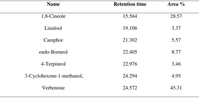
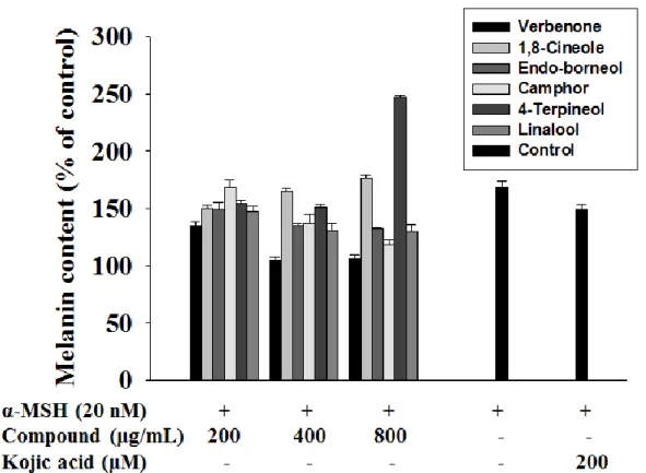
The chemical composition of Rosmarinus officinalis L. hydrosol was analyzed by GC-MS with the results shown in Table 1. The major components in the hydrosol are verbenone (45.31%) which consist of 1,8-Cineole (28.57%), endo-Borneol (8.77%), Camphor (5.57%), 3-Cyclohexene-1-methanol (4.95%), 4-Terpineol (3.46%), Linallool (3.37%). To identify an active compound(s) in Rosmarinus officinalis L. hydrosol that inhibits melanogenesis activation, compound was melanin content assay (Fig. 1). The verbenone of Rosmarinus officinalis L. hydrosol strongly reduced melanin content in B16F10 cell.
13
Table 1 Composition of rosemary hydrosol from Jeju Island.
Name Retention time Area %
1,8-Cineole 15.564 28.57 Linalool 19.106 3.37 Camphor 21.302 5.57 endo-Borneol 22.405 8.77 4-Terpineol 22.976 3.46 3-Cyclohexene-1-methanol, 24.294 4.95 Verbenone 24.572 45.31
14
Figure 1. Effect of the components of Rosmarinus officinalis L. on cellular melanin synthesis in B16F10 cells. The cells were treated to α-MSH (20nM) in the presence of
Rosmarinus officinalis L. hydrosol compounds (0-800 μg/mL) or kojic acid (200mM) for 72 h. The cellular melanin content were determined and are reported as percentages. Each percentage in the treated cells is reported relative to that in the control cells. kojic acid was used as a positive control for tyrosinase inhibition.
15
3.2. Effect of verbenone on cell viability in B16F10 cells
The effect of the verbenone on the viability of B16F10 cell lines was determined using an MTT-based assay(52). Briefly, exponential-phase cells were collected and transferred to a 96-well plates (2×10⁴ cell/ml). The cells were then incubated for 24 h, 48 h and 72 h with various concentrations (0-5mM) of the verbenone. Verbenone exhibited no cytotoxic effect on B16F10 cells at concentrations ranging from 0 to 2.5 mM (Fig. 2).
16
Figure 2. Effect of verbenone on cell viability in B16F10 cells. Cells were seeded, incubated for 24 h and then incubated with the indicated concentrations of verbenone for additional 72 h. Viability was determined on the basis of MTT reduction. B16F10 mouse melanoma cells were treated with increasing doses of verbenone for varying lengths of time (24-72 h). compared to the control. Values are the mean ± SD of three independent experiments. *
17
3.3. Effect of verbenone on melanin biosynthesis
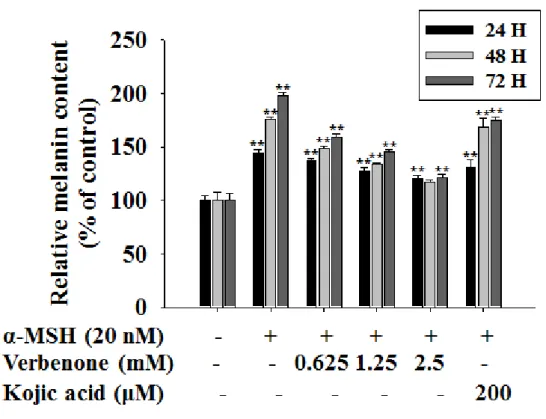
Melanin content determination The melanin content of cells was determined using the method described(53) with some modifications. Briefly, 2×10⁴ cells were seeded onto 100-mm dish and incubated overnight. After incubation, the medium was replaced with sample containing medium and incubated for 24 h, 48 h and 72 h. To determine the effect of verbenone on melanin synthesis, the cells were then exposed to 20 nM α-MSH in the presence of 0-2.5 mM verbenone or 200 µM kojic acid, which are representative tyrosinase inhibitors. As shown in Fig. 3, the cellular melanin content of the 2.5 mM verbenone-treated cells were significantly reduced below the levels of the kojic acid- verbenone-treated cells.
18
Figure 3. Effect of verbenone on cellular melanin synthesis in B16F10 cells. The cells
were treated to α-MSH (20nM) in the presence of verbenone (0-2.5 mM) or kojic acid (200 mM) for 72 h. The cellular melanin content were determined and are reported as percentages. Each percentage in the treated cells is reported relative to that in the control cells. kojic acid was used as a positive control for tyrosinase inhibition. Values are the mean ± SD of three independent experiments. *P<0.05, **P<0.01, compared to the control.
19
3.4. Effect of verbenone on cellular or cell-free system of the tyrosinase activity
To determine the effect of verbenone on intracellular tyrosinase activity, the cells were then exposed to 20 nM α-MSH in the presence of 0-2.5 mM verbenone or 200 μM kojic acid, which are representative tyrosinase inhibitors. As shown in Fig. 4a, the cellular tyrosinase activity of the 2.5 mM verbenone-treated cells were significantly reduced below the levels of the kojic acid- treated cells. Consistent with verbenone suppression of melanin biosynthesis by B16F10 cells via inhibition of tyrosinase activity. To examine whether verbenone could affect tyrosinase activity in cell-free system, we compared verbenone with kojic acid and observed that verbenone was absolutely ineffective in suppressing mushroom tyrosinase activity (Fig. 4b). Results of our study apparently indicated that the inhibitory effect of verbenone on melanogenesis was not due to its direct inhibition of tyrosinase.
20
Figure 4. Effect of verbenone on cellular or cell-free system of the tyrosinase activity.
The cells were treated to α-MSH (20 nM) in the presence of verbenone (0-2.5 mM) or kojic acid (200 mM) for 72 h. The cellular tyrosinase activity (a) were determined and are reported as percentages. Each percentage in the treated cells is reported relative to that in the control cells. The mushroom tyrosinase activity (b) was determined and is as percentages. kojic acid was used as a positive control for tyrosinase inhibition. Values are the mean ± SD of three independent experiments. *P<0.05, **P<0.01, compared to the control.
21
3.5. Effect of verbenone on the levels melanogensis protein in B16F10 cells
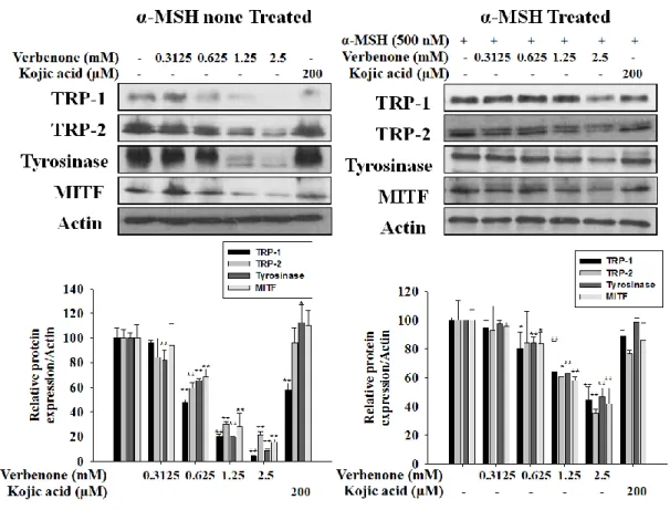
To explore the mechanism responsible for the decreased pigmentation, the expression levels of TRP-1, TRP-2, tyrosinase and MITF were examined by Western blot(Fig. 5) . When cells are not treated with α-MSH, the level of tyrosinase are dramatic decrease a verbenone 1.25 mM. treatment, whereas the level of TRP-1,-2, and MITF are gradually decrease in a concentration dependent manner. When cells are treated with α-MSH, the level of TRP-1,2 Tyrosinase. MITF all decreased gradually on a dose dependent manner.
22
Figure 5. Effects of verbenone on melanogenic protein level in B16F10 cells. B16F10
cells were treated presence of verbenone (2.5 mM) or kojic acid (200 μM) and/or α-MSH (500 nM) in the for 72 h. The expression levels of the tyrosinase, TRP-1, TRP-2 and MITF proteins were determined by Western blot, respectively. The data represent at least three independent experiments. Relative density ratios of each protein over actin, respectively, are shown. Values are the mean ± SD of three independent experiments. *P<0.05, **P<0.01, compared to
23
3.6. Effect of verbenone on the levels of melanogenic mRNA in B16F10 cells
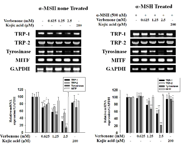
To explore the mechanism responsible for the decreased pigmentation, the mRNA expression levels of TRP-1, TRP-2, tyrosinase and MITF after verbenone treatment. As shown in Fig. 6, TRP-1, TRP-2, tyrosinase mRNA expressions were clearly reduced after verbenone treatment but the level of MITF mRNA was not changed. These results suggest that verbenone reduced melanin synthesis through protein degradation of MITF in B16F10 mouse melanoma cells.
24
Figure 6. Effects of verbenone on melanogenic mRNA expressions level in B16F10 cells.
B16F10 cells were treated presence of verbenone (2.5 mM) or kojic acid (200 μM) and/or α-MSH (500 nM) for 72 h. The expression levels of the tyrosinase, TRP-1, TRP-2 and MITF mRNA were determined by RT-PCR analysis, respectively. The data represent at least three independent experiments. Relative density ratios of each mRNA over GAPDH, respectively, are shown.
25
3.7. Effect of verbenone on phosphorylation of p38, ERK and Akt in B16F10 cells
Recently, the MAPKs and the Akt intracellular signal kinase family have been reported to play an important role in melanogenesis (58). We therefore investigated the effect of verbenone on phosphate Akt, ERK and p38 MAPK in an attempt to further elucidate the molecular mechanisms. As shown in Fig. 7a, the phospho-p38 MAPK was significantly reduced after 12 min of verbenone treatment. On the other hand, phospho-Akt was significantly enhanced at after 12 hr , whereas no effect was observed for phospho-ERK. As shown in Fig. 7b, phospho-p38 MAPK was significantly reduced 0.625 mM or more of verbenone treat. On the other hand, phospho-Akt was enhanced by concentration dependent, whereas no effect was observed for phospho-ERK In addition, verbenone treatment did not seem to significantly alter the expression levels of total p38 MAPK, ERK, and Akt. Values are the mean ± SD of three independent experiments. *P<0.05, **P<0.01, compared to the control.
27
Figure 7. Verbenone induces activation of Akt or inactivation of p38, but not of ERK
B16F10 cells were treated with 2.5 mM of verbenone and/or 500 nM of α-MSH and for the indicated time. Whole cell lysates were then subjected to western blot analysis using antibodies against concentration-dependent manner (a) and time-dependent (b) of Akt, p-Akt, ERK, p-ERK, p38 and p-p38. Equal protein loading was confirmed using an anti-actin antibody. Values are the mean ± SD of three independent experiments. *P<0.05, **P<0.01, compared
28
3.8. Effect of verbenone on intracellular ROS Generation
Direct evaluation of intracellular ROS levels is a very good indication of the oxidative damage to living cells (59). The formation of intracellular ROS was measured in FACS of fluorescence by DCF. As shown in Fig 8b, increased ROS generation in cultured B16F10 cells submitted to an oxidative stress by H2O2 (200 μM) was completely inhibited by
pre-treatment with 0-2.5 mM of verbenone for 1 h. These results suggest that verbenone strongly inhibits the generation of ROS induced by H2O2 in cultured B16F10 cells.
29
Figure 8. Verbenone on the ROS level decrease effect in B16F10 cell. Effect of
verbenone on the ROS level in B16F10 cells. Cells were incubated with 0-2.5 mM verbenone for 1 h before the addition of H2O2 (200 μM, 4 h) and then the ROS level was measured by fluorescent probe DCFH-DA staining using fluorescence microscope (a) and FACS (b).
Values are the mean ± SD of three independent experiments. *P<0.05, **P<0.01, compared to the
30
3.9. Effect of verbenone on the level of antioxidant enzyme mRNA in B16F10 cells
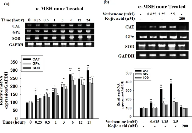
As a counterpart to ROS-generating enzymes, several anti-oxidative enzymes are involved to preserve the balance of ROS availability within vascular cells. And we also the antioxidant enzyme mRNA level was measured in RT-PCR (Fig. 9). The Fig. 8 showed that the transcription level of the CAT, GPx, SOD gene was time-dependent and concentration dependent increased in the B16F10 cells.
31
Figure 9. Verbenone on the antioxidant enzyme expression level in B16F10 cells. Effect
of verbenone on mRNA expressions of CAT, GPx and SOD in B16F10 cells. On concentration-dependent manner (a) and time-dependent (b) total cellular mRNA was extracted and subjected to RT-PCR. CAT, GPx and SOD mRNA levels were quantified and normalized with GAPDH. Values are the mean ± SD of three independent experiments. *P<0.05,
**
32
4. Discussion
Verbenone, an active compound present in Rosmarinus officinalis L. extract Recent studies have revealed that Verbenone possesses antioxidant activities(51) anti-inflammatory(60) and Antibacterial activity(61). However, despite many published results, the effect of verbenone on melanogenesis has not been reported. In the present study, we investigated the effect of verbenone on melanin synthesis and the molecular mechanism involved in the process in B16F10 mouse melanoma cells.
First, we showed that verbenone inhibited melanin synthesis in a concentration-dependent manner without cytotoxicity (0-2.5 mM) in B16F10 cells and that the extent of inhibition was comparable between treatment with verbenone and with kojic acid (200 μM). Second, because tyrosinase is the key enzyme involved in melanogenesis(62, 63) we measured the effect of verbenone on tyrosinase activity. Interestingly, we found that verbenone decreased tyrosinase activity in B16F10 cells, but it did not affect mushroom tyrosinase activity in a cell-free assay system.
MITF plays a critical role in melanogenesis, as the major transcriptional regulator of tyrosinase (64-66). Decreased MITF gene expression is known to lead to the downregulation of melanocyte differentiation markers (67). Our results show that the activation of Akt and inactivation of p38 after verbenone treatment is correlated with the phosphorylation and degradation of MITF. In accordance with reduced MITF, tyrosinase and TRP-1 protein were also reduced.
The ERK pathway regulates cell proliferation and differentiation in many types of cells(68, 69). This pathway is also involved in the regulation of melanin synthesis. It has been reported that activation of MEK/ERK leads to phosphorylation of MITF at serine 73, resulting in its degradation(70, 71).The Akt pathway plays an important role in cell growth regulation and apoptosis inhibition(72, 73). In addition, activation of the Akt pathway reduces tyrosinase transcription and melanogenesis in B16F10 cells(74, 75). So far, many natural and synthetic agents have been found to inhibit melanogenesis in B16F10 cells through activation of the ERK and/or the Akt pathways. For example, baicalein, cimicifuga heracleifolia, and KHG22394 (a synthesized chemical) were found to decrease melanin synthesis by activating
33
ERK or Akt(76-78). On the other hand, phosphorylation of p38 can activate MITF, which may transcriptionally regulate the expression of tyrosinase, tyrosinase-related protein 1 (TRP-1), and tyrosinase-related protein 2 (TRP-2), thus inducing melanin production (79). Therefore, we are interested in the effects of verbenone on Akt, ERK and p38 activities. We found that verbenone induced Akt and reduced p38, but not ERK activation. Moreover, the suppressive effect of verbenone on melanin synthesis was diminished by a prior inhibition of the Akt and reduced-p38 pathway. These results suggest that Akt activation and p38 inactivation by verbenone plays an important role in the compound’s anti-melanogenic effect in B16F10 cells.
Besides increasing pigmentation, α-MSH reduces UV-induced oxidative DNA damage by inhibiting the generation of hydrogen peroxide and enhances the repair of DNA photoproducts (80,81)Activation of MC1R up-regulates the expression of DNA repair genes (82). UVR also up-regulates the expression of α-MSH receptors, amplifying the melanogenic effect of exogenous α-MSH in a dose-dependent manner in vivo and in cell culture systems (83). UVA-mediated augmentation of melanogenesis was correlated to depletion of SOD and catalase activities and GSH content as well as aggravation of ROS production in lightly pigmented melanocytes (82, 84). On the other hand, promotion of antioxidant defenses such as GSH level and GPx activity and decrease in ROS/ RNS formation have been found to associate with reduction of melanogenesis by inhibiting tyrosinase activity in cultured melanoma cells and human melanocytes (85, 86). SOD, Cat and GPx is the key enzyme involved in antioxidant we measured the effect of verbenone on SOD, Cat, GPx mRNA level. Interestingly, we found that verbenone increased mRNA level of SOD, Cat and GPx in B16F10 cells
In conclusion, verbenone might inhibit the melanogenesis-related signal pathways, including the inhibition of TRP-1,TRP-2, tyrosinase, MITF signaling and/or activation of Akt signaling or inhibition p38 MAPK signaling. And we found that verbenone increased mRNA level of SOD, Cat and GPx in B16F10 cells In addition, cytotoxicity was not observed up to 2.5 mM concentration(Fig. 1 a). Hence, verbenone can be may contribute used safely to make hypopigmenting agents. In addition, verbenone can to the hypopigmenting effect, if it is used with another hypopigmenting agent which acts with a different mechanism.
34
REFERENCES
1. Plonka P M, Passeron T, Brenner M et al. Exp Dermatol 2009: 18: 799-819.
2, Hu D N. Photochem Photobiol 2008: 84: 645-649.
3. Larsson B S. Pigment Cell Res 1993: 6: 127-133.
4. Kameyama K, Sakai C, Kuge S et al. Pigment Cell Res 1995: 8: 97-104
5. Fang D, Kute T, Setaluri V. Pigment Cell Res 2001: 14: 132-139
6. Hearing V J. J Inverst Dermatol 1999: 4: 24-28
7. Jimbow K, Fitzpatrick T B, Wick M et al. Academic Press, New York 1979: 131-163
8. Naish-Byfield S, Riley P A. Pigment Cell Res 1998: 11: 94-97.
9. Levy C, Khaled M, Fisher DE. Trends Mol Med 2006: 12: 406-414.
10. Kono M, Dunn I S, Durda P J et al. Mol Cancer Res 2006: 4: 779-792.
11. Kim D S, Park S H, Kwon S B et al. Pigment Cell Res 2006: 19: 146-153.
12. Kawano M, Matsuyama K, Miyamae Y et al. Exp Dermatol 2007: 16: 977-984.
13. Oka M, Nagai H, Ando H et al. J Invest Dermatol 2000: 115: 699-703.
14. Lee J, Jung K, Kim Y S et al. Life Sci 2007: 81: 249-254.
15. Seger R, Krebs EG. FASEB J 1995: 9: 726-735.
35
17. Jiang Z, Xu J, Long M et al. J Life Sic 2009: 85: 345-350.
18. Singh SK, Sarkar C, Mallick S et al. Pigment Cell Melanoma Res 2005: 18: 113-121.
19. Saha B, Singh S K, Sarkar C et al. Pigment Cell Melanoma Res 2006: 19: 595-605.
20. Hirata N, Naruto S, Ohguchi K et al. Bioorg Med Chem 2007: 15: 4897-4902.
21. Corre S, Galibert M D. Pigment Cell Melanoma Res 2005: 18: 337-348.
22. Tsang T F, Ye Y, Tai W C S et al. J Ethnopharmacol 2012: 141: 622-628.
23. Ye Y, Chu J H, Wang H et al. J Ethnopharmacol 2010: 132: 533-535.
24. Kim Y J, Uyama H. Cell Mol Life Sci 2005: 62: 1707-1723.
25. Seo S Y, Sharma V K, Sharma N, J Agric Food Chem 2003: 51: 2837-2853.
26. Funayama M, Arakawa H, Yamamoto R et al. Biosci Biotechnol Biochem 1995: 59: 143-144.
27. Kim Y J, Uyama H. Cell Mol Life Sci 2005: 62: 1707-1723.
28. Nakagawa M, Kawai K, Kawai K. Contact Dermatitis 1995: 32: 9-13.
29. Kim S, Jung S H, Cho C W. Pharm Res 2008: 31: 1363-1368.
30. Halliwell B. Lancet 1994: 344: 721-724.
31. Finkel T, Holbrook N. Nature 200: 408: 239-247.
32. Wlaschek M, Briviba K, Stricklin G P et al. J Invest Dermatol 1995: 104: 194-198.
36
34. Yasui H, Sakurai H. Exp Dermatol 2003: 12: 655-661.
35. Adriana M, Ojeda S, Catalina M et al. Food Control 2013: 31: 189-195
36. Romano C S, Abadi K, Repetto V et al. Food Chem 2009: 115: 456-461
37. Gibriel A Y, Al-Sayed H M A, Rady A H et al. J Food Saf 2013: 33: 222-228
38. Monika S, Monika L, Marta P et al. Molecules 2013: 18: 9334-9351
39. Jiang Y, Wu N, F Y J et al. Environ Toxicol Phar 2011: 32: 63-68
40. Gonzalo V, Susana M, Margarita G V et al. J Supercrit Fluids 2013: 79: 101-108
41. Ngo S N, Williams D B, HEad R J. Crit Rev Food Sci Nutr 2011: 51: 946-954
42. Ahmad H H, Hamza AH, Hassan AZ et al. Int J Pharm Pharm Sci 2013: 5: 164-170
43. Afonso M S, Silva A M D O, Carvalho E B et al. J Nutr Metab 2013: 10: 19
44. Lee C Y, Kim K M, Son H S. Korean J. Food Sci Technol 2013: 45: 501-507
45. Cordeiro A M T M, Medeiros M L, Santos N A et al. J Therm Anal Calorim 2013: 113: 889-895
46. Sagdic O. Food Sci Technol 2003; 36; 467-473.
47. Sagdic O, Ozcan M. Food Control 2003; 14; 141-143.
48. Tajkarimi M M, Ibrahim S A, Cliver D O. Food Control 2010; 21; 1199–1218
49. Boyraza N, Özcan M. Fitoterapia 2005; 76; 661–665
37
Product Letters 2013; 27; 2219-2223
51. Ju C, Song S M, Hwang S Y et al. Bioorg Med Chem Lett 2013: 23: 5421-5425
52. Carmichael J, DeGraff W G, Gazdar A F, Minna J D et al. Cancer Res 1987; 47; 936-942)
53. Hosoi J, Abe E, Suda T et al. Cancer Res 1985; 45; 1474–1478)
54. Tomita Y, Maeda K, Tagami H. Pigment Cell Res 1992; 5; 357–361)
55. Lee C S, Park M Y, Han J W, Lee J H et al. J Invest Dermatol 2013; 133; 1063-1071
56. Lim H K, Kim Y W, Lee D H, Kim Cho S M et al. J Appl Biol Chem 2007; 50; 78-84
57. Wang Z, Yang X, Yang S, Ren G et al. Arch Toxicol 2011; 85; 1407-1417
58. Singh S K, Sarkar C, Mallick S et al. Pigment Cell Res 2005: 18: 113-121
59. Wang H, Joseph J A. Free radical. Bio. Med. 1999: 27: 612-616
60. Kuo C F, Su J D, Chiu C H, Peng C C et al. J Agr Food Chem 2011: 59: 3674-3685
61. Bernardes W A, Lucarini R, Tozatti M G, Bocalon Flauzino L G et al. Z Naturforsch C 2010: 65: 588-593
62. del Marmol V, Beermann F. FEBS Lett 1996; 381; 165-168.
63. Inagaki H, Bessho Y, Koga A, Hori H. Gene 1994; 150; 319-324.
64. Bentley N J, Eisen T, Goding C R. Mol Cell Biol 1994; 14; 7996 -8006.
65. R Busca, R Ballotti. Pigment Cell Res. 2000; 13; 60-69.
38
67. Jimenez-Cervantes C, Martinez-Esparza M, Perez C, Daum N et al. J Cell Sci. 2001; 114; 2335-2344
68. Cowley S, Paterson H, Kemp P, Marshall C J. Cell 1994; 77; 841-852.
69. Sale E M, Atkinson P G, Sale G J. EMBO J 1995; 14; 674-684.
70. Wu M, Hemesath T J, Takemoto C M, Horstmann M A et al. Genes Dev 2000; 14; 301-312.
71. Goodall J, Carreira S, Denat L, Kobi D et al Cancer Res 2008; 68; 7788–7794.
72. Ahmad S, Singh N, Glazer R I. Biochem Pharmacol 1999; 58; 425–430.
73. Tang Y, Zhou H, Chen A, Pittman RN et al. J Biol Chem 2000; 275; 9106–9109.
74. Khaled M, Larribere L, Bille K, Ortonne J P et al. J Invest Dermatol 2003; 121; 831–836.
75. Khaled M, Larribere L, Bille K, Aberdam E et al. J Biol Chem 2002; 277; 33690–33697.
76. Li X, Guo L, Sun Y, Zhou J et al. Int J Mol Med 2010; 25; 923–927.
77. Kim D S, Jeong YvM, Park IvK, Hahn HvG et al. Biol Pharm Bull 2007; 30; 180–183.
78. Jang J Y, Lee J H, Kang B W, Chung K T et al Exp Dermatol 2009; 18; 232–237.
79. Ye Y, Chu J H, Wang H, Xu H et al. J Ethnopharmacol 2010; 132; 533–535
80. Bohm M, Wolff I, Scholzen T E et al. J Biol Chem 2005; 280; 5795–5802
81. Kadekaro AL, Kavanagh R, Kanto H et al. Cancer Res 2005; 65; 4292–4299
12564-39
12570
83. Slominski A, Tobin D J., Shibahara S, Wortsman J. Physiol Rev 2004; 84; 1155–1228
84. Baldea I, Mocan T, Cosgarea R. Exp Oncol 2009; 31; 200-208
85. Benathan M. Arch Dermatol Res 1997; 289; 341-346.
40
ABSTRACT IN KOREAN
Verbenone 은 로즈마리(Rosmarinus officinalis L.) hydrosol 추출물의 메인
활성화합물이다. 본 연구에서 우리는 verbenone 이 B16F10 세포에서의 melanin synthesis 에 관여하는 분자 메커니즘에 대해 밝혀내었다. 2.5 mM 의 verbenone 을 처리한 B16F10 세포에서 melanin content 와 intracellular tyrosinase activity 를 긍정적 대조군으로 사용한 kojic acid 에 비해 현저하게 저해하는 현상을 확인하였으며, 반대로 mushroom tyrosinase activity 는 감소하지 않는 것을 확인하였다. 이를 통하여 verbenone 의 anti-melanogenesis 는 tyrosinase 의 효소활성을 직접적으로 저해하는 것이 아니라는 것을 알 수 있었다. 또한 western blot 과 RT-PCR 을 통해 MITF 와 TRP-1, TRP-2, Tyrosinase 의 단백질과 mRNA 수준의 변화를 관찰한 결과 모든 melanogenesis 관련 단백질의 수준을 억제하며, mRNA level 에서 다른 melanogenic mRNA 의 수준은 농도의존적으로 감소 시키나, MITF 의 mRNA 수준은 변화되지 않는 것을 확인하였다. 이를 통해 verbenone 이 MITF 의 degradation 을 통하여 anti-melanogenic effect 를 나타내는 것을 알 수 있었다. 또한 상위 signaling pathway 를 확인하기 위해 Akt, ERK, p38 의 phosphor-form 의 변화를 관찰하였다. 이를 통해 p38 의 감소와 AKT 의 증가를 통해 anti-melanogenic effect 를 나타내는
것을 확인하였다. 또한, H2O2 에 의해 증가한 ROS 수준의 감소와 antioxidant
enzyme 의 mRNA 수준의 감소를 나타내는 것을 통해 항산화능 역시 확인하였다. 따라서 verbenone 은 미백과 항산화 관련 화장품과 색소 침착 등의 치료에 유용한 화합물이 될 것이다.
41