Introduction
Spinal pain is a common patient complaint, affecting 80%–90% of individuals at least once in their lifetime [1,2]. There are various causes of spinal pain, such as spinal degeneration, trauma, inflam- mation, infection, and deformities. In clinical practice, spinal de- generation (herniated disc or spinal stenosis) and trauma are the most common causes of spinal pain [3-5]. To alleviate spinal pain, conservative treatments, including rest, physiotherapy (e.g., heat therapy, traction therapy, and manual therapy), injections, ortho- ses, and medication, are used before the surgical treatment [6-8].
Although the clinical application of orthoses is debated because of potential complications associated with long-term use, such as muscle weakness and joint contracture, its short-term use is known to improve pain and disability during the treatment period without significant adverse effects [9-11].
Effectiveness of orthoses for treatment in patients with spinal pain
Yoo Jin Choo, Min Cheol Chang
Department of Physical Medicine and Rehabilitation, Yeungnam University College of Medicine, Daegu, Korea
Spinal pain is a common patient complaint in clinical practice. Conservative treatment methods include oral medication, physical therapy, injections, and spinal orthoses. The clinical application of orthoses is debated because of potential complications associated with long-term use, such as muscle weakness and joint contracture. We reviewed the orthoses most frequently used to man- age spinal pain. We review the use of soft cervical and Philadelphia collars, lumbosacral corsets, and thoracolumbosacral orthosis to manage spinal pain. Spinal orthoses can help reduce pain by protecting the muscles and joints of the injured spinal region, preventing or correcting malfor- mations, and limiting trunk flexion, extension, lateral flexion, and rotation. The short-term use of spinal orthoses is known to improve pain and disability during the treatment period without sig- nificant adverse effects. Spinal orthoses are expected to alleviate pain and improve patients’ life- style.
Keywords: Conservative treatment; Orthotic devices; Pain; Spine
Yeungnam Univ J Med 2020;37(2):84-89 https://doi.org/10.12701/yujm.2020.00150
Received: March 11, 2020 Revised: March 16, 2020 Accepted: March 17, 2020 Corresponding author:
Min Cheol Chang
Department of Physical Medicine and Rehabilitation, Yeungnam University College of Medicine, 170 Hyeonchung-ro, Nam-gu, Daegu 42415, Korea
Tel: +82-53-620-4682 Fax: +82-53-625-3508 E-mail: [email protected]
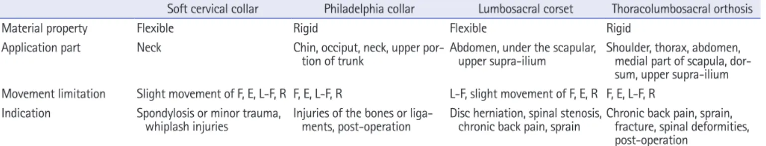
In this study, we reviewed the following types of orthoses most frequently used to manage spinal pain: soft cervical and Philadel- phia collars, lumbosacral corset, and thoracolumbosacral orthosis (Table 1).
Soft cervical collar
The soft cervical collar is comprised of a soft foam material, a fab- ric covering the foam, and a Velcro strap (Fig. 1) [12,13]. The strap is mostly fastened at the back but can also be placed at the front, depending on the user’s preference. Patients wearing a soft cervical collar can experience feelings of warmth and psychologi- cal comfort owing to the fabric sheathing [14]. However, soft cer- vical collars cannot significantly restrict the cervical spine’s range of motion, thus falling short in providing sufficient structural sup- port [12,14]. Therefore, they are used to manage muscle pain and
Copyright© 2020 Yeungnam University College of Medicine
This is an Open Access article distributed under the terms of the Creative Commons Attribution Non-Commercial License (http://creativecommons.org/licenses/by-nc/4.0/) which permits unrestricted non-commercial use, distribution, and reproduction in any medium, provided the original work is properly cited.
spasms due to spondylosis or minor trauma and as an initial treat- ment for whiplash injuries. According to a previous study, the soft cervical collar is recommended to be worn for 2 weeks [11].
Muzin et al. [11] reported no side effects (e.g., muscle weak- ness) associated with the use of soft cervical collars for fewer than 10 days. Mealy et al. [15] assessed 61 patients with acute cervical whiplash injuries following the use of the soft cervical collar for 2 weeks. The patients were divided into the following two groups:
one that progressively combined exercise with use of the soft cer- vical collar and the other that performed exercise without use of the soft cervical collar. Eight weeks later, the visual analog scale (VAS) score from 0 to 10 (with 10 indicating unsustainable pain and 0 indicating absence of pain) and range of motion of the cer- vical spine (i.e., flexion, extension, lateral flexion, and rotation) were obtained. The group using the soft cervical collar reported a higher reduction in both the intensity of pain and range of motion of the cervical spine [15]. Furthermore, Rosenfeld et al. [16] in- vestigated 97 patients with whiplash injuries by dividing them into two groups as follows: (1) those who wore soft cervical col- lars within 96 hours from the injury and were treated after 2 weeks and (2) those who did not use the collar and were treated using
the same protocol. In addition, the VAS scores for pain were ob- tained for both the groups. After 6 months, the VAS scores de- creased by 3 points in the group that used the soft cervical collars and only by 1.5 points in the group that did not use the soft cervi- cal collar.
Philadelphia collar
The Philadelphia collar, which usually comprises of a solid plastic sheet, limits a greater range of movements compared to the soft cervical collar (Fig. 2) [12]. It is vertically reinforced from the chin to the manubrium in the front and shaped to cover the area from the external protuberance of the occipital bone to the upper part of the spine of the scapula at the back. The anterior part of the Philadelphia collar has a hole for tracheostomy; therefore, the user’s chin has to be aligned with its center [17,18]. Furthermore, the inner side of the Philadelphia collar is lined with a replaceable padding, which permits good hygiene and causes less irritation to the skin. The Philadelphia collar slightly reduces the load on the spine by promoting the correct posture at the cervical spine and plays a role in limiting the cervical flexion/extension, lateral flex- Fig. 1. Soft cervical collar. The subject is wearing soft cervical collar.
Table 1. Characteristics of spinal orthoses
Soft cervical collar Philadelphia collar Lumbosacral corset Thoracolumbosacral orthosis
Material property Flexible Rigid Flexible Rigid
Application part Neck Chin, occiput, neck, upper por-
tion of trunk Abdomen, under the scapular,
upper supra-ilium Shoulder, thorax, abdomen, medial part of scapula, dor- sum, upper supra-ilium Movement limitation Slight movement of F, E, L-F, R F, E, L-F, R L-F, slight movement of F, E, R F, E, L-F, R
Indication Spondylosis or minor trauma,
whiplash injuries Injuries of the bones or liga-
ments, post-operation Disc herniation, spinal stenosis,
chronic back pain, sprain Chronic back pain, sprain, fracture, spinal deformities, post-operation
F, flexion; E, extension; L-F, lateral flexion; R, rotation.
ions and rotation [18,19]. Nonetheless, some pressure may be ap- plied on the clavicle by the Philadelphia collar. Considering that excessive pressure can cause discomfort or pressure sores, special attention is required for users with sensitive skin [18,20]. The
Philadelphia collar can be used to treat injuries of the bones and ligaments in the mid-cervical spine region and for postsurgical stabilization. In addition, it can be used instead of the halo ortho- sis to stabilize upper cervical fractures (Jefferson and hangman’s Fig. 2. Philadelphia collar. The subject is wearing Philadelphia collar.
Fig. 3. Lumbosacral corset. The subject is wearing lumbosacral corset.
Fig. 4. Thoracolumbosacral orthosis. The subject is wearing thoracolumbosacral orthosis.
fractures) and fractures of the odontoid process [11].
According to a study by Beavis [14], hard cervical collars are ef- fective in limiting the motion of the cervical spine (level of move- ment reduction: flexion 69%, extension 34%, left lateral flexion 22%, right lateral flexion 34%, leftward rotation 50%, and right- ward rotation 48%). Similarly, Muzin et al. [11] suggested that hard cervical collars were effective for the initial management of trauma (i.e., to prevent cervical instability). Finally, Motiei-Lan- groudi and Sadeghian [21] studied 11 patients with a C2 fracture who used the Philadelphia collar either until the bone completely recovered or until the neck pain disappeared. After a follow-up of 21 months, the patients reported no neurological symptoms or deficits, a mean VAS score of 2, and recovery of their lifestyle be- fore the injury.
Lumbosacral corset
The lumbosacral corset is comprised of soft materials. It encloses the trunk and pelvis and has a string or hook to adjust the circum- ference. When necessary, a canvas, nylon mesh, or coil spring is used to increase its capability to provide support (Fig. 3). The posterior support column is made of semirigid or soft plastic ma- terials and is molded to the shape of the patient’s body. It is insert- ed into a corset, which confers rigidity to the support, thus limit- ing hyperextension of the spine and reducing spinal lordosis [22- 25]. In the groin region, straps can be attached to prevent the movement of the corset. The upper margin of the anterior surface of the corset is positioned 1.3 cm (1/2 inch) below the xiphoid process, and the lower margin is located 2.5–3 cm (1 inch) above the pubic symphysis. Furthermore, while the upper margin of the posterior surface is 2.5 cm (1 inch) below the inferior angle of the scapula, the lower margin is located at the most prominent part of the hip [18].
The lumbosacral corset limits several movements in the frontal plane. It is less rigid at the pelvis, allowing movements in both the sagittal and transverse planes. Moreover, the corset applies pres- sure on the abdomen; therefore, the intraabdominal pressure in- creases, which reduces stress on the spine and load on the spinal disc and extensors. Furthermore, it enhances the user’s perception of proprioception. The lumbosacral corset can be used in cases of disc herniation, spinal stenosis, chronic back pain, pelvic fracture, and sprain of the lumbosacral spine [23-27].
Kim [28] investigated 69 patients who used the lumbosacral corset to treat a herniated disc or sprain in the lumbar spine re- gion. Physical examinations (e.g., straight leg raising [SLR] and gait analysis) were performed, and the intensity of pain and clini- cal outcomes of using the lumbosacral corset were determined
based on the criteria suggested by Stauffer and Coventry [29].
While the results of both SLR and gait analysis were “poor” in 63.77% and 59.4% of the patients, respectively, prior to use of the lumbosacral corset, improvements were noticed following its use (i.e., 84.06% and 85.50% of the patients reported a greater than
“fair” result). These findings suggest that the use of the lumbosa- cral corset is effective in reducing pain and improving the activi- ties of daily living. In 2001, Prateepavanich et al. [30] measured the claudication distance and pain score (VAS) in 21 patients with lumbar spinal stenosis without wearing a lumbosacral corset, and a week later with a lumbosacral corset, evaluated again and com- pared the results. As a result, a significant difference between the two groups, an average claudication distance was 393.2 m when wearing a lumbosacral corset and 314.6 m when not wearing a lumbosacral corset, and a mean value of pain score was 4.7 when wearing a corset and 5.9 when not wearing a lumbosacral corset.
This result showed that the effects of lumbosacral corset in lum- bar spinal stenosis.
Thoracolumbosacral orthosis
The thoracolumbosacral orthosis (TLSO) can be of the following two types based on the type of material used: soft and hard. In ad- dition, it is classified based on the location and presence of struc- tural elements as flexion adjustable, flexion/extension adjustable, flexion/extension/lateral flexion adjustable, and flexion/exten- sion/lateral flexion/rotation adjustable. It is fabricated on the principle of three-point pressure. TLSO can be used to treat chronic back pain, sprains and fractures of the thoracic or lumbar spine, and spinal deformities, and for postsurgical management of the spine [26,31].
TLSO is widely used in current clinical practice (Fig. 4). Owing to its light and breathable mesh fabric, it provides a comfortable fit. Furthermore, ergonomically designed plastic panels provide high stability through the application of abdominal pressure and insertion of the back panels. In addition, TLSO is adjustable through the connection of two elastic straps so as to fit the shape of the patient’s body. It has a shoulder strap to prevent it from slid- ing out of place.
Jacobs et al. [32] assessed 15 patients with an osteoporotic ver- tebral compression fracture. Following the use of a semirigid TLSO for 6 weeks, their VAS scores and quality of life (measured using the Quality of Life Questionnaire of the European Founda- tion for Osteoporosis) were evaluated. The outcomes were de- creased mean VAS scores (i.e., from 5 to 2 points) and improve- ments in pain (38%), physical function (42%), social function (21%), and health perception (16%).
Conclusion
Several patients with spinal pain are encountered in clinical prac- tice. Spinal orthoses are expected to alleviate pain and improve patients’ lifestyle. Nevertheless, studies on the clinical efficacy of orthoses are neither quantitatively nor qualitatively sufficient to reach a solid conclusion. Therefore, additional investigations are required to issue guidelines on the appropriate use of spinal or- thoses. Our study is limited in that we reviewed only most com- monly used orthoses, accordingly more various orthoses should be reviewed in the future study.
Acknowledgments
Conflicts of interest
No potential conflict of interest relevant to this article was report- ed.
Author contributions
Conceptualization: YJC, MCC; Data curation: YJC, MCC; For- mal analysis: MCC; Methodology: YJC, MCC; Investigation:
YJC; Resources: YJC; Supervision: MCC; Visualization: YJC, MCC; Writing-original draft: YJC, MCC; Writing-review & edit- ing: YJC, MCC.
Additional information
The model provided written informed consent for the use and publication of his photographs.
ORCID
Yoo Jin Choo, https://orcid.org/0000-0002-3820-2279 Min Cheol Chang, https://orcid.org/0000-0002-7629-7213
References
1. Balague F, Mannion AF, Pellise F, Cedraschi C. Non-specific low back pain. Lancet 2012;379:482–91.
2. Salekzamani Y, Mirzaee S, Shakouri SK, Nezami N. Pain reliev- ing effect of thermoplastic lumbosacral orthosis with adjustable posterior pad in chronic non-specific low back pain. Iran Red Crescent Med J 2011;13:903–5.
3. Jang SH, Chang MC. Follow-up of at least five years after lum- bar transforaminal epidural steroid injection for radicular pain due to lumbar disc herniation. Ann Palliat Med 2020;9:116–8.
4. Do KH, Kim TH, Chang MC. Effects of interlaminar epidural steroid injection in patients with moderate to severe lumbar central spinal stenosis. Ann Palliat Med 2020;9:163-8.
5. Yang JY. The pathogenesis and medical treatment of spondylo- genic pain. Asian Spine J 2010;4:57–63.
6. Lurie J, Tomkins-Lane C. Management of lumbar spinal steno- sis. BMJ 2016;352:h6234.
7. van Tulder MW, Koes BW, Bouter LM. Conservative treatment of acute and chronic nonspecific low back pain. A systematic re- view of randomized controlled trials of the most common inter- ventions. Spine (Phila Pa 1976) 1997;22:2128–56.
8. van Tulder MW, Koes B, Malmivaara A. Outcome of non-inva- sive treatment modalities on back pain: an evidence-based re- view. Eur Spine J 2006;15(Suppl 1):S64–81.
9. Azadinia F, Ebrahimi Takamjani E, Kamyab M, Parnianpour M, Cholewicki J, Maroufi N. Can lumbosacral orthoses cause trunk muscle weakness? A systematic review of literature. Spine J 2017;17:589–602.
10. Bible JE, Biswas D, Whang PG, Simpson AK, Rechtine GR, Grauer JN. Postoperative bracing after spine surgery for degen- erative conditions: a questionnaire study. Spine J 2009;9:309–
16.
11. Muzin S, Isaac Z, Walker J, Abd OE, Baima J. When should a cervical collar be used to treat neck pain? Curr Rev Musculo- skelet Med 2008;1:114–9.
12. Barati K, Arazpour M, Vameghi R, Abdoli A, Farmani F. The ef- fect of soft and rigid cervical collars on head and neck immobili- zation in healthy subjects. Asian Spine J 2017;11:390–5.
13. Richter D, Latta LL, Milne EL, Varkarakis GM, Biedermann L, Ekkernkamp A, et al. The stabilizing effects of different orthoses in the intact and unstable upper cervical spine: a cadaver study. J Trauma 2001;50:848–54.
14. Beavis A. Cervical orthoses. Prosthet Orthot Int 1989;13:6–13.
15. Mealy K, Brennan H, Fenelon GC. Early mobilization of acute whiplash injuries. Br Med J (Clin Res Ed) 1986;292:656–7.
16. Rosenfeld M, Gunnarsson R, Borenstein P. Early intervention in whiplash-associated disorders: a comparison of two treat- ment protocols. Spine (Phila Pa 1976) 2000;25:1782–7.
17. Ghorbani F, Kamyab M, Azadinia F, Hajiaghaei B. Open-design collar vs. conventional Philadelphia collar regarding user satis- faction and cervical range of motion in asymptomatic adults.
Am J Phys Med Rehabil 2016;95:291–9.
18. Kim JH, Park YS, Song JC, Shin HS, Chang YC. Prosthetics &
orthotics. 3rd ed. Seoul (KR): Topmed; 2006.
19. Kaufman WA, Lunsford BR, Lunsford TR, Lance LL. Compar- ison of three prefabricated cervical collars. Orthot Prosthet 1985;39:21–8.
20. Sparke A, Voss S, Benger J. The measurement of tissue interface pressures and changes in jugular venous parameters associated with cervical immobilization devices: a systematic review.
Scand J Trauma Resusc Emerg Med 2013;21:81.
21. Motiei-Langroudi R, Sadeghian H. C2 body fracture: report of cases managed conservatively by Philadelphia collar. Asian Spine J 2016;10:920–4.
22. Morrisette DC, Cholewicki J, Logan S, Seif G, McGowan S. A randomized clinical trial comparing extensible and inextensible lumbosacral orthoses and standard care alone in the manage- ment of lower back pain. Spine (Phila Pa 1976) 2014;39:1733–
42.
23. Rizzone K, Gregory A. Using casts, splints, and braces in the emergency department. Clin Pediatr Emerg Med 2013;
14:340–8.
24. Sullivan MS, Mayhew TP. The effect of lumbar support belts on isometric force production during a simulated lift. J Occup Re- habil 1995;5:131–43.
25. Terai T, Yamada H, Asano K, Nawata A, Iwasaki T, Henmi T, et al. Effectiveness of three types of lumbar orthosis for restricting extension motion. Eur J Orthop Surg Traumatol 2014;24(Sup- pl 1):S239–43.
26. Agabegi SS, Asghar FA, Herkowitz HN. Spinal orthoses. J Am Acad Orthop Surg 2010;18:657–67.
27. Schroeder S, Rossler H, Ziehe P, Higuchi F. Bracing and sup-
porting of the lumbar spine. Prosthet Orthot Int 1982;6:139–
46.
28. Kim MH. A biomechanical effectiveness of corset and back brace for low back pain syndrom. Phys Ther Korea 1996;3:59–
66.
29. Stauffer RN, Coventry MB. Anterior interbody lumbar spine fu- sion: analysis of Mayo Clinic series. J Bone Joint Surg Am 1972;54:756–68.
30. Prateepavanich P, Thanapipatsiri S, Santisatisakul P, Somshevita P, Charoensak T. The effectiveness of lumbosacral corset in symptomatic degenerative lumbar spinal stenosis. J Med Assoc Thai 2001;84:572–6.
31. Vander Kooi D, Abad G, Basford JR, Maus TP, Yaszemski MJ, Kaufman KR. Lumbar spine stabilization with a thoracolumbo- sacral orthosis: evaluation with video fluoroscopy. Spine (Phila Pa 1976) 2004;29:100–4.
32. Jacobs E, Senden R, McCrum C, van Rhijn LW, Meijer K, Wil- lems PC. Effect of a semirigid thoracolumbar orthosis on gait and sagittal alignment in patients with an osteoporotic vertebral compression fracture. Clin Interv Aging 2019;14:671–80.