Effects of Macrolide and Corticosteroid in Neutrophilic Asthma Mouse Model
Tai Joon An, M.D.
1, Chin Kook Rhee, M.D., Ph.D.
2, Ji Hye Kim, M.S.
1, Young Rong Lee, M.D.
3, Jin Young Chon, M.D., Ph.D.
3, Chan Kwon Park, M.D.
1and Hyoung Kyu Yoon, M.D., Ph.D.
11
Division of Pulmonary, Allergy and Critical Care Medicine, Department of Internal Medicine, Yeouido St. Mary’s Hospital, College of Medicine, The Catholic University of Korea, Seoul,
2Division of Pulmonary, Allergy and Critical Care Medicine, Department of Internal Medicine, Seoul St. Mary’s Hospital, College of Medicine, The Catholic University of Korea, Seoul,
3
Department of Anesthesiology and Pain Medicine, Yeouido St. Mary’s Hospital, College of Medicine, The Catholic University of Korea, Seoul, Korea
Background: Asthma is a disease of chronic airway inflammation with heterogeneous features. Neutrophilic asthma is corticosteroid-insensitive asthma related to absence or suppression of T
H2 process and increased T
H1 and/or T
H17 process. Macrolides are immunomodulatory drug that reduce airway inflammation, but their role in asthma is not fully known. The purpose of this study was to evaluate the role of macrolides in neutrophilic asthma and compare their effects with those of corticosteroids.
Methods: C57BL/6 female mice were sensitized with ovalbumin (OVA) and lipopolysaccharides (LPS). Clarithromycin (CAM) and/or dexamethasone (DXM) were administered at days 14, 15, 21, 22, and 23. At day 24, the mice were sacrificed.
Results: Airway resistance in the OVA+LPS exposed mice was elevated but was more attenuated after treatment with CAM+DXM compared with the monotherapy group (p<0.05 and p<0.01). In bronchoalveolar lavage fluid study, total cells and neutrophil counts in OVA+LPS mice were elevated but decreased after CAM+DXM treatment. In hematoxylin and eosin stain, the CAM+DXM-treated group showed less inflammation additively than the monotherapy group. There was less total protein, interleukin 17 (IL-17), interferon g, and tumor necrosis factor a in the CAM+DXM group than in the monotherapy group (p<0.001, p<0.05, and p<0.001). More histone deacetylase 2 (HDAC2) activity was recovered in the DXM and CAM+DXM challenged groups than in the control group (p<0.05).
Conclusion: Decreased IL-17 and recovered relative HDAC2 activity correlated with airway resistance and inflammation in a neutrophilic asthma mouse model. This result suggests macrolides as a potential corticosteroid- sparing agent in neutrophilic asthma.
Keywords: Neutrophils; Asthma; Macrolides; Adrenal Cortex Hormones; Th17 Cells; Inflammation; Histone Deacetylases
Address for correspondence: Hyoung Kyu Yoon, M.D., Ph.D.
Division of Pulmonary, Allergy and Critical Care Medicine, Department of Internal Medicine, Yeouido St. Mary’s Hospital, College of Medicine, The Catholic University of Korea, 10 63-ro, Yeongdeungpo-gu, Seoul 07345, Korea
Phone: 82-2-3779-2213, Fax: 82-2-780-3132, E-mail: [email protected]
Received: Sep. 6, 2017, Revised: Nov. 18, 2017, Accepted: Nov. 21, 2017, Published online: Dec. 21, 2017
cc
It is identical to the Creative Commons Attribution Non-Commercial License (http://creativecommons.org/licenses/by-nc/4.0/).
Copyright © 2018
The Korean Academy of Tuberculosis and Respiratory Diseases.
All rights reserved.
Introduction
Asthma is a common chronic airway disorder characterized by variable symptoms, airway obstruction, airway inflamma- tion, and airway remodeling
1. Classically, mast cells, T helper type II cells (T
H2 cells), and eosinophils play roles in asthma by cytokine-based airway inflammation
2. Asthma has a com- plex syndrome of many different clinical features without pre- dominance, and there are many types, such as non-T
H2 cell‒
related, neutrophilic, and paucigranulocytic asthma
3.
The burden of asthma is continuously increasing; low qual- ity of life, disability, morbidity, and socioeconomic issues have been emerging worldwide. Refractory or persistent asthma has been the main focus in previous studies
4-8. Asthma can be categorized according to type of underlying inflammation, and much refractory asthma is neutrophilic. Neutrophilic asthma is non-allergic asthma that often does not respond to inhaled or systemic corticosteroids
9.
There is some proposed pathobiology of corticosteroid insensitivity in neutrophilic asthma. First, the T
H2 process of neutrophilic asthma was absent or suppressed; corticoste- roids suppressed eosinophil and T-lymphocyte inflammation but did not prevent neutrophil accumulation
10. Instead, T helper 17 cells (T
H17) were pivotal and linked with neutrophil- ic asthma
11,12. Second, corticosteroid insensitivity correlated with histone deacetylase-2 (HDAC2) activity and expression, which suggests that drugs that restore HDAC activity can re- duce neutrophil recruitment in asthma
13.
Macrolides are antibiotics with immunomodulatory properties
14,15. They may be effective in patients with severe neutrophilic asthma, but there is a lack of evidence for using macrolides for asthma
16-18.
In this study, we set a neutrophilic asthma model, describe the possibility of macrolides as a therapeutic agent for severe and refractory neutrophilic asthma, and compare the effec- tiveness of macrolides, corticosteroids and concurrent treat- ment in the neutrophilic asthma mouse model.
Materials and Methods
1. Animal and experimental model
In a previous study, we developed eosinophilic asthma and neutrophilic asthma mouse models. Intraperitoneal ovalbu- min (OVA) sensitization induced T
H2-derived eosinophilic inflammation in the mouse model
19-21. After the eosinophilic asthma mouse model was sensitized, using lipopolysaccha- rides (LPS) shifted the airway inflammation from eosinophil- predominant to neutrophil-predominant and highly cor- related with levels of interleukin (IL)-17A and T
H17 cells
22. Modifying previous studies, we sensitized 6-week-old female C57BL/6 mice (Orient Inc., Seongnam, Korea) with peritoneal injection of 100 µg of OVA grade V (Sigma-Aldrich, St. Louis, MO, USA) that was dissolved in 2 mg aluminum hydroxide (Sigma-Aldrich) at days 0 and 7. Mice were exposed to intra- nasal 50 µg OVA at days 14, 15, 21, 22, and 23 to induce eo- sinophilic inflammation. We used intranasal 10 µg LPS from Escherichia coli O11:B4 (Sigma-Aldrich) to induce neutro- philic inflammation at days 21, 22, and 23.
2. Drug challenges
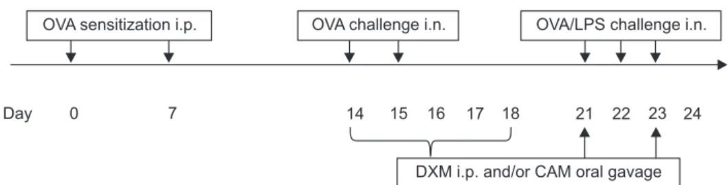
Figure 1 describes the drug challenges in the neutrophilic asthma mouse model. We randomly allocated the mice to the following groups: (1) control, (2) OVA+LPS+0.5% carboxymeth- yl cellulose (CMC), (3) OVA+LPS+clarithromycin (CAM) (10 mg/kg), (4) OVA+LPS+dexamethasone (DXM; 1 mg/kg)+CMC, and (5) OVA+LPS+CAM (10 mg/kg)+DXM (1 mg/kg).
The control group comprised five mice, and the drug-chal- lenge groups comprised seven. We used 0.5% CMC (Sigma- Aldrich) as a vehicle to compare with other groups. The mice were exposed to peritoneal injection of 1 mg/kg DXM (Sigma- Aldrich) and 10 mg/kg CAM (Sigma-Aldrich) via oral gavage every 24 hours on days 14, 15, 16, 17, 18, 21, 22, and 23. The mice were sacrificed at day 24. All animal protocols were ap- proved by the Animal Subjects Committee of the Catholic University of Korea.
Day 0 7 14 15 16 17 18 21 22 23 24
OVA sensitization i.p. OVA challenge i.n. OVA/LPS challenge i.n.
DXM i.p. and/or CAM oral gavage
Figure 1. Schematic outline of the neutrophilic asthma mouse model. Mice were sensitized via OVA on days 0 and 7 by i.p. injection and chal-
lenged through inhalation of OVA on days 14, 15, 21, 22, and 23. For neutrophilic asthma, mice were additionally challenged with LPS on days
21, 22, and 23. DXM by i.p. injection and CAM via oral gavage were challenged every 24 hours on days 14, 15, 16, 17, 18, 21, and 23. OVA: oval-
bumin; i.p.: intra peritoneally; LPS: lipopolysaccharide; DXM: dexamethasone; CAM: clarithromycin; i.n: intranasally.
3. Airway responsiveness
We calculated airway hyperresponsiveness by respiratory system resistance (Rrs) with methacholine (Mch; Sigma- Aldrich) in the FlexiVent system (SCIREQ, Montreal, QC, Can- ada)
23,24. Mice were exposed to nebulized phosphate-buffered saline (PBS) for 3 minutes for baseline Rrs value, and we as- sessed Rrs after challenging them with increasing concentra- tions of Mch (6.25, 12.5, 25, and 50 mg/mL; Sigma-Aldrich) in PBS using an Aerosonic ultrasonic nebulizer (DeVilbiss, Som- erset, PA, USA). We averaged and expressed the Rrs measured during each 3-minute sequence for each Mch concentration.
4. Bronchoalveolar lavage
The trachea was exposed and cannulated with silicone tubing attached to a 22-gauge needle on a 1-mL tuberculin syringe. We withdrew bronchoalveolar lavage (BAL) fluid after instilling 0.8 mL cold sterile PBS through the trachea into the lung. We cytospun the recovered BAL fluid for 10 minutes at 1,000 ×g at 4°C and stored supernatant at –80°C until required for further analysis. We counted total cells in the BAL fluid us- ing a hemocytometer. After we adjusted the number of cells to 1×10
6/mL, we centrifuged 50 µL aliquots of BAL fluid onto microscope slides for 5 minutes at 43 g and stained the slides with Diff-Quick (Sysmax, Kobe, Japan). We obtained the per- centages of macrophages, eosinophils, lymphocytes, and neu- trophils in the BAL fluid by counting 500 leucocytes in ran- domly selected fields of the slides under a light microscope.
5. Lung tissue preparation for histopathology
After the mice were killed, their lungs were inflated, fixed in 4% paraformaldehyde for 24 hours, and embedded in paraffin wax. We cut sections at a thickness of 4 µm using a microtome and subjected the deparaffinized tissue sections to hematoxy- lin and eosin (H&E) staining for microscopic examination (magnification of ×200).
6. Measurement of cytokines
We measured the concentrations of IL-5 and IL-17 in the BAL fluids and IL-13, interferon g (IFN-g), and tumor necrosis factor a (TNF-a) in the lung homogenates using an enzyme- linked immunosorbent assay kit (R&D Systems, Minneapolis, MN, USA). The assay sensitivities were 7, 5, 2, and 7.21 pg/mL, respectively. We conducted the protocol according to manu- facturer’s instructions.
7. Western blot analysis
We disrupted separated lung tissues that had been immedi- ately frozen in liquid nitrogen using a Polytron homogenizer
(Pellet pestles cordless motor; Sigma) and centrifuged them.
We purified the proteins from the supernatant and assessed the concentrations using the Bradford method. We sepa- rated the protein samples by 10% sodium dodecyl sulfate- polyacrylamide gel electrophoresis and transferred them to a polyvinylidene difluoride membrane (Amersham Pharmacia Biotech, Little Chalfont, UK). After blocking with 5% skimmed milk (Difco, BD, San Jose, CA, USA), we incubated the mem- brane overnight with an anti-HDAC2 antibody (1:1,000, rabbit monoclonal; Abcam, Cambridge, UK). We washed the mem- brane three times with TBST and incubated it with a second- ary antibody for 2 hours. Horseradish peroxidase-conjugated goat anti rabbit IgG (1:2,000, Santa Cruz Biotech, Dallas, TX, USA) was used as the secondary antibody. We detected the target proteins using the ECL Western Blotting Analysis Sys- tem (Thermo Fisher Scientific, Rockford, IL, USA). We either exposed the membrane to X-ray film or analyzed it with an LAS 3000 (Fujifilm, Tokyo, Japan) image analyzer using Multi Gauge v. 3.0 software. We also quantified the HDAC2 and ß- actin band intensities with Multi Gauge.
8. HDAC2 activity
We measured HDAC2 activity using the colorimetric EpiQuik HDAC2 Activity Assay Kit (Epigentek, Brooklyn, NY, USA). We incubated the nuclear extracts with specific sub-
6
4
2
0 Airwayresistance(Rrs) (cmHOsec/mL)2
PBS 6.25 12.5 25 50
Methacholine (mg/mL)
*
ControlO+L O+L+C O+L+D O+L+D+C
***
**
* ***
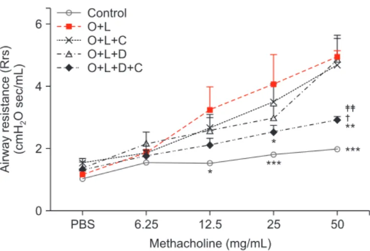
Figure 2. Airway responsiveness to methacholine by forced oscil- lation technique. Airway responsiveness was expressed in terms of respiratory systemic Rrs. Baseline Rrs value was assessed after 3 minutes of nebulized PBS. Rrs was measured in response to in- creasing challenges of methacholine dose (6.25 mg/mL, 12.5 mg/
mL, 25 mg/mL, and 50 mg/mL) in control, O+L, O+L+C, O+L+D, and O+L+D+C group. Rrs was increased after OVA+LPS challenge.
Rrs of combination of CAM+DXM group was significantly changed compared to the CAM or DXM group especially at 50 mg/mL of methacholine challenge. O+L vs. all (*p<0.05, **p<0.01, ***p<0.001), O+L+C vs. O+L+D+C (
†p<0.05), O+L+D vs. O+L+D+C (
‡‡p<0.01). Rrs:
resistance; PBS: phosphate buffered saline; O: ovalbumin; L: lipo-
polysaccharide; C: clarithromycin; D: dexamethasone.
strate for 90 minutes at 37°C, followed by capture antibody for 60 minutes, and detection antibody for 30 minutes at room temperature. We measured absorbance at 450 nm in a micro- elisa reader.
9. Statistical analysis
All data are expressed as the means±SEM, and we used GraphPad Prism 5.01 (GraphPad Software, San Diego, CA, USA) for the data analysis. We performed the statistical analy- ses using one-way ANOVA and t tests and considered p<0.05 to indicate statistical significance.
Results
1. Combination therapy of macrolide and corticosteroid attenuates airway resistance in the neutrophilic asthma mouse model
The mice in the OVA+LPS group showed greater airway
resistance after they received 12.5 mg/mL of Mch than did the control group (p<0.05) (Figure 2). CAM or DXM monotherapy group did not decrease Rrs after 12.5, 25, or 50 mg/mL of Mch, but the OVA+LPS-challenged mice treated with CAM+DXM showed decreased Rrs at 25 and 50 mg/mL of Mch challenge (p<0.05 and p<0.01). The change was significant compared with findings in the CAM and DXM monotherapy groups, es- pecially after 50 mg/mL of Mch (p<0.05 and p<0.01).
2. Combination therapy of macrolide and corticosteroid reduces lung inflammation in the neutrophilic asthma mouse model
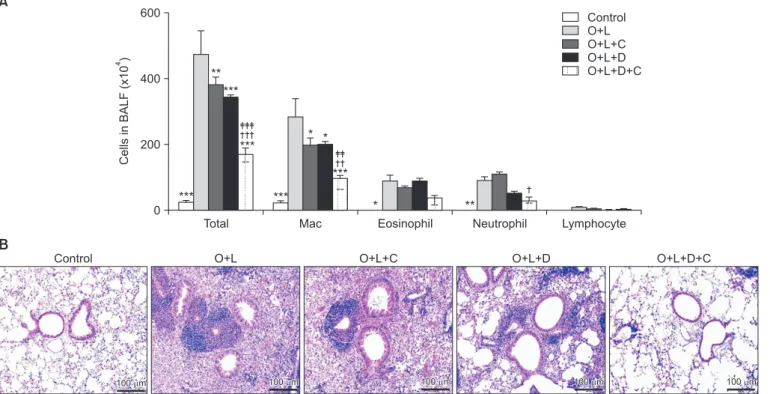
In BAL fluid, total cell counts were higher in OVA+LPS- challenged mice (p<0.001) (Figure 3A). These mice that were treated with CAM+DXM revealed lower total cell counts (p<0.001) compared with those in the monotherapy groups.
We conducted differential cell counts to evaluate reduced inflammation in the pathways based on microscopic morpho- logical criteria. In OVA+LPS-challenged mice, alveolar neu- trophil counts percentage was 20.7% and eosinophil counts
A
Control O+L O+L+C O+L+D O+L+D+C
100 m
100 m 100 m100 m 100 m100 m 100 m100 m 100 m100 m
B
600
400
200
0
CellsinBALF(x10)4
Total Mac Eosinophil Neutrophil Lymphocyte
Control O+L O+L+C O+L+D O+L+D+C
***
***
**
***
***
* *
***
* **
Figure 3. Combination therapy of macrolide and corticosteroid inhibited lung inflammation in murine model of neutrophilic asthma. (A)
BALF analysis was performed to evaluate lung inflammation. The number of total cell counts, eosinophil, and neutrophil was significantly
increased after OVA+LPS challenge. The number of total cell counts was significantly more decreased in CAM+DXM group compared to
decrease in CAM or DXM group. Neutrophils in combination of CAM+DXM treatment group was decreased and it was not seen in CAM
or DXM group. The number of eosinophils was not decreased in CAM, DXM, and CAM+DXM group. (B) Accumulation of inflammatory
cells improved significantly more in CAM+DXM group compared to DXM group in H&E staining. O+L vs. all (*p<0.05, **p<0.01, ***p<0.001),
O+L+C vs. O+L+D+C (
†p<0.05,
††p<0.01,
†††p<0.001), O+L+D vs. O+L+D+C (
‡‡p<0.01,
‡‡‡p<0.001). BALF: bronchoalveolar lavage fluid; O: ovalbu-
min (OVA); L: lipopolysaccharide (LPS); C: clarithromycin (CAM); D: dexamethasone (DXM).
percentage was 18.6%, which is higher compared to control group (0.2% and 0%). The numbers of alveolar neutrophils, eosinophils, and lymphocytes did not decrease with CAM or DXM monotherapy, but with the two together, the numbers of neutrophils decreased (16.3%, p<0.05) but not eosinophils (16.3%). On H&E staining, inflammatory cell accumulation improved in the DXM monotherapy group. In the CAM+DXM group, accumulation of inflammation cells improved more than it did in the monotherapy groups (Figure 3B).
3. Macrolides have more potent effects than corticosteroids on attenuating T
H17 inflammation in the neutrophilic asthma mouse model
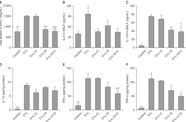
OVA+LPS challenge increased the total protein amounts in the BAL fluid (p<0.01). After treatment with DXM monother-
apy and a combination of CAM+DXM, total protein amounts in the BAL fluid decreased (p<0.001). Protein amounts were lower in the combination group than in the DXM monothera- py group, but difference was not statistically significant (Figure 4A).
T
H2 inflammation marker (IL-5 and IL-13) and T
H17 in- flammation marker (IL-17A) were induced after OVA+LPS challenge in the mouse model (p<0.05; Figure 4B, C and p<0.001; Figure 4D); IFN-g, and TNF-a increased also (p<0.01) (Figure 4E, F). After treatment with CAM only, IL-5 and IL-13 decreased (p<0.05 for both). After treatment with DXM, total protein (p<0.001) (Figure 4A) and IL-17A decreased (p<0.01) (Figure 4B, C). We also noted decreases in the CAM+DXM group. IFN-g and TNF-a decreased with CAM+DXM, but there was no attenuation in either the CAM or the DXM monother- apy groups (p<0.05) (Figure 4E, F).
A
2,0001,500
1,000
500
TotalproteininBALF(mg/mL) 0
** *** ***
Contro l
O+L
O+L+C O+L+D O+L+D+C
B
12090
60
30
0
IL-5inBALF(pg/mL)
* *
O+L
O+L+C O+L+D O+L+D+C
C
10080
60
40
20
0
IL-17AinBALF(pg/mL)
*
** *
O+L
O+L+C O+L+D O+L+D+C
D
15
10
5
0
IL-13(pg/mgprotein)
***
*
O+L
O+L+C O+L+D O+L+C+D
E
150
100
50
0
IFN-(pg/mgprotein)
**
*
O+L
O+L+C O+L+D O+L+D+C
0
TNF-a(pg/mgprotein)
**
*
O+L
O+L+C O+L+D O+L+C+D 150
100
50 Contro
l
Contro l
Contro l
Contro l
Contro l
F
Figure 4. T
H17 inflammation was attenuated by combination of macrolide and corticosteroid. Total protein, T
H2 inflammation, T
H17 inflam- mation, and proinflammatory cytokine in BALF analysis was increased after OVA+LPS challenge. (A) The number of total protein in BALF was decreased in DXM and CAM+DXM group. (B) IL-5 in BALF, marker of T
H2 inflammation, was not significantly decreased in DXM or CAM+DXM group. (C) IL-17, marker of T
H17 inflammation, was significantly decreased in DXM and CAM+DXM group. (D) IL-13, marker of T
H2 inflammation, was evaluated in lung tissue. There was no significant decrease in DXM or CAM+DXM group. (E, F) Proinflammatory cy- tokine was decreased in combination of CAM+DXM and it was not decreased in monotherapy of CAM or DXM. O+L vs. all (*p<0.05, **p<0.01,
***p<0.001), O+L+C vs. O+L+D+C (
†p<0.05,
†††p<0.001). BALF: bronchoalveolar lavage fluid; O: ovalbumin (OVA); L: lipopolysaccharide (LPS);
C: clarithromycin (CAM); D: dexamethasone (DXM); IL: interleukin; IFN-g: interferon g; TNF-a: tumor necrosis factor a.
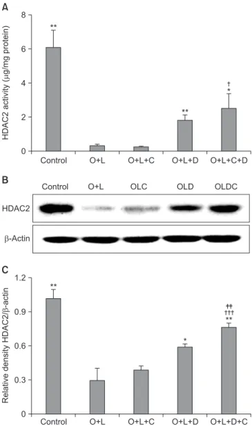
4. Macrolides act on HDAC2 activity and have potent effects with corticosteroids
HDAC activity is correlated to corticosteroid activity and de- creases in many cases of corticosteroid insensitivity
25. HDAC2 activity in OVA+LPS challenged mice was lower than the ac-
tivity in the control group (p<0.01) (Figure 5A) and it was not recovered after CAM treatment. HDAC2 activity was more recovered in the DXM-challenged group than in the control group (p<0.01) (Figure 5A). The CAM+DXM-challenged group recovered more HDAC2 activity than either the control or the CAM-challenged groups (p<0.05 for both) (Figure 5A).
Relative density analysis of HDAC2/β-actin showed signifi- cant differences. In the OVA+LPS-challenged mice, the rela- tive density of HDAC2/β-actin was lower than in the control group (p<0.01) (Figure 5C); it was not significantly higher in the CAM-challenged group than in the control group. In the DXM-challenged group, relative density of HDAC2/β-actin was greater than that in the control group (p<0.05) (Figure 5C), and it was greater in the CAM+DXM-challenged group than in the DXM group (p<0.01) (Figure 5C).
Discussion
We developed a neutrophilic asthma mouse model by chal- lenging OVA+LPS to C57BL/6 female mice, and we found neutrophilia and eosinophilia in the OVA+LPS murine model.
We demonstrated that macrolide and corticosteroid treat- ment reduced airway inflammation through attenuating airway resistance in the neutrophilic asthma mouse model.
A combination of macrolide and corticosteroid reduced the neutrophil counts in BAL fluid but not eosinophil counts.
T
H17 inflammation marker was attenuated by corticosteroid, and the macrolide potentiated the effect of the corticosteroid.
Pro-inflammatory cytokines did not decrease with macrolide alone or corticosteroid alone but with the combination of the two. The combination of macrolide and corticosteroid recov- ered relative HDAC2 activity that accounted for improvement in corticosteroid insensitivity.
Neutrophilic asthma is characterized by sputum neutro- philia, low lung function, more trapped air, and thicker airway walls than non-neutrophilic asthma; little is understood about its related molecular elements and pathophysiology
12,26,27. It has remained controversial whether neutrophilia is an inde- pendent driving component and synergistic factor with eo- sinophilia or a consequence of corticosteroid therapy.
Airway resistance and airway inflammation on H&E stain were not attenuated by macrolide alone or corticosteroid alone but with the combination of the two, the neutrophil count decreased. This demonstrates that macrolide and cor- ticosteroid together decreased airway resistance and inflam- mation by attenuating neutrophilic inflammation.
Neutrophilic asthma often responds less to inhaled cortico- steroids or to high-dose of systemic corticosteroids. There is no proven mechanism of corticosteroid insensitivity in neu- trophilic asthma. Characteristics of neutrophilic asthma, such as more fixed airway obstruction and thicker airway walls, may be related to unresponsiveness to corticosteroids. Previ- A
86
4
2
0
HDAC2activity(g/mgprotein)
**
*
**
Control O+L O+L+C O+L+D O+L+C+D
B
HDAC2
-Actin
Control O+L OLC OLD OLDC
1.2
0.9
0.6
0.3
0
RelativedensityHDAC2/-actin
**
*
Control O+L O+L+C O+L+D O+L+D+C
**
C
Figure 5. Macrolide has potent effects of recovering HDAC2 activ- ity with corticosteroid. (A) HDAC2 activity was decreased after OVA+LPS challenge. It was not recovered after CAM treatment, but it was recovered after DXM or CAM+DXM treatment. (B) DXM or DXM+CAM group showed higher density than OVA+LPS or CAM group in western blot analysis. (C) Relative density of HDAC2/
β-actin was well correlated these tendency and CAM+DXM group was further increased in density of HDAC2/β-actin than DXM group. O+L vs. all (*p<0.05, **p<0.01), O+L+C vs. O+L+D+C (
†p<0.05,
†††