정제봉독의 멜라닌 생성 억제 효과
한상미#,†·김정민†·이광길·박관규*·장영채*
국립농업과학원 농업생물부, *대구가톨릭대학교 의과대학 (Received June 6, 2012; Revised July 26, 2012; Accepted August 1, 2012)
Inhibitory Effects of Purified Bee Venom on Melanin Synthesis
SangMi Han#,†, JungMin Kim†, KyungGill Lee, KwanKyu Park* and YoungChae Chang*
Department of Agricultural Biology, National Academy and Agriculturual Science, Suwon 441-100, Korea
*Catholic University of Daegu School of Medicine, Daegu 705-718, Korea
Abstract — To further access honeybee (Apis mellifera L.) venom (BV) as a cosmetic ingredient and potential external treatment for topical use, we investigated its ability to inhibit tyrosinase activity and melanin biosynthesis on melanogenesis in B16F1 melanoma cells. We found that BV increased the cell viability in B16F1 melanoma cell and BV (0.01~1µg/ml) inhibited melanin synthesis in with 10 nM α-melanocyte-stimulating hormone (α-MSH) for 48 h. In addition, we used reverse transcription-polymerase chain reaction and western blotting for me melanogenesis-related genes such as tyro- sinase to examine the mechanisms underlying the inhibitory effects of BV on melanogensis. BV inhibited direct tyrosinase activity, which decreased melanin synthesis in α-MSH stimulated B16F1 melanoma cells. Thease findings suggest that BV induces the downregulation of melanogenesis by inhibiting tyrosinase activation.
Keywords □ bee venom, B16F1 melanoma cell, melanin, tyrosinase
피부는 신체의 최외각에 위치하며 자외선의 영향을 가장 많이 받는 기관으로 자외선으로부터 피부를 보호하기 위해 멜라닌 색 소가 생성된다.1) 그러나, 피부의 지속적인 자외선의 노출은 피부 의 광노화 뿐 아니라 기미, 주근깨 및 검버섯과 같은 색소침착을 유발하는데 이는 피부 표피층에 존재하는 멜라닌이 피부 표면으 로 침착되어 나타난다.2)멜라닌 합성은 자외선의 자극 뿐만 아 니라, 다양한 cytokines과 growth factors 등에 의해서 자극을 받 는다.3) 이들 가운데, α-melanocyte-stimulating hormone(α- MSH)는 가장 많은 연구가 진행된 호르몬으로, 멜라닌 생성 세 포 막에 존재하는 melanocortin receptor 1(MC1R)이라는 수용 체와 결합을 통해 cAMP 의 생성을 자극한다. 또한 이 반응은 microphthalmia associated transcription factor(MITF)의 발현을 증가시켜, 결국 멜라닌 합성을 자극하게 된다.4,5)
멜라닌은 피부 표피층에 존재하는 멜라닌 생성세포의 멜라노 좀(melanosome)이라는 특정 기관에서 합성되는데, 멜라노좀에 는 멜라닌 합성에 관여하는 주요 효소인 tyrosinase, tyrosinase
related protein(TRP)-1과 TRP-2 등을 함유하고 있다.6)특히, tyrosinase는 멜라닌 합성에서 가장 먼저 작용하는 속도조절효소 로써, tyrosine을 L-3,4-dihydroxyphenylalanine(DOPA)으로 전환 시키고, 또 DOPA를 dopaquinone으로 전환시키는 역할을 한다.
이렇게 형성된 dopaquinone은 TRP-2라는 효소에 의해 촉매되어 5,6-dihydroxyindole-2-carboxylic acid(DHICA)로 전환되고 이는 다시 TRP-1 효소의 산화 작용에 의해 indole-5-6-quinone carboxylic acid로 전환, 최종 산물인 멜라닌을 합성한다.6,7) 멜라닌은 갈색, 흑색을 띠는 eumelanin과 노랑, 적색을 띠는 pheomelanin으로 구분되어지며 tyrosinase는 두 가지 형태인 eumelanin과 pheomelanin 합성에 영향을 미치는 것으로 알려져 있다.6)따라서, 멜라닌 합성 억제에 대한 연구는 tyrosinase의 발 현 억제에 대한 효능 평가가 일차적으로 이루어져야 한다.8)
최근 천연 물질에 대한 관심이 증가하면서 이에 대한 많은 연 구가 수행되고 있다. 순수 천연물질이면서 강력한 항균, 항염증 효과를 갖는 봉독은 부작용과 잔류에 대한 위험성이 적어 봉침 요법으로 오래전부터 관절염, 통풍 등의 질환에 사용되어 오고
있다.9,10)봉독은 꿀벌(Apis mellifera L.)의 일벌 독낭에 저장되
어 있는 자기방어물질로서 다양한 성분이 복합적으로 구성되어 있으며, 주 성분인 멜리틴(melittin)은 항염증11,12)과 항균작용,13)
#본 논문에 관한 문의는 저자에게로 (전화) 031-290-8510 (팩스) 031-290-8516 (E-mail) [email protected]
†제1저자와 제2저자의 논문에 대한 기여도가 동일함.
종설
강력한 진통작용,14)면역증강15)등의 역할을 한다. 봉독은 그람 음 성 및 양성균에 대해 모두 항균작용을 보이나, 특히 그람 양성균 에서 더 강한 것으로 알려져 있다.16) 특히, 봉독은 여드름 유발 균인 Propionibacterium acnes와 피부에 염증을 유발하는 피 부 상재 균주인 Staphylococcus epidermidis, Staphylococcus pyogens 및 Staphylococcus aureus에 강한 항균력을 갖고 있는 것으로 확 인되었다.17)또한 봉독은 자외선과 상처로 손상된 피부세포의 재 생을 촉진한다고 밝혀져 있어 화장품의 원료로 사용되고 있다.18) 이에, 본 연구에서는 화장품의 원료로 사용되고 있는 국내산 정제봉독이 피부 미백에 효과를 갖고 있는지를 알아보고자 하였 다. 국내에서 사육되는 꿀벌에 전기충격법을 이용한 봉독채집장 치를 사용하여 채취한 봉독으로부터 이물질을 제거한 정제봉독 을 mouse melanoma cell로부터 유래한 B16F1 세포에서 멜라 닌 합성과 관련된 주요 효소인 tyrosinase를 억제시키는 미백효 과를 조사하였다.
실험방법
시료
본 실험에서 사용한 정제봉독은 국내산 꿀벌로부터 봉독채집 장치를 이용하여 채집된 봉독을 봉독의 간이정제방법19)으로 정 제된 정제봉독을 구입하여 사용하였다(청진바이오텍, 한국). 구 입한 정제봉독은 실험에 사용하기 전까지 -20oC에서 보관하였으 며, PBS로 용해시킨 후 사용하였다.
B16F1 세포 배양 및 생존율 측정
마우스 흑색 B16F1 세포주는 한국세포주은행(서울, 한국)에서 구입하여 사용하였으며, 세포는 10% fetal bovine serum(Gibco BRL, MD, USA), 1% penicillin/streptomycin(Gibco BRL, MD, USA)이 첨가된 Dulbecco's modified Eagle's medium (DMEM) 배지에 5% CO2 배양기에서 37oC로 배양하였다. 정제 봉독과 멜라닌 합성을 유발하기 위해 사용되는 α-melanocyte stimulating hormone(MSH, Sigma, USA)에 대한 B16F1 세포 생존율을 측정하기 위하여 3-(4,5-dimethythiazol-2-yl)-2-5- diphenytetrazolium bromide(MTT) 환원 방법은 Chung 등20)의 방법을 변형하여 측정하였다. B16F1 세포를 96 well plate에 5×10³cells/ml 농도로 분주하여 24시간 배양 후 각각 다양한 농 도로 회석한 정제봉독과 α-MSH를 처리하였다. 24시간 동안 배 양 후 배지를 제거한 다음 50 µl MTT 용액을 첨가하여 3시간 배양하고 DMSO 200 µl를 처리한 다음 570 nM의 흡광도로 측 정하였다.
멜라닌 정량
멜라닌 정량은 Hosoi21)의 방법을 변형하여 측정하였다. 6 well
plate에 2×10⁴cells/ml 농도로 B16F1 세포를 배양한 후, 10 nM 의 MSH가 함유된 배지로 교체하였다. 48시간 후, 다양한 농도 의 봉독이 함유된 배지를 세포에 처리하여 24시간 동안 배양한 후 세포를 수집하여 1200 rpm에서 10분간 원심분리하였다. 상층 액을 제거한 후 pellet에 500 µl lysis buffer(50 mM Sodium phosphate pH 6.5, 1% Triton X-100, 2 mM PMSF)을 더하여 용해시킨 후 14,000 rpm 15분간 원심분리하여 pellet을 얻었다.
여기에 200 µl의 10% DMSO가 첨가된 1 N NaOH를 넣고 80oC 에서 1시간 동안 반응시킨 후 405 nm에서 흡광도를 측정하였다.
멜라닌 양은 멜라닌 표준품(Sigma, USA)으로부터 얻은 값을 기 준으로 이용하여 정하였다.
Tyrosinase mRNA 발현 측정
미백관련 인자 tyrosinase 발현에 미치는 영향을 보기 위하여 B16F1세포를 100 mm tissue culture dish에 2×10⁴cells/ml가 되도록 분주하고 24시간 동안 배양하였다. 배지를 제거한 후 10 nM의 MSH가 함유된 배지로 교체하여 48시간 동안 배양 한 후 봉독을 처리하였다. 봉독 처리 후 24시간 배양한 다음 세포 를 수집하였다. Total RNA 추출은 Total RNA Purification kit(Invitrogen, USA)를 이용하여 분리하였다. Total RNA 1 µg이 cDNA 합성에 사용되었으며 RT Premix kit(Oligo dT primer) (iNtRON Biotechnology, USA)를 사용하여 45oC에서 1시간, 95oC에서 5분 방치하여 reverse transcriptase를 불활성화 시켰다.
합성된 cDNA는 PCR Premix kit(i-Taq)(iNtRON Biotechnology, USA)를 이용하여 PCR을 실시하였다. PCR 조건은 94oC에서 5 분(1 cycle), 94oC 에서 30초, 56oC 에서 30초, 그리고 72oC에서 1분(30 cycles), 72oC에서 10분(1 cycle)으로 조절하였다. 실험에 사용한 primer 서열은 다음과 같다; mouse tyrosinase, 5'- GGCCAGCTTTCAGGCAGAGGT-3'(forward) and 5'-TGGTG- CTTCATGGGCAAAATC-3'(reverse) and mouse glyceraldehydes- 3-phosphate dehydrogenase(GAPDH), 5'-ACCACAGTCCATG- CCATCAC-3'(forward) and 5'-TCCACCACCCTGTTGCTGTA- 3'(reverse). PCR 산물은 1.5% agarose gel 의 전기영동을 통해 발현정도를 확인하였으며 각 band의 강도는 Quantity One(Bio- Rad, USA)에 의해 정량하였다.
Tyrosinase 단백질 발현 측정
미백관련 인자 tyrosinase 발현에 미치는 영향을 보기 위하여 B16F1세포를 100 mm tissue culture dish에 2×10⁴cells/ml가 되도록 분주하고 24시간 동안 배양하였다. 배지를 제거한 후 10 nM의 MSH가 함유된 배지로 교체하여 48시간 동안 배양 한 후 봉독을 처리하였다. 봉독 처리 후 24시간 배양한 다음 세포 를 수집하였다. Protease inhibitor cocktail 을 포함한 cell lysis buffer를 사용하여 세포를 용해시키고 12,000 rpm에서 20분간 원
심분리하여 얻은 단백질은 Bio-rad protein assay kit를 사용하여 단백질을 정량하였다. 10% SDS-PAGE gel을 이용하여 전기영 동하고 이를 PVDF membrane으로 이전시켰다. 1% tween 20 이 함유된 tris buffer saline 에 5% skim milk를 첨가하여 tyrosinase와 β-actin(abCam, USA)을 희석하였으며 4oC에서 반 응시켰다. 다음 날, horseradish peroxidase가 결합된 항체를 1 시간 반응시킨 후 ECL로 발색시켰다. 각 band의 강도는 Quantity One(Bio-Rad, USA)에 의해 정량하였다.
통계처리
실험에서 얻어진 결과의 통계적 유의성은 SPSS(18.0 version., USA) 통계 프로그램을 이용하여 분석하였으며, 그 결과는 평균
±표준오차(mean±SEM)로 표시하였다. 대조군과 실험군간의 평 균에 대한 유의성은 analysis of variance(ANOVA)의 Duncan's multiple range test를 이용하여 p<0.05에서 검증하였다.
실험결과 및 고찰
세포의 생존율
본 실험에 사용할 봉독의 농도는 MTT assay를 통하여 세포 독성을 살펴보았다. 또한 멜라닌 생성을 촉진하는 봉독은 α-MSH 의 처리농노는 10 nM에서 멜라닌 생성이 가장 많았으며 세포독 성을 유발하지 않았다(Data not shown). 10 nM의 α-MSH가 처 리된 B16F1 melanoma 세포에 다양한 농도의 봉독을 10 µg, 1µg, 100 ng, 10 ng, 1 ng으로 처리하였으며, 그 결과 봉독 10 µg 농도는 세포의 생존율이 48%로 현저히 낮게 나타났으며, 봉독
1µg부터 1 ng의 농도는 각각 99.9%, 103.2%, 109.7% 그리고 105.5%의 세포의 생존율을 보였다(Fig. 1). 따라서 1 µg 이하의 봉독은 B16F1의 생존에 영향을 미치지 않는 것으로 확인되었다.
이러한 결과를 바탕으로 본 실험에서는 10 nM의 α-MSH로 멜 라닌 형성을 촉진시킨 B16F1 세포에 1 µg 이하의 봉독을 처리 한 후 멜라닌 합성 및 tyrosinase의 단백질 및 mRNA 발현에 미 치는 영향을 조사하였다.
멜라닌 생성 억제 효과
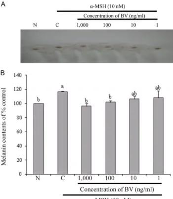
멜라닌은 세포 내의 소기관인 ribosome에서 tyrosinase라는 효 소의 생합성에에서 합성되기 시작한다. 이 효소의 작용으로 아 미노산의 일종인 tyrosine에서 몇 단계의 합성을 거쳐, 기저층의 멜라닌 세포 melanin site라는 흑색소포 표면에 침착하여 검고 갈색의 작은 멜라닌 입자가 만들어 진다.1)화장품의 원료로 사 용되고 있는 봉독의 멜라닌 합성에 미치는 영향을 살펴보기 위 해 B16F1 melanoma 세포에서의 멜라닌 생합성 저해활성을 측 정하였다(Fig. 2). Fig. 2A에서 보는 바와 같이 봉독 1 µg, 100 ng 처리는 α-MSH 처리군에 비교하여 세포내의 멜라닌 합성이 크
Fig. 1− Cell viability of BV on B16F1 melanoma cells by MTT assay. The cells were stimulated with 10 nM α-MSH for 48 hr and the medium was exchanged with fresh medium containing or not containing various concentrations of BV for 24 hr. Data are presented as mean±SEM of three independent experiments. N is cell only. Different letters indicate a significant difference with p<0.05.
Fig. 2− Effect of BV on melanin synthesis in B16F1 melanoma cells. The cells were stimulated with 10 nM α-MSH for 48 hr and the medium was exchanged with fresh medium containing or not containing various concentrations of BV for 24 hr. Data are presented as mean±SEM of three independent experiments. (A) Photograph of cell pellets.
(B) Synthesized melanin contents. N is cell only. Different letters indicate a significant difference with p<0.05.
게 억제되었으며 α-MSH를 처리하지 않은 정상 대조군과 비슷 한 수준이었다. 봉독 10 ng과 1 ng 처리는 멜라닌 합성을 억제하 는 경향을 보였으나, 군간 유의성은 나타나지 않았다(Fig. 2A).
또한 멜라닌 합성 억제효과를 육안으로 확인하기 위해 세포를 수 거하기 전 세포의 형태를 관찰하였다(Fig. 2B). 사진에서 보는 바 와 같이 α-MSH 처리군은 매우 진하게 멜라닌 생성 정도를 확 인할 수 있었으나, 정상대조군과 100 ng 및 1 µg 봉독 처리군에 서 멜라닌 생성을 거의 확인 할 수 없었다. 그러나 1 ng 및 10 ng 농도로 봉독을 처리한 군에서는 멜라닌 합성이 진하게 나타났다.
이러한 결과로 α-MSH에 의해 멜라닌 합성이 촉진된 B16F1 세 포에서 100 ng 이상의 농도에서 봉독이 맬라닌 합성을 효과적으 로 억제하는 것으로 확인되었다.
Tyrosinase의 단백질 발현 억제 효과
멜라닌 생합성 신호전달 체계에는 매우 다양한 신호전달물질 이 관여하고 있다. 멜라닌은 몇 가지 신호전달 기전을 통하여 합 성되는데, 그 중 cAMP/PKA 경로가 멜라닌 합성의 주요 경로로 서 UV에 피부가 노출되었을 때 맬라닌 세포의 cAMP가 증가되 고 하류 신호전달 물질인 PKA를 활성화시키며, CREB를 거쳐 MITF의 발현을 증가시켜, tyrosinase의 합성을 촉진시켜 멜라닌 합성을 증가시킨다.8)봉독의 멜라닌 억제효과 검증을 위한 멜라
닌 합성 관련인자인 tyrosinase 단백질 발현 정도를 측정하였다 (Fig. 4). 1µg과 100 ng의 농도로 처리한 결과, 봉독이 tyrosinase 발현이 크게 감소되는 것으로 확인되었다. 이들 처리군에서의 발 현량은 α-MSH 처리군에 비해 각각 36.1%, 31.7% 정도 발현이 감소되었으며 이는 정상 대조군과 비슷한 수준이었다. 이는 봉 독이 tyrosinase 발현 억제를 통하여 멜라닌 생성을 감소시키는 것으로 사료된다. 그러나 봉독 10 ng과 1 ng 처리는 mRNA 및 단백질 발현에 군간 유의성이 없었다(Fig. 4).
Tyrosinase의 mRNA 발현 억제 효과
봉독이 멜라닌 합성 관련 단백질인 tyrosinase의 발현 억제가 확인됨에 따라 tyrosinase mRNA 발현에 미치는 영향을 측정하 였다. 그 결과, 1 µg과 100 ng의 농도로 봉독을 처리한 군에서 tyrosinase mRNA 발현이 크게 억제되는 것으로 확인되었으며, 발현량이 α-MSH 처리군에 비해 각각 56%, 46% 정도 감소되었 다. 이는 정상 대조군과 비슷한 수준이었다(Fig. 4).
Tyrosinase는 전사인자인 microphthalmia associated trans- cription factor(MITF)에 의해 조절되는데, MITF는 멜라닌 생성 관련 유전자의 발현(tyrosinase, TRP-1과 2)을 활성화시켜 멜라 닌 생성을 증가시키고 멜라닌 생성세포의 증식 및 생존을 조절
Fig. 4− Effect of BV on melanin synthesis related enzyme tyrosinase protein expression in B16F1 melanoma cells by Western blot. The cells were stimulated with 10 nM α-MSH for 48 hr and the medium was exchanged with fresh medium containing or not containing various concentrations of BV for 24 hr. Data are presented as mean±SEM of three independent experiments. (A) Western blot analysis of protein for tyrosinase and β-actin. (B) he relative band intensity of the enzymes was normalized by β-actin.
Different letters indicate a significant difference with p<
0.05.
Fig. 3− Effect of BV on melanin synthesis related enzyme tyrosinase mRNA expression in B16F1 melanoma cells by RT-PCR.
The cells were stimulated with 10 nM α-MSH for 48 hr and the medium was exchanged with fresh medium containing or not containing various concentrations of BV for 24 hr. Data are presented as mean±SEM of three independent experiments. (A) RT-PCR analysis of mRNA for tyrosinase and GAPDH. (B) The relative band intensity of the enzymes was normalized by GAPDH. Different letters indicate a significant difference with p<0.05.
한다고 알려져있다.22-24)또한, 멜라닌 생성 자극 호르몬인 α- MSH 역시 MITF 발현을 자극하는 것으로 알려져 있다.25,26) α- MSH는 cAMP 신호전달기전을 자극하는데, 활성화된 cAMP는 cAMP responsive element binding protein과 cAMP responsive element domain 과의 결합을 자극하여 최종적으로, MITF의 발 현을 촉진시킨다.27,28) Oh 등은 α-MSH으로 멜라닌 합성이 유도 된 B16F1 melanoma 세포에 고무나무추출물을 처리한 결과, MITF의 발현 억제와 함께 tyrosinase의 발현 또한 감소함을 보 고하였다.29)현재 미백제로 가장 많이 사용되는 알부틴은 L- tyrosine과 경쟁적으로 작용하는 저해제이며, 코직산은 tyrosinase 의 활성 부위의 cooper를 chelating하여 tyrosine에서 DOPA로 그리고 DOPA에서 DOPA quinone으로 진행되는 과정을 저해한 다고 알려져 있다.4,5)한편, tyrosinase glycosylation 저해제에 대 한 보고들에 의하면 tyrosinase가 carbohydrate moieties 측면에 서 T1, T2, T3로 다르며, B16 melanoma 세포에서 glucosamine 과 tunicamycine 같은 물질들은 tyrosinase의 glycosylation 반응 을 저해야 멜라닌 합성이 완전히 억제될 수 있음을 보고하였다.
그리고 ER processing enzyme인 aglucosidase I, II의 저해제인 NB-DNJ가 B16 melanoma 세포의 활성을 억제하며 비처리군에 비해 2%의 멜라닌만이 합성되었다고 보고하여 tyrosinase glycosylation 저해제에 대한 연구들이 활발히 이루어지고 있음 을 알 수 있다.29) 비록, 본 실험에서는 cAMP 의존에 의한 MITF 의 발현 정도는 측정하지 않았으나, 봉독이 tyrosinase 발현 억 제를 통해 멜라닌 합성이 감소되는 것으로 확인되었다. 따라서 정제봉독의 미백효과를 확인하였고, 천연 미백 소재로 활용될 수 있을 것으로 기대되며 향후 cAMP 의존에 의한 MITF 의 발현 기작에 대한 연구와 동물실험을 통한 미백효과 검증이 필요하다.
결 론
본 연구는 화장품의 원료로 사용되고 있는 정제봉독의 멜라닌 생성 억제에 미치는 영향에 대해 살펴보기 위하여, α-MSH로 멜 라닌 합성이 유도된 B16F1 melanoma 세포에서 멜라닌 생성 억 제, tyrosinase의 단백질 및 mRNA 발현 억제 효과에 대해 연구 하였다. 그 결과, 봉독 1 µg 및 100 ng 처리는 α-MSH 처리군에 비해 멜라닌 합성을 억제하였으며, 멜라닌 합성 주요효소인 tyrosinase의 mRNA 및 단백질 발현 역시 효과적으로 억제하였 다. 이는, 봉독이 tyrosinase의 발현 억제를 통하여 멜라닌 합성 억제효과를 나타내는 것으로 판단되며, 추후 미백화장품 개발을 위한 기능성 물질로서의 가능성을 제시한다.
감사의 말씀
본 연구는 농촌진흥청 차세대바이오그린21사업(과제번호:
PJ008130012012)에 의하여 수행되었으므로 감사를 드립니다.
참고문헌
1) Urabe, K., Nakayama, J. and Hori, Y. : Mixed epidermal and dermal hypermelanoses. In "The pigmentary System:
Physiology and Pathophysiology", eds. Norlund, J. J., Boissy, R. E., Hearing, V. J., King, R. A., Ortonne, J. P., Oxford University Press, New York, p. 909 (1998).
2) Chang, T. S. : An updated review of tyrosinase inhibitors. Int.
J. Mol. Sci. 10, 2440 (2009).
3) Imokawa, G. : Autocrine and paracrine regulation of melanocytes in human skin and in pigmentary disorders.
Pigment. Cell Res. 17, 96 (2004).
4) Ando, H., Kondoh, H., Ichihashi, M. and Hearing, V. J. : Approaches to identify inhibitors of melanin biosynthesis via the quality control of tyrosinase. J. Invest. Dermatol. 127, 751 (2007).
5) Solano, F., Briganti, S., Picardo, M. and Ghanem, G. : Hypopigmenting agents: an updated review on biological, chemical and clinical aspects. Pigment. Cell Res. 19, 550 (2006).
6) Hearing, V. J. : Biochemical control of melanogenesis and melanosomal organization. J. Investig. Dermatol. Symp. Proc. 4, 24 (1999).
7) del Marmol, V. and Beermann, F. : Tyrosinase and related proteins in mammalian pigmentation. FEBS Lett. 381, 165 (1996).
8) Mallick, S., Singh, S. K., Sarkar, C., Saha, B. and Bhadra, R. : Human placental lipid induces melanogenesis by increasing the expression of tyrosinase and its related proteins in vitro.
Pigment. Cell Res. 18, 25 (2005).
9) 김민정, 박상동, 이아람, 김경호, 장준혁, 김갑성 : 쥐의 Collagen 유발 관절염의 활액에서 단백분해효소의 활성 및 유리기 손상에 미치는 봉독약침의 억제효과. 대한침구학회지 19, 161 (2002).
10) Kim, H. W., Kwon, Y. B., Ham, T. W., Roh, D. H., Yoon, S. Y., Lee, H. J., Han, H. J., Yang, I. S., Beitz, A. J. and Lee, J. H. : Acupoint stimulation using bee venom attenuates formalin- induced pain behavior and spinal cord fos expression in rats. J.
Vet. Med. Sci. 65, 349 (2003).
11) Piek, T. : Venoms of the Hymenoptera. Academic Press, London (1986)
12) Habermann, E. and Reiz, K. G. : On the biochemistry of bee venom pep-tides, melittin and apamin. Biochemistry 343, 192 (1965).
13) Fennell, J. F., Shipman, W. H. and Cole, L. J. : Antibacterial action of a bee venom fraction (melittin) against a penicillin- resistant Staphylococcus and other microorganisms. Res. Dev.
Tech. Rep. 5, 1 (1967).
14) Curcio-Vonlanthen, V., Schneider, C. H., Frutig, K., Blaser, K.
and Kalbacher, H. : Molecular parameters in melittin immunogenicity. J. Pept Sci. 3, 267 (1997).
15) Rudenko, S. V. and Nipot, E. E. : Modula-tion of melittin- induced hemolysis of erythocytes. Biokhimiia. 61, 2116 (1996).
16) Han, S. M., Lee, K. G., Yeo, J. H., Kweon, H. Y., Woo, S. O., Lee, I. K., Lee, M. L., Lee, M. Y., Baek, H. J. and Bae, K. H. : Studies on the antimicorbial effect of collected bee venom using electric shock method (I). Korean J. Apiculture 20, 53 (2005).
17) Han, S. M., Lee, K. G., Yeo, J. H., Baek, H. J. and Park, K. K. : Antibacterial and anti-inflammatory effects of honeybee (Apis mellifera) venom against acne-inducing bacteria. J. Med. Plant.
Res. 4, 459 (2010).
18) Han, S. M., Lee, K. G., Yeo, J. H., Kim, W. T. and Park, K. K. : Biological effects of treatment of an animal skin wound with honeybee (Apis mellifera L.) venom. J. Plast. Reconstr. Aesthet.
Surg. 64, e67 (2011).
19) 한상미, 이광길, 여주홍, 우순옥, 권해용 : 봉독의 간이 정제 방 법, 대한민국특허 10-075881 (2007).
20) Chung, M. J., Walker, P. A., Brown, R. W. and Hogstrand, C. : ZINC-mediated gene expression offers protection against H2O2-induced cytotoxicity. Toxicol. Appl. Pharmacol. 205, 225 (2005).
21) Hosoi, J., Abe, E., Suda, T. and Kuroki, T. : Regulation of melanin synthesis of B16 mouse melanoma cells by 1 alpha, 25-dihydroxyvitamin D3 and retinoic acid. Cancer Res. 45, 1474 (1985).
22) Hodgkinson, C. A., Moore, K. J., Nakayama, A., Steingrímsson, E., Copeland, N. G., Jenkins, N. A. and Arnheiter, H. : Mutations at the mouse microphthalmia locus are associated
with defects in a gene encoding a novel basic-helix-loop-helix- zipper protein. Cell. 74, 395 (1993).
23) Lowings, P., Yavuzer, U. and Goding, C. R. : Positive and negative elements regulate a melanocyte-specific promoter.
Mol. Cell. Biol. 12, 3653 (1992).
24) Yavuzer, U., Keenan, E., Lowings, P., Vachtenheim, J., Currie, G. and Goding, C. R. : The Microphthalmia gene product interacts with the retinoblastoma protein in vitro and is a target for deregulation of melanocyte-specific transcription.
Oncogene. 10, 123 (1995).
25) Bertolotto, C. , Abbe, P., Hemesath, T. J., Bille, K., Fisher, D. E., Ortonne, J. P. and Ballotti, R. : Microphthalmia gene product as a signal transducer in cAMP-induced differentiation of melanocytes. J. Cell Biol. 142, 827 (1998).
26) Bertolotto, C., Buscà, R., Abbe, P., Bille, K., Aberdam, E., Ortonne, J. P. and Ballotti, R. : Different cis-acting elements are involved in the regulation of TRP1 and TRP2 promoter activities by cyclic AMP: pivotal role of M boxes (GTCATGTGCT) and of microphthalmia. Mol. Cell Biol. 18, 694 (1998).
27) Roesler, W. J., Park, E. A. and McFie, P. J. : Characterization of CCAAT/enhancer-binding protein alpha as a cyclic AMP- responsive nuclear regulator. J. Biol. Chem. 273, 14950 (1998).
28) Karin, M. : Signal transduction from the cell surface to the nucleus through the phosphorylation of transcription factors.
Curr. Opin. Cell Biol. 6, 415 (1994).
29) Oh, M. J., Hamid, M. A., Ngadiran, S., Seo, Y. K., Sarmidi, M. R. and Park, C. S. : Ficus deltoidea (Mas cotek) extract exerted anti-melanogenic activity by preventing tyrosinase activity in vitro and by suppressing tyrosinase gene expression in B16F1 melanoma cells. Arch. Dermatol. Res. 303, 161 (2011).