The clinical and radiographic findings of lung cancer have been well established many journals. Even if the radiographic findings of lung cancer show a typical pattern, the specific cell type of lung cancer sometimes needs to be determined prior to a pathological diagnosis. For example, the usual finding of a squamous cell carcinoma is similar to other cancer types such as an adenocarcinoma or a small cell carcinoma but with a lower incidence. Therefore, it should not be used to make a diagnosis of the cell type prior to a pathological diagnosis. Many unusual findings of lung cancer, so called atypical pattern have been reported, but atypical findings are widely accepted. The more important thing is not to diagnose a specific cell type of cancer but to differentiate it from other benign conditions such as tuberculosis, fungal infections or organizing pneumonia. This paper presents typical information of the cell type of lung cancer along with the atypical radiographic findings. (Tuberc Respir Dis 2005; 58: 554-561)
Key words : Lung neoplasms, Radiography, Diagnosis 경희대학교 의과대학 진단방사선과학교실
성 동 욱
Radiological Findings of Lung Cancer: Focus on Atypical Pattern
Dong-Wook Sung, M.D.
Diagnostic Radiology Kyung Hee University Hospital, Seoul, Korea
Address for correspondence : Dong-Wook Sung, M.D.
Department of Diagnostic Radiology
Kyung Hee University Hospital #1 Hoeki-dong, Dongdaemun-gu, Seoul, 130-702, Korea Phone : 02-958-8616 Fax : 02-968-0787 E-mail : [email protected]
서 론
폐암의 임상적 및 전형적 방사선 소견에 관한 설명 은 잘 정리되어 여러 책이나 논문에서 소개되고 있다1,2. 전형적 소견으로 방사선 검사에서 나타나는 경우, 폐 암의 세포 성분까지 추측할 수 있으나, 전형적인 소견 이 어느 한 가지 세포 형태의 폐암에서만 관찰되는 경 우는 없으므로 세포 성분까지 말하려 하는 오류는 범 하여서는 안 된다. 비정형적인 소견은 그 빈도도 낮고, 어느 경우에는 감염성 질환의 특징적 방사선 소견으 로 나타나는 경우도 있어, 폐암을 방사선학적으로 진 단하려 할 때는 폐암이 아니지 않을 까 하는 의문과, 역으로 감염성 질환의 특징적 방사선 소견이더라도 혹시 폐암이 이렇게 나타나지 않을 까 하는 염두를 항 상 생각하며 진단하는 습관을 가져야 한다. 특히 조금 깊게 폐암의 각 세포별 특징을 알게 되는 경우에 세포 성분까지 진단하려고 하는 과오를 범하기 쉽다. 폐암 의 진단에서는 세포 성분을 맞추려는 노력보다는 다
른 감별 진단에 더 치중하여야 한다. 여기서는 전형적 인 소견의 기술과 더불어, 놓치기 쉬운 비전형적인 소 견을 중심으로 기술한다.
폐암의 분류
WHO에 의해 ① 편평상피세포암(squamous cell carcinoma) ② 선암(adenocarcinoma) ③ 소세포암(small cell carcinoma) ④ 대세포암(large cell carcinoma)의 네가지 형태의 세포 형태로 구분하고, 이들이 전체 폐 암의 90-95 %를 차지한다(Table 1)3. 일반적으로 6:4 의 비율로 우측폐에 많이 발생하며, 상엽의 전방엽에 호발한다.
폐암에서 조직학적 세포 형태를 나누는 것은 그 예 후에는 중요한 인자는 아니나 치료에 중요한 역할을 하기 때문이다. 예후는 세포 형태보다는 병기판정에 의해 결정된다.
편평상피세포암 1. 중심성 종괴
중심성 기관지에서 발생하는 경우가 65 % 정도이
Table 1. Histologic classification of lung tumors*
(1) Squamous carcinoma (epidermoid carcinoma) Variant : Spindle cell (squamous) carcinoma (2) Small cell carcinoma
a. Oat cell carcinoma b. Intermediate cell type c. Combined oat cell carcinoma (3) Adenocarcinoma
a. Acinar adenocarcinoma b. Papillary adenocarcinoma c. Bronchiolo-alveolar carcinoma d. Solid carcionma with mucus formation (4) Large cell carcinoma
Variants :
a. Giant cell carcinoma b. Clear cell carcinoma (5) Adenosquamous carcinoma (6) Carcinoid tumor
(7) Bronchial gland carcinomas a. Adenoid cystic carcinoma b. Mucoepidermoid carcinoma c. Others
(8) Others
* WHO classification, 1981
Figure 1. Squamous cell carcinoma in a 50-year-old man. CT shows multiple nodular thickening on the posterior wall of the right upper lobe bronchus. Chest PA(not shown) was normal, and a malignancy was detected on sputum cytology.
며, 방사선학적으로나 임상적으로 종괴를 발견하지 못해도 객담 검사에서 암세포를 발견할 수 있다(Fig.
1). 기관을 막고 있는 종괴에 의해 이차적 소견이 나타 나며, 여기에는 무기폐, 폐쇄성 폐렴이 보인다. 대개의 경우 공기-기관지 음영 (air-bronchogram)이 폐쇄성 폐렴의 내부에 보이지 않게 되며, 이것으로 원발 종괴 를 확인하지 못하더라도 결핵성이나, 감염성 폐쇄와 감별을 할 수 있다. 재발성 폐렴은 흉부X선에서 종양 을 발견할 수 없어도 기관지내 병변을 감별하기위해 기관지내시경이 필요할 수 있다(Fig. 2) 폐쇄성 폐렴 을 치료하지 않으면 공동이 생길 수 있다. 그 외 기관 지 확장증, 점액 충전(mucoid impaction), 체크밸브에 의한 병변측의 공기 충만이 나타날 수 있다. 상엽을 막는 폐암의 경우 Golden's S sign을 관찰할 수 있고, 페암에 의한 골 전위로 인해 골 파괴, 종격동 구조물 의 침범, 상대정맥의 압박, 폐쇄, 늑간신경의 침범으로 인한 횡격막 거상이 나타날 수 있다. 중심상 종괴와 그 이하 부위의 폐쇄성 폐렴을 일으키는 질환은 폐암 뿐 만 아니라, 기질성 폐렴(organizing pneumonia), 폐 방선균증(pulmonary actinomycosis), 결핵 때문에 나 타날 수 있어, 항상 위의 질환들을 감별해야 한다(Fig.
3). 종괴의 변연부는 교미침 모양(spiculation)을 할 수
있다. 이는 악성, 양성에서 모두 관찰할 수 있으나4, Han 등5의 보고에 의하면 폐암에서 보이는 교미침 모 양의 길이는 폐암에서 평균 5.7mm, 결핵성육아종에 서는 13.8mm로 의미있는 차이가 보이며, 그 숫자는 차이가 없다고 하였다. 폐암과 결핵성 육아종을 감별 하는 한 가지 방법이 될 수 있겠다.
2. 변연부 종괴
편평상피세포암의 30-40 %에서 폐변연부에 위치 한다. 폐암 중에서 가장 크기가 커질 수 있다. 가끔 변 연부에서 종괴 형태를 가지지 않고 결핵성 형태, 변연 부 무기폐 모양으로 흉막을 따라 옆으로 넓게 퍼지는 경우가 있는데, 이런 경우 항상 폐암의 가능성을 생각 해야 한다(Fig. 4). 주위의 골파괴가 있으면 진단에 도 움이 된다. 다른 폐암과는 달리 공동형성을 잘 한다.
특히 상엽에 발생시 공동형성을 잘 한다. Chaudhuri 등6의 보고에 의하면 공동을 형성한 100예의 폐암 중 82예가 편평상피세포암 이었다. 공동의 벽은 두껍고 불규칙한 내부 벽을 가진다. 공동내로 돌출하는 종양 을 형성할 수 있어 이를 mural nodule이라 한다. 드물
Figure 2. Squamous cell carcinoma with pneumonia in a 68-year-old man.
A. Chest PA shows an ill-defined opacity on the right lower lung zone, impressed initially pneumonia.
B. Follow up PA after 3 weeks with antibiotic treatment shows consolidation still remaining.
C. CT shows an intraluminal mass on the right lower lobe bronchus, and distal postobstructive pneumonia.
Figure 3. Variable causes of a similar pattern of a mass like lesion with distal obstructive pneumonitis.
A, B. Actinomycosis in a 45-year-old man.
C. Organizing pneumonia in a 62-year-old man.
D. Bronchial aspergillosis in a 78-year-old man.
A B C
Figure 4. Peripheral type of lung cancer.
A. CT shows a necrotic mass(squamous cell carcinoma) on the left lower lobe superior segment. The adjacent rib is destroyed. The mass is broad based to the thoracic wall. It has grown mainly lateral rather than into the lung parenchyma.
B, C. CT of a 71-year-old man shows irregular peripheral air-space consolidation on the right lower lobe.
Air-bronchograms can be seen within the opacity and it broad based to the thoracic wall. It was confirmed as an adenocarcinoma with percutaneous needle aspiration cytology.
A B
Figure 5. Cavitary change in a squamous cell carcinoma in a 70-year-old woman.
A. CT shows a well defined mass with total inner necrosis on the right upper lobe.
B. CT of the lung setting after 5 days shows a cavitary change in the mass. Its inner wall is smooth, but focal irregular thickening on the medial inferior wall indicates the tumor site.
게 공동의 벽 두께가 아주 얇아 기관지원성낭종(bro
nchogenic cyst)과 감별이 필요하다. 이는 과다한 괴 사 때문이다(Fig. 5).
3. Pancoast Tumor
Pancoast tumor 혹은 상구종양(superior sulcus tumor) 의 가장 흔한 형태이며 흉막을 가로질러 흉벽, 늑골 파괴, 후두 신경 마비, 팔신경얼기(brachial plexus)를 침범할 수 있다. 단순흉부X-선 촬영에서 골파괴를 잘 인지하지 못하는 경우가 많으므로 양쪽 폐첨부 흉막 비후의 차이가 5 mm 이상이면 주의해야 하며 반드시
CT로 폐암의 존재를 확인해야 한다.
4. 전이
원격전이는 잘 하지 않으나, 부검에서는 약 25 % 에서 발견된다. 주로 간, 부신, 신장, 뼈에 전이된다.
선 암
전체 폐암의 25-30 %를 차지하며 대개 변연부 종 괴의 형태 (60-70%)로 나타난다. 편평상피세포암의 감소에 비해 증가 추세에 있다. 이는 여성에서의 선암
A B C
B
D A
C
A B
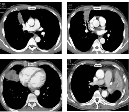
Figure 6. Adenocarcinoma in a 57-year-old woman.
CT(A, B) shows enlargement of the mediastinal, hilar and subcarinal lymph nodes, which has a similar pattern to that of a small cell carcinoma. Radiographically, there is an absence of a primary parenchymal mass.
발생이 증가하기 때문이다.
1. 변연부 종괴
선암의 60-70 %가 변연부 형태이며, 변연부 종괴의 30-40 %가 선암이다. 비호흡기 증상으로 뼈통증, 허 약, 소화불량, 식욕부진이 잘 나타나며, 종괴의 크기가 작아도 초기에 전이를 잘 한다. 선암은 이전에 존재한 폐의 만성 질환, 외부의 독소, 진폐 분진, 화학 가스, 석면폐, 마이코박테리아, 진균, 바이러스와도 밀접한 관계가 있다.
방사선 소견은 둥글거나 타원형을 이루고 대개 4 cm 이내이나, Woodring 등7은 4 cm 이상도 50 % 보 고하고 있다. 종괴의 변연부가 불규칙하거나, 불분명 할 수도 있다. 방사형 햇무리(corona radiata) 혹은 햇 살모양(sunburst appearance)를 취할 수 있는데 이는 종괴의 침윤 혹은 결절 주변의 결합조직 반응 때문이 다. 종괴의 내부에 공기-기관지 음영이 보일 수 있 다. 그 외 소견으로 다발성 폐종괴를 형성할 수 있 으며, 석회화는 대부분 주변의 석회를 침식한 경우 로 변연부에 치우쳐 있다. 수년에 걸쳐 천천히 자랄 수 있으며 혹은 몇 년간 변연부 종괴로 변화가 없는 경우도 있다. 2-3년간 변화가 없으면 대개의 경우, 양성이라 판정하는 경우가 있는데, 아무리 양성의 소견이 보인다 하더라도 악성의 가능성을 잊어버려 서는 안 된다4.
2. 중심성 종괴
중심성 종괴의 선암이 최근 증가하여 30%에 이르 고 있다. 이는 여성에서 선암의 발생빈도가 증가하 고, 이 세포 형태의 조직병리학적 분류 방법이 변화 한 데 기인한 것이다. 중심성인 경우, 편평세포상피 암 혹은 소세포암과 같은 방사선 소견을 보일 수 있다 (Fig. 6).
3. 전이
조기에 원격 전이를 잘 하고, 흉막 침범을 잘 한다.
흔한 전이 위치로서 종격동, 폐분부, 부신, 간, 뇌신경 계, 골수 들이다. Sider 등8의 보고에 의하면 단일 폐결 절이 있으면서, 종격동에는 림프절 종대가 없고, 오히 려 흉곽 외에 전이가 있는 경우, 선암이 가장 흔한 세 포 형태(67%)라 하였다. 한 가지 알아둘 것은 흉곽 밖 에 악성 종양이 있는 환자에서 단일 폐결절이 보이면, 원발암이 두경부, 방광, 유방, 자궁경부, 식도, 위, 난 소이면 이 단일 결절은 전이암보다는 원발성 폐암 이 따로 생긴 가능성이 높으며, 원발암이 salivary gland, 부신, 대장, 신장, 갑상선, 자궁, 흉선인 경우 에는 단일폐결절이 전이암 혹은 원발성 폐암일 경 우가 반반이다. 그러나 원발암이 melanoma, 육종, 고환암인 경우는 폐에 생긴 단일 결절은 전이암 가 능성이 높다9.
A B
Figure 7. Scar cancer in a 71-year-old man.
A. Chest PA shows a diffuse patchy and mottled air-space consolidations on both lungs, which were confirmed to be tuberculosis.
B. Chest PA after 14 years from A shows a complete resolution of previous tuberculosis, but an irregular mass on the left mid-lateral lung zone, and mediastinal lymph nodes enlargement. The mass on the left lower lung zone was confirmed to adenocarcinoma.
A B
Figure 8. Solid type of a bronchioloalveolar cell carcinoma.
A. CT shows a mass on the right lower lobe with inner bubbly lucencies.
B. CT of another patient shows a mass with inner air-bronchograms.
4. 반흔암(scar carcinoma)
오래 지속되는 폐실질 섬유화 혹은 육아종에서 발 생한 종괴를 말한다(Fig. 7). 반흔암이라 진단하기 위 해선 조직병리학적으로 hyalined fibrosed tissue 내에 고 형 부위가 존재해야 하며, 고밀도의 elastic fiber가 관찰되 며, antracotic 물질이 발견되고, 육안적인 반흔의 흔적이 존 재해야 반흔암이라 진단할 수 있다. George 등10의 보고에 의 하면, 반흔암 19예 중 선암이 11예(58%), 편평상피세포암 3 예(16%), 소세포암 3예(16%) 이었고, 반흔암이 아닌 경우 는 편평상피세포암이 52%로 가장 많았다.
세기관지폐포암 (BAC)
선암의 한 조직학적 형태이며, 전체 폐암의 1.5-6.5
%를 차지한다. 말초세기관지 혹은 폐포벽에서 기원하 여 폐조직을 간질로 해서 주변부로 퍼져나가는 특징 이 있으며(lepidic growth), 이때 버팀간질반응(stromal response)은 적어 폐포의 구조물은 정상을 유지한다.
젊은이에서 잘 발생하고 비흡연자에서도 발생한다.
폐실질의 반흔화나 미만성간질성염증, 섬유화와도 관 련이 있다. 방사선 소견은 각기 전혀 다른 종괴의 모 습을 보인다. 단일 결절형이 가장 흔하며(43 %), 예후
A B
Figure 10. Large cell carcinoma in 59-year-old man.
A. Chest PA shows a small nodule on the right upper lobe.
B. CT shows a lobulated and fine spiculated nodule on the right upper lobe.
A B C D
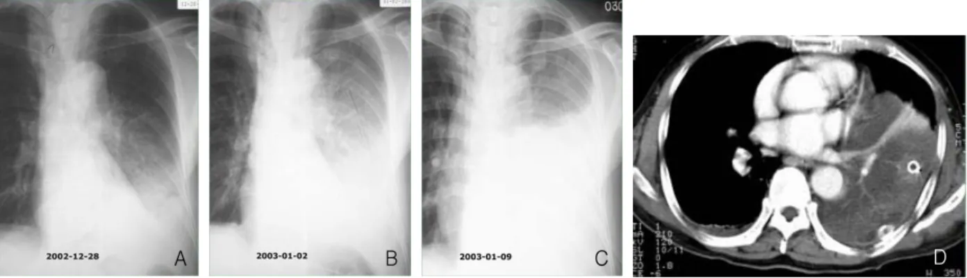
Figure 9. Rapid progression of a bronchioloalveolar cell carcinoma(BAC).
A. Initial chest PA shows a homogeneous air-space consolidation on the left lower lobe, impressed pneumonia.
B. Follow up PA after 4 days, the consolidation is more aggravated, suggesting an aggravation of the pneumonia with a parapneumonic effusion. Therefore, a chest tube was inserted.
C. PA after 7 days from B shows more aggravation of the consolidation.
D. CT shows a low density consolidation on the left lingular and lower lobes. The density is different from that of pneumonia. The CT angiogram sign and focal enhanced parenchymal collapse within the consolidation, which was confirmed to be the pneumonic type of BAC.
도 좋고, 가끔 수 년 동안 크기가 변화없이 아주 천천 히 자란다. 종양으로 폐포가 채운다 하더라도 기관지 는 정상으로 통해있어 종괴의 내부에 공기-기관지 음 영이 보인다. 혹은 가성 공동(pseudocavity), 공기방울 음영(bubbly lucency)가 나타난다(Fig. 8). 종격동 림 프절 종대, 전이는 거의 없다. 대엽성 폐렴 형태로 30%에서 나타나며, 내부에 공기-기관지 음영, 공기- 폐포 음영, CT 혈관 징후(CT angiogram sign) 들이 나타난다. CT에서 저음영으로 나타나는 데, 이는 암 세포와 과다한 점액의 분비에 기인한 것이다. 경계가 분명하거나 불분명한 결절(27 %)이 다발성으로 양측 폐에 나타날 수도 있다. 작은 결절 내에 공동을 형성 할 수 있고 결절 주위에 간유리 음영(ground-glass opacity)이 보일 수도 있다. 흉막삼출은 약 8-10 %에 서 발생되며 종격동 림프절 종대는 비교적 드물며 그 외 무기폐, 기흉, 공동형성, 흉곽 외 전이를 발생할 수 있다. 흔히 종괴 내에 공기-기관지 음영이 보이면 세 기관지폐포암 혹은 림프종(lymphoma)이라 진단할 수 있는데, 결핵성 육아종, 기질적 폐렴에서도 종괴 형태 를 지니며 그 내부에 공기-기관지 음영이 존재할 수 있으므로 주의해야 한다. 다른 형태의 폐암에서도 마 찬가지이나, 세기관지폐포암은 그 진행이 마치 급성 폐렴처럼 변화하여 나타날 수 있다. 이 때에도 CT를 촬영하여 폐경화의 모양이 정상적 폐렴과는 다른 것
으로 세기관지폐포암을 진단할 수 있다(Fig. 9).
대세포암
전체 폐암의 15 %를 차지하며 빨리 자라며 70 %에 서 변연부에서 발생한다. 선암보다 크기가 크며 종괴 의 경계가 불분명하고 소엽성을 이룬다. 공동은 드물 고 전이는 말기에 나타난다. 두가지의 아형이 있는데 심한 흡연자에 잘 나타나고 가끔 CEA (carcinoembryonic antigen)가 나타나는 거대세포암종(giant cell carcinoma) 과 많은 양의 글리코겐을 함유한 투명세포암종(clear
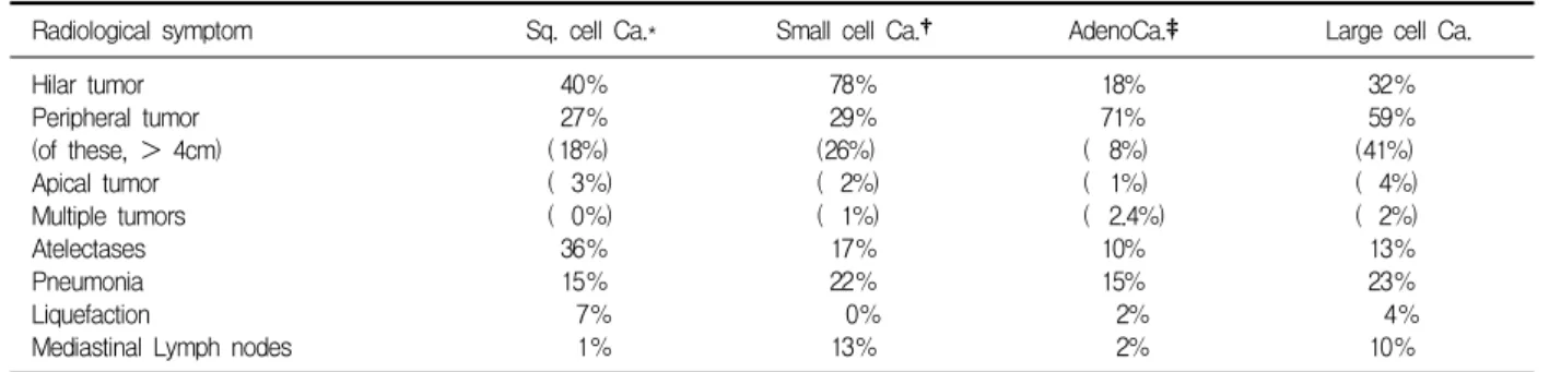
Table 2. Radiographic patterns based on cell type*
Radiological symptom Sq. cell Ca.* Small cell Ca.† AdenoCa.‡ Large cell Ca.
Hilar tumor Peripheral tumor (of these, > 4cm) Apical tumor Multiple tumors Atelectases Pneumonia Liquefaction
Mediastinal Lymph nodes
40%
27%
( 18%) ( 3%) ( 0%) 36%
15%
7%
1%
78%
29%
(26%) ( 2%) ( 1%) 17%
22%
0%
13%
18%
71%
( 8%) ( 1%) ( 2.4%)
10%
15%
2%
2%
32%
59%
(41%) ( 4%) ( 2%) 13%
23%
4%
10%
Note : *Squamous cell carcinoma. †Small cell carcinoma. ‡Adenocarcinoma.
±Large cell carcinoma
(from R.G. Fraser, J.A.P. Pare: Diseases of the Chest, Saunders, Philadelphia 1983)
cell carcinoma)이 있다. 이름처럼 외형적으로 큰 폐 암을 생각하여서는 안 되며, 크기가 작을 수도 있다는 것을 염두에 두어야 한다(Fig. 10).
소세포암 (귀리세포암종, oat cell carcinoma)
폐암의 20-30 %를 차지하며 가장 예후가 나쁘다.
조기에 원격 전이를 잘해 주위 림프절, 혈관을 빨리 침범하여 폐 증상이 나타나기 전에 전신에 퍼진다. 종 괴가 Kulchitsky-type 세포에서 기원하여 내분비성 병변을 잘 동반한다. 특히 inappropriate secretion of adenocorticotropic hormone (ACTH) (Cushing syn
drome), Eaton-Lambert syndrome (근위 근육 허약, proximal muscle weakness)이 동반한다. 방사선 소견 으로 동측의 폐분부나 종격동 림프절 전이로 인해 중 심성 종괴로 나타나고, 림프절 종대는 병변의 발견시 80%에서, 부검에서는 100%에서 관찰된다. 실제 중심 성 종괴의 70-80%가 소세포암이다. 원발성 폐암이 작 고, 커진 종격동 림프절이 원발성 폐암과 융합되어 CT에서도 커진 종격동 림프절 만 관찰되는 경우가 많 다. 변연부 종괴로 나타나는 경우는 14% 정도이며, 이 때는 매우 악성이 높으며 미분화된 세포로 구성되어 있다. 약 40%에서 흉막 삼출을 동반한다. 종괴가 단지 한쪽 흉곽만을 침범한 경우를 제한적 형태(limited type)라 하며 수술로 치유가 가능하다. 뇌전이는 진단 시 10-15 %에서 보고되고, 골수 전이는 50 % 까지 이
다. 폐암의 병리조직학적 세포형에 따른 방사선 소견 을 정리하면 표 2와 같다.
참 고 문 헌
1. Sider L. Radiographic manifestation of primary bro
nchogenic carcinoma. Radiol Clin North Am 1990;28:
583-97.
2. Grippi MA. Clinical aspects of lung cancer. Semin Roentgenol 1990;25:12-24.
3. Yesner R. Histopathology of lung cancer. Semin ult
rasound CT MR 1988;9:4-26.
4. Erasmus JJ, Connolly JE, McAdams HP, Roggli VL.
Solitary pulmonary nodules: part I. morphologic ev
aluation for differentiation of benign and malignant lesions. Radiographics 2000;20:43-58.
5. Han TI, Sung DW, Lim SJ, Yoon Y. Spiculation of lung mass on CT: carcinoma vs. tuberculoma. J Ko
rean Radiol Soc 1994;31:63-7.
6. Chaudhuri MR. Primary pulmonary cavitating car
cinoma. Thorax 1973;28:354-66.
7. Woodring JH, Stelling CB. Adenocarcinoma of the lung: a tumor with a changing pleomorphic character.
Am J Roentgenol 1983;140:657-64.
8. Sider L, Horejs D. Frequency of extrathoracic metas
tasis from bronchogenic carcinoma in patients with normal-sized hilar and mediastinal lymph nodes on CT. Am J Roentgenol 1988;151:893-5.
9. Quint LE, Park CH, Iannettoni MD. Solitary pulmo
nary nodules in patients with extrapulmonary neo
plasms. Radiology 2000;217:257-61.
10. Bakris GL. Mulopulos GP, Korchik R, Ezdinli EZ, Ro J, Yoon BH. Pulmonary scar carcinoma. Cancer 1983;52:493-7.