• 검색 결과가 없습니다.

Effects of Resuscitative Endovascular Balloon Occlusion of the Aorta in Neurotrauma: Three Cases

N/A
N/A
Protected

Academic year: 2021

Share "Effects of Resuscitative Endovascular Balloon Occlusion of the Aorta in Neurotrauma: Three Cases"

Copied!
6
0
0

로드 중.... (전체 텍스트 보기)

전체 글

(1)

Received: September 7, 2020 Accepted: September 17, 2020 Trauma and InJury

Correspondence to Ye Rim Chang, M.D., Ph.D.

Department of Trauma Surgery, Trauma Center, Dankook University Hospital, 201 Manghyang-ro, Dongnam-gu, Cheonan 31116, Korea

Tel: +82-41-550-3066 Fax: +82-41-550-0039 E-mail: [email protected]

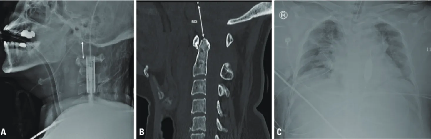
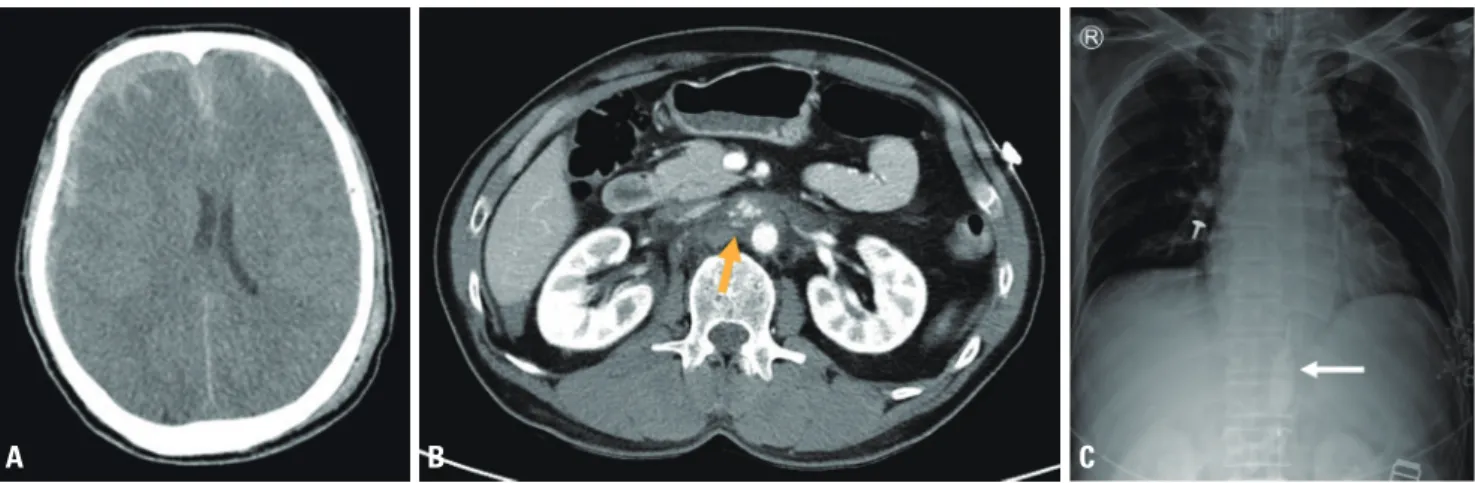
Effects of resuscitative Endovascular Balloon occlusion of the aorta in neurotrauma: Three Cases

Dong Hun Kim, M.D., M.S.

1

, Ye Rim Chang, M.D., Ph.D.

1

, Jung-Ho Yun, M.D., M.S.

2

1

Department of Trauma Surgery, Trauma Center, Dankook University Hospital, Cheonan, Korea

2

Department of Neurosurgery, Trauma Center, Dankook University Hospital, Cheonan, Korea

Resuscitative endovascular balloon occlusion of the aorta (REBOA) is widely performed as an adjunct to resuscitation or bridge to definitive control of non-compressible torso hemorrhage in patients with hemorrhagic shock. It is a crucial adjunct for the mainte- nance of cerebral and coronary perfusion during resuscitation. However, in polytrauma patients with concomitant neurotrauma, such as traumatic brain injury (TBI) or spinal cord injury, the physiological effects of REBOA are unclear. In this report on REBOA performed in a clinical setting for polytrauma patients with spinal cord injury or TBI, the physiological effects of REBOA in neurotrauma are reviewed.

Keywords: Trauma; Spinal cord; Brain; Balloon occlusion; Resuscitation

INTRODUCTION

Resuscitative endovascular balloon occlusion of the aorta (REBOA) is performed

as a bridging therapy for non-compressible torso hemorrhage (NCTH) in patients

with hemorrhagic shock [1]. It is also performed for patients with neurogenic shock

and non-traumatic cardiac arrest to support the central aortic pressure necessary for

cerebral and coronary perfusion [2,3]. However, when hemorrhagic shock occurs in

patients with concomitant traumatic brain injury (TBI) or spinal cord injury (SCI),

the effect of REBOA on the concomitant neurotrauma is unclear. In this report, we

present three patients, including one with cervical SCI and NCTH and 2 with TBI and

NCTH, who underwent REBOA, and review the effects of REBOA on the physiology

(2)

of neurotrauma.

CASE REPORT

Case 1

A 67-year-old woman had a traffic accident, and cardiac arrest was noted at the accident scene and at a local hospi- tal with return of spontaneous circulation (ROSC) after 7 and 14 minutes of cardiopulmonary resuscitation (CPR), respectively. Upon arrival from the local hospital, she was comatose and intubated, her pupils were 4 mm in size and fixed, and her vital signs were as follows: blood pres- sure, 79/64 mmHg; pulse rate, 128 bpm; and body tem- perature, 36.0°C. Focused Assessment with Sonography for Trauma (FAST) did not show the lung sliding sign in the right hemithorax or intra-abdominal fluid collection.

A lateral-view radiograph of the cervical spine revealed suspected atlanto-occipital dislocation (AOD) (Fig. 1A).

Computed tomography (CT) at the local hospital showed grade I spleen contusion, retroperitoneal hematoma around the left kidney, multiple rib fractures with hemo- pneumothorax in the right hemithorax, and AOD with a 25-mm basion-dental interval (BDI) and prevertebral hematoma (Fig. 1B). Despite volume resuscitation with crystalloid and blood transfusions, hypotension with a systolic blood pressure (SBP) less than 70 mmHg persist- ed. Therefore, Zone I REBOA using a 7-Fr balloon cathe-

ter (RESCUE Balloon

TM

, Tokai Medical Products, Aichi, Japan) was performed (Fig. 1C), and her blood pressure increased temporarily and then decreased despite 40-mL full balloon inflation and high-dose vasopressor infusion.

Cardiac arrest recurred 8 minutes after balloon inflation, and ROSC was not achieved in 36 minutes of CPR.

Case 2

A 58-year-old man sustained blunt trauma from an 8-m fall. His Glasgow Coma Scale score was 3. His pupils had a size of 6 mm on light reflex and were fixed. Upon arrival, his blood pressure and pulse rate were 98/60 mmHg and 92 bpm respectively, and he had multiple facial abrasions.

FAST showed no intraabdominal fluid collection, but 20-minute serial FAST showed a small amount of fluid in the Morison pouch and perisplenic space. A CT scan revealed subdural hematoma (SDH) in the right hemi- sphere with a vague boundary between white and gray matter consistent with brain edema (Fig. 2A), multiple rib fractures, and central inframesocolic retroperitoneal hematoma in the para-aortic area beneath the left renal vein with small fluid collection in the abdominal cavity (Fig. 2B). After the CT scan, the patient became hemody- namically unstable with a blood pressure of 61/38 mmHg and a pulse rate of 91 bpm. Zone I REBOA was then per- formed using a RESCUE Balloon

TM

(Fig. 2C). Following balloon inflation with 15 mL of saline, his blood pressure and pulse rate increased to 101/60 mmHg and 112 bpm,

Fig. 1. (A) Lateral-view radiograph of the cervical spine showing a distraction-type AOD. (B) Computed tomography scan showing AOD with a 25-mm basion-dental interval and a thick prevertebral hematoma. (C) Chest radiograph showing the positioning of the balloon catheter in the descending thoracic aorta. AOD: atlanto-occipital dislocation, BDI: basion-dental interval.

A b C

(3)

respectively. Simultaneous craniectomy and laparotomy were then performed. A central inframesocolic retroper- itoneal hematoma and superficial liver and spleen lacera- tions with a small intraperitoneal hematoma were found during crash laparotomy. As the retroperitoneal hemato- ma was approached and controlled by midline infrarenal aortic exposure, it was possible to complete weaning from REBOA. However, the bleeding from the liver and spleen lacerations were not easily controlled even though the injuries were minor. During laparotomy, the SDH was evacuated and severe brain edema was observed by cra- niectomy. Since the brain injury was virtually irreversible, both surgical procedures were finished without further

steps, and laparotomy was finished with temporary ab- dominal closure. Postoperatively, the patient remained hemodynamically unstable despite volume resuscitation and high-dose vasopressor infusion, and he died 341 min- utes after arrival.

Case 3

A 72-year-old man was admitted after a motorcycle acci- dent. Upon arrival, he was comatose, and his pupils were 3 mm in size and fixed. He had a blood pressure of 71/54 mmHg, a pulse rate of 120 bpm, a respiratory rate of 12 breaths/minutes, and a hemoglobin level of 9.5 g/dL. He had multiple facial lacerations and cerebrospinal fluid

Fig. 2. (A) Brain computed tomography (CT) scan showing subdural hemorrhage and subarachnoid hemorrhage in the right hemisphere with midline shifting and diffuse brain edema. (B) Abdominal CT scan showing para-aortic hematoma with contrast extravasation beneath the left renal vein (arrow) and fluid collection in the abdominal cavity. (C) Chest radiograph showing that the positioning of the balloon catheter (arrow) had been checked in the abdominal aorta, and the catheter was then adjusted 7 cm in the cranial direction for proper positioning in the thoracic aorta in Zone I.

A b C

Fig. 3. (A) Brain computed tomography (CT) scan showing subdural hemorrhage and subarachnoid hemorrhage in both hemispheres with diffuse brain edema. (B) Abdominal CT scan showing spleen laceration with contrast extravasation (arrows) and fluid collection in the perisplenic space.

(C) Chest radiograph showing proper positioning of the balloon catheter in the descending thoracic aorta.

A b C

(4)

leakage from the nose and left ear. FAST did not initially show intra-abdominal fluid collection, but 10-minute serial FAST showed a small amount of fluid in the sple- norenal recess. Since the patient responded to volume resuscitation, he could undergo imaging evaluation. A CT scan revealed subarachnoid hemorrhage and SDH in both hemispheres with basal skull fracture and pneumoceph- alus (Fig. 3A), a grade III spleen laceration with contrast extravasation and perisplenic fluid collection (Fig. 3B), and multiple rib fractures with hemopneumothorax in the left hemithorax. Shortly after the imaging evaluation, hypotension gradually developed even though volume resuscitation and low-dose vasopressor infusion were continued. With his blood pressure at 48/26 mmHg and pulse rate at 89 bpm, Zone I REBOA using a RESCUE Balloon

TM

was performed 55 minutes after arrival (Fig. 3C).

After 11-mL balloon inflation, his blood pressure and pulse rate rose to 127/76 mmHg and 128 bpm, respective- ly. Since a poor outcome was expected due to severe brain edema, craniectomy was performed prior to laparotomy.

Craniectomy revealed severe brain edema after hematoma removal and was completed with suspension of the crash laparotomy. Weaning from REBOA was completed in the

intensive care unit about 1 hour after the operation and replaced with vasopressor infusion. The patient remained hemodynamically unstable despite high-dose vasopressor infusion and died 4 hours after the operation.

The injury severity scores of patients 1, 2, and 3 were 50, 38, and 43, respectively. The procedural timepoints with regards to REBOA and the clinical features of the three patients are shown in Table 1.

DISCUSSION

REBOA is a bridging modality for patients with hemor- rhagic shock that is aimed at maintaining cerebral and coronary perfusion, while reducing distal bleeding, until hemorrhage is controlled. It is widely performed in cases of postpartum hemorrhage, upper gastrointestinal bleed- ing, pelvic surgery, trauma, ruptured aortic aneurysm, and non-traumatic cardiac arrest [2,4]. The main indica- tion for REBOA in trauma settings is NCTH with hemor- rhagic shock including abdominal or pelvic hemorrhage [1]. However, traumatic accidents often involve complex injury patterns. Patients often suffer polytrauma, in which a neurological injury such as TBI or SCI is among the most common factors. NCTH with concomitant trau- matic neurological injury is not known to be a contraindi- cation of REBOA. Thus, it is important to understand the effects of REBOA on NCTH in patients with hemorrhagic shock and concomitant neurological injury.

There are few reports and no consensus on REBOA as an adjunct for patients with neurogenic shock resulting from traumatic spine injury. Gray and Dieudonne [3]

reported a case of successful REBOA in a patient with neurogenic shock secondary to a comminuted fracture at C5. In case 1, high-level SCI secondary to AOD might have resulted in brainstem injury unresponsive to com- plete occlusion of aortic flow, which is termed complete REBOA. A BDI ≥16 mm or AOD with a neurological deficit is especially associated with mortality [5]. Further, in an animal study on Zone I REBOA using a primate model, it was reported that a long duration of balloon in- flation during REBOA may result in spinal cord ischemia [6]. Therefore, we should keep in mind that patients with traumatic SCI may be more vulnerable to ischemic and Table 1. Critical time courses and clinical features of patients

with regards to REBOA

Parameters Case 1 Case 2 Case 3

Door-to-femoral puncture time (minutes) 33 84 55 Door-to-balloon inflation time (minutes) 40 91 66 Maximal volume of balloon inflation (mL) 40 15 20 SBP before balloon inflation (mmHg) 62 61 48 SBP after balloon inflation (mmHg) 66 105 127 Maximal SBP during REBOA (mmHg) 66 109 140 Total duration of aortic occlusion (minutes) 40 125 138 Hemoglobin level on arrival (g/dL) 7.8 14.3 10.7 Lactate level on arrival (mmol/L) 8.6 3.3 3.6

INR on arrival 1.70 1.17 1.26

24-hour transfusion units required (RBC:FFP) 8:5 15:10 21:15 Door-to-operating room time (minutes) - 106 82

Door-to-death time (minutes) 90 341 396

FFP: fresh frozen plasma, INR: international normalized ratio, RBC: red

blood cells, REBOA: resuscitative endovascular balloon occlusion of the

aorta, SBP: systolic blood pressure.

(5)

reperfusion injury from REBOA.

Hemorrhagic shock in trauma patients often occurs with concomitant TBI; therefore, it is important to un- derstand the relationship between aortic occlusion and cerebral physiology following TBI to maintain optimal physiology of the vital organs. Aortic occlusion increases coronary and cerebral perfusion [7]. According to a re- ported smooth U-shaped correlation between SBP and TBI outcomes, outcomes can improve as SBP increases up to 135 mmHg [8]. Thus, supra-physiological blood pressure and carotid blood flow created by aortic occlu- sion may worsen cerebral edema, increase the intracranial pressure, or exacerbate intracranial hemorrhage [9-11].

Moreover, as Zone I REBOA immediately increases blood pressure substantially more than Zone III REBOA, this may have important implications for cerebral function and perfusion goals, particularly in patients with concom- itant TBI [12]. In cases 2 and 3, partial REBOA (pREBOA) in zone I was performed, and resuscitation was successful, but severe brain edema secondary to TBI before REBOA contributed to rapid worsening of shock and created a situation that was difficult to reverse with surgical inter- vention.

In animal studies, pREBOA performed to allow titrated and controlled low-volume aortic flow distal to the site of occlusion resulted in physiological patterns similar to baseline, with smaller increases in the intracranial pres- sure and proximal mean arterial pressure, and did not aggravate TBI, although prolonged pREBOA may worsen the severity of shock [9,13].

REBOA for NCTH in patients with concomitant neu- rotrauma such as TBI and SCI has several challenges, in- cluding insufficient clinical and preclinical data and a lack of consensus on simultaneous operations for brain and torso hemorrhage. Extensive studies are needed to deter- mine the optimal use of REBOA in neurotrauma patients with hemorrhagic shock or other shock states.

REFERENCES

1. Napolitano LM. Resuscitative endovascular balloon occlusion of the aorta: indications, outcomes, and training. Crit Care Clin 2017;33:55-70.

2. Daley J, Morrison JJ, Sather J, Hile L. The role of resuscitative endovascular balloon occlusion of the aorta (REBOA) as an adjunct to ACLS in non-traumatic cardiac arrest. Am J Emerg Med 2017;35:731-6.

3. Gray S, Dieudonne B. Resuscitative endovascular balloon oc- clusion of the aorta as an adjunct in a patient with neurogenic shock. Cureus 2018;10:e3375.

4. Morrison JJ, Galgon RE, Jansen JO, Cannon JW, Rasmussen TE, Eliason JL. A systematic review of the use of resuscitative endo- vascular balloon occlusion of the aorta in the management of hemorrhagic shock. J Trauma Acute Care Surg 2016;80:324-34.

5. Cooper Z, Gross JA, Lacey JM, Traven N, Mirza SK, Arbabi S.

Identifying survivors with traumatic craniocervical dissociation:

a retrospective study. J Surg Res 2010;160:3-8.

6. Eliason JL, Myers DD, Ghosh A, Morrison JJ, Mathues AR, Durham L, et al. Resuscitative endovascular balloon occlusion of the aorta (REBOA): zone I balloon occlusion time affects spinal cord injury in the nonhuman primate model. Ann Surg 2019 Jun 7. doi: 10.1097/SLA.0000000000003408 [Epub ahead of print].

7. Bailey ZS, Cardiff K, Yang X, Gilsdorf J, Shear D, Rasmussen TE, et al. The effects of balloon occlusion of the aorta on cerebral blood flow, intracranial pressure, and brain tissue oxygen ten- sion in a rodent model of penetrating ballistic-like brain injury.

Front Neurol 2019;10:1309.

8. Butcher I, Maas AI, Lu J, Marmarou A, Murray GD, Mush- kudiani NA, et al. Prognostic value of admission blood pressure in traumatic brain injury: results from the IMPACT study. J Neurotrauma 2007;24:294-302.

9. Johnson MA, Williams TK, Ferencz SE, Davidson AJ, Russo RM, O’Brien WT Sr, et al. The effect of resuscitative endovascular balloon occlusion of the aorta, partial aortic occlusion and ag- gressive blood transfusion on traumatic brain injury in a swine multiple injuries model. J Trauma Acute Care Surg 2017;83:61- 70.

10. Norii T, Crandall C, Terasaka Y. Survival of severe blunt trauma patients treated with resuscitative endovascular balloon oc- clusion of the aorta compared with propensity score-adjusted untreated patients. J Trauma Acute Care Surg 2015;78:721-8.

11. Uchino H, Tamura N, Echigoya R, Ikegami T, Fukuoka T. “RE- BOA” - Is it really safe? A case with massive intracranial hem- orrhage possibly due to endovascular balloon occlusion of the aorta (REBOA). Am J Case Rep 2016;17:810-3.

12. Wasicek PJ, Li Y, Yang S, Teeter WA, Scalea TM, Hu P, et al. Ex-

(6)

amination of hemodynamics in patients in hemorrhagic shock undergoing resuscitative endovascular balloon occlusion of the aorta (REBOA). Injury 2019;50:1042-8.

13. Williams AM, Bhatti UF, Dennahy IS, Graham NJ, Nikolian VC,

Chtraklin K, et al. Traumatic brain injury may worsen clinical outcomes after prolonged partial resuscitative endovascular bal- loon occlusion of the aorta in severe hemorrhagic shock model.

J Trauma Acute Care Surg 2019;86:415-23.

수치

Fig. 1. (A) Lateral-view radiograph of the cervical spine showing a distraction-type AOD
Fig. 2. (A) Brain computed tomography (CT) scan showing subdural hemorrhage and subarachnoid hemorrhage in the right hemisphere with midline  shifting and diffuse brain edema

참조

관련 문서

Herein, we share simulation video clips, including those of a vascular circuit model for simulation; sheath preparation; long guidewire and balloon

Herein, we report a case in which a 7-Fr REBOA catheter was deployed in a pediatric patient using a 7-Fr sheath via the common femoral artery, help- ing to achieve

As an optimal guideline for the treatment of SMV injury has not yet been established, the purpose of this report is to review the order of priority in DCS for patients

When determining the occlusion zone during the REBOA procedure, factors such tortuosity of the aorta, co-occurring minor chest injuries, and the severity of shock must

For residents and specialists who performed the REBOA procedures successfully in both attempts, the time for completion of the procedure was shorter at the second attempt compared

Aortic Valve Replacement Using Balloon Catheter for Thoracic Endovascular Aortic Repair to Patient with Calcified Aorta.. In Sook Kim,