Received: September 17, 2020 Revised: September 24, 2020 Accepted: September 24, 2020 Journal of
Trauma and InJury
CASE REPORT
J Trauma Inj 2020;33(3):191-194 https://doi.org/10.20408/jti.2020.0054
Correspondence to
Chan Yong Park, M.D., Ph.D.
Department of Trauma Surgery, Wonk- wang University Hospital, 895 Mu- wang-ro, Iksan, 54538, Korea Tel: +82-63-859-1188 Fax: +82-63-858-3922 E-mail: [email protected]
http://www.jtraumainj.org eISSN 2287-1683
pISSN 1738-8767
Copyright © 2020 The Korean Society of Trauma
This is an Open Access article distributed under the terms of the Creative Commons Attribution Non-Commercial License (http://creativecommons.org/licenses/by-nc/4.0/) which permits unrestricted noncommercial use, distribution, and reproduction in any medium, provided the original work is properly cited.
merit of Zone III resuscitative Endo- vascular occlusion of the aorta under real-Time fluoroscopy in Hybrid Er:
a Case of rEBoa in Traumatic Cardiac arrest
Sung Do Lee, M.D.
1, Seungwoo Chung, M.D.
2, Young Jun Ki, M.D.
3, Sang Hyun Seo, M.D.
4, Chan Yong Park, M.D., Ph.D.
5,61
Department of Emergency Medicine, Wonkwang University Hospital Regional Trauma Center, Iksan, Korea
2
Department of Surgery, Hanyang University Hanmaeum Changwon Hospital, Changwon, Korea
3
Division of Acute Care Surgery, Department of Surgery, Asan Medical Center, University of Ulsan College of Medicine, Seoul, Korea
4
Department of Radiology, Wonkwang University Hospital Regional Trauma Center, Iksan, Korea
5
Department of Trauma Surgery, Wonkwang University Hospital, Iksan, Korea
6
Department of Traumatology, Wonkwang University College of Medicine, Iksan, Korea
Resuscitative endovascular balloon occlusion of the aorta (REBOA) is a novel technique to maintain proximal arterial pressure. It is important to locate the balloon catheter correctly in performing REBOA but it is inaccurate to check the catheter position by ex- ternal measurement. Even if the position of the catheter is initially confirmed by X-ray, it is difficult to determine the location of the catheter that changes according to various situations. We performed REBOA under real-time fluoroscopy and could maintain the catheter in correct position under various situations.
Keywords: REBOA; Catheters; Position; Fluoroscopy
192
https://doi.org/10.20408/jti.2020.0054Journal of Trauma and Injury Volume 33, Number 3, September 2020
INTRODUCTION
Resuscitative endovascular balloon occlusion of the aorta (REBOA) can be useful for temporary hemorrhage con- trol as a bridge technique to definitive hemostasis. RE- BOA can maintain proximal arterial pressure, but it caus- es organ ischemia due to aortic occlusion [1-3]. In order to reduce these complications, it is important to position the catheter balloon correctly, but it is currently difficult to accurately confirm the location of the catheter balloon in most trauma bays. REBOA under real-time fluorosco- py in hybrid ER for a patient with traumatic cardiac arrest has not yet been reported in the English literature.
CASE REPORT
A 50-year-old male visited the emergency room af- ter falling from a height of 14 m. Upon arrival at the emergency room, the patient was in cardiac arrest and returned of spontaneous circulation after 2 minutes of cardiopulmonary resuscitation. The patient’s blood pressure was 64/42 mmHg. In the arterial blood gas analysis test, PH value was 6.99 and hemoglobin was 4.9 g/dL.
Computed tomography taken in a previous hospital showed unstable pelvic bone fracture, 5th lumbar spine compression fracture and multiple extravasation in the both internal iliac arteries. In addition, multiple extrava-
Fig. 1. Abdomen computed tomography taken in a previous hospital showed (A) unstable pelvic bone fracture, (B) active bleeding from psoas
muscle, (C) 5th lumbar spine compression fracture (axial view), (D) 5th lumbar spine compression fracture (sagittal view).
A B C D
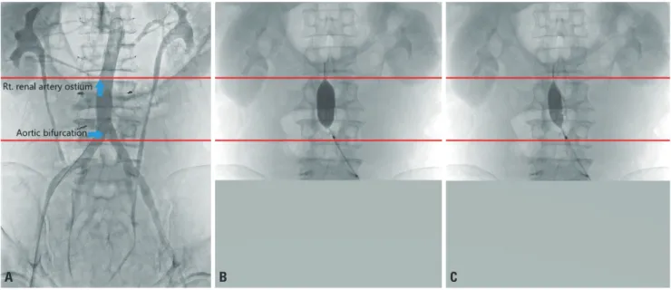
Fig. 2. These are fluoroscopy images of REBOA and Zone III is marked with two red lines. The length of Zone III was 7.5 cm in this patient, and the
length of fully inflated balloon was 5.5 cm. We could locate balloon catheter in Zone III correctly under real-time fluoroscopy. (A) Aortic Zone III (from lowest renal artery to aortic bifurcation). (B) Fully inflated balloon catheter. (C) Partially inflated balloon catheter.
A B C
193
http://www.jtraumainj.org
Sungdo Lee, et al. Merit of REBOA under Fluoroscopy
sation in the both psoas muscle was noted (Fig. 1).
Fluid resuscitation and transfusion were performed, but the patient’s blood pressure was not recovered. We insert- ed a balloon catheter through the left common femoral artery and located the catheter in Zone III (Fig. 2). We maintained total occlusion for the first 3 minutes, then performed angioemolization alternately between patial inflation and total deflation under real-time fluoroscopy according to the patient’s blood pressure and other situa- tions.
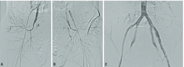
First, for both internal iliac arterial bleeding, emboli- zation was performed as the concept of damage control interventional radiology (Fig. 3). However, since the patient’s blood pressure was not stabilized, additional embolization was performed on the bleeding of multiple lumbar arteries.
After the procedure, the patient’s blood pressure was recovered to 96/67 mmHg and hemoglobin level returned to 10.6 g/dL by emergency transfusion and the patient was admitted to the trauma intensive care unit. The patient underwent two orthopedic surgeries during the hospital- ization period and was discharged without any complica- tion dew to ischemic injury 72 days after hospitalization.
DISCUSSION
REBOA is a novel device approved by the Food and Drug
Administration (FDA) in 2017 as an alternative to resusci- tative thoracotomy [4]. It is very effective to get proximal arterial pressure but distal ischemia is one of main com- plications. So most of the REBOA guidelines recommend that REBOA should be placed at Zones I and III, depend- ing on the location of the hemorrhage or hemodynamic status [5-7] and placement at Zone II should be always avoided for preservation of gastrointestinal perfusion [8].
Although the position of the balloon is important, it is not easy to inflate at the exact position when REBOA is performed with blind technique using external measure- ment. And there is a study that showed that the accuracy of the position was 12.5% when REBOA was performed targeting Zone III with blind technique [9].
In this case, the length of Zone III was 7.5 cm. We used a 7-French aortic occlusion catheter (RESCUE BALLOON
®, Tokai Medical Products, Aichi, Japan) and when the bal- loon was fully inflated, the length was 5.5 cm. Since the length of Zone III is not that long compared to the length of the balloon, it would have been difficult to accurately position the balloon through external measurement. We were able to stably maintain the correct position of the balloon in various situations under real-time fluoroscopy.
In this case, even if the balloon was located on the 1 cm proximal side, the ostium of the right renal artery could be blocked, causing ischemic injury.
As in this case, when REBOA is performed under re- al-time fluoroscopy, we can confirm the correct position
Fig. 3. Angiography under real-time fluoroscopy showed the following findings. (A) Active bleeding from the right internal iliac artery, (B) active bleed-