Received: June 3, 2020 Revised: July 14, 2020 Accepted: July 17, 2020 Trauma and InJury
Correspondence to Dong Hun Kim, M.D.
Department of Trauma Surgery, Trauma Center, Dankook University Hospital, 201 Manghyang-ro, Dongnam-gu, Cheonan 31116, Korea
Tel: +82-41-550-7119 Fax: +82-41-550-0039 E-mail: [email protected]
Successful damage Control resuscita- tion with resuscitative Endovascular Balloon occlusion of the aorta in a Pediatric Patient
Yoonjung Heo, M.D.
1, Sung Wook Chang, M.D.
2, Dong Hun Kim, M.D.
11
Department of Trauma Surgery, Trauma Center, Dankook University Hospital, Cheonan, Korea
2
Department of Thoracic and Cardiovascular Surgery, Trauma Center, Dankook University Hospital, Cheonan, Korea
Resuscitative endovascular balloon occlusion of the aorta (REBOA) is considered an emerging adjunct therapy for profound hemorrhagic shock, as it can maintain tempo- rary stability until definitive repair of the injury. However, there is limited information about the use of this procedure in children. Herein, we report a case of REBOA in a pediatric patient with blunt trauma, wherein the preoperative deployment of REBOA played a pivotal role in damage control resuscitation. A 7-year-old male patient experi- enced cardiac arrest after a motor vehicle accident. After 30 minutes of cardiopulmo- nary resuscitation, spontaneous circulation was achieved. The patient was diagnosed with massive hemoperitoneum. REBOA was then performed under ongoing resusci- tative measures. An intra-aortic balloon catheter was deployed above the supraceliac aorta, which helped achieved permissive hypotension while the patient was undergoing surgery. After successful bleeding control with small bowel resection for mesenteric avulsion, thorough radiologic evaluations revealed hypoxic brain injury. The patient died from deterioration of disseminated intravascular coagulation. Although the pa- tient did not survive, a postoperative computed tomography scan revealed neither remaining intraperitoneal injury nor peripheral ischemia correlated with the insertion of a 7-Fr sheath. Hence, REBOA can be a successful bridge therapy, and this result may facilitate the further usage of REBOA to save pediatric patients with non-compressible torso hemorrhage.
Keywords: Shock, hemorrhagic; Balloon occlusion; Aorta; Child; Wounds and injuries
INTRODUCTION
Hemorrhage is the most substantive contributor to poten- tially preventable mortality from trauma [1]. The interval between injury and definitive control of the bleeding focus is critical in reducing the incidence of preventable death. Resuscitative endovascular balloon occlusion of the aorta (REBOA) is an emerging adjunct therapy for pro- found hemorrhagic shock, as it can maintain temporary stability until definitive repair of the injury. Compared to resuscitative thoracotomy, REBOA is a less invasive procedure that allows earlier control of hemorrhage. Ac- cording to the American Association for the Surgery of Trauma Aortic Occlusion for Resuscitation in Trauma and Acute Care Surgery registry, compared to resuscita- tive thoracotomy, REBOA has a survival benefit for pa- tients with non-compressible truncal injury who undergo Zone 1 REBOA [2]. However, clinicians are still hesitant in implementing REBOA in children due to the immature anatomy of their vasculature, the unavailability of com- mercial catheters with small calibers, and insufficient data supporting its use. Herein, we report a case in which a 7-Fr REBOA catheter was deployed in a pediatric patient using a 7-Fr sheath via the common femoral artery, help- ing to achieve successful damage control resuscitation.
CASE REPORT
A 7-year-old male patient with no previous significant medical history experienced cardiac arrest after a motor vehicle accident, during which he was a backseat pas- senger. Initially, the patient was transported to a nearby hospital by an emergency medical service team. After 30 minutes of cardiopulmonary resuscitation, spontaneous circulation was achieved. He presented with profound hemorrhagic shock due to sustained blunt trauma in the left chest wall and the abdomen. The first responders per- formed endotracheal intubation and closed thoracostomy for left hemopneumothorax. An unfavorable outcome was expected due to suspected hypoxic brain damage caused by a long cardiac arrest time. However, the patient was still brought to our regional level 1 trauma center for the best possible treatment considering his age. The pre-hospital time from injury to arrival in the trauma center was 120 minutes.
Upon arrival, the patient was in extremis, with a blood pressure of 44/19 mmHg and minimal arterial pulsation.
The Glasgow Coma Scale score was 3. According to the assessment of light reflex, the patient’s pupils were fixed with a size of 6 mm. The primary assessment revealed a seat belt sign and severe distention in the lower abdomen.
Despite optimal resuscitation with crystalloid and blood
Fig. 1. (A) Position of the 7-Fr balloon catheter (arrow) in aortic Zone 1. (B, C) Preoperative and postoperative placement of the balloon catheter via the
left femoral artery.
A b C
transfusions, the patient did not have any hemodynamic response. The initial arterial blood gas analysis revealed a pH of 6.8, lactate of 12.2 mmol/L, and hemoglobin level of 3.8 g/dL. Only a small volume of blood drained through the indwelling chest tube. Thus, other major bleeding foci were suspected. Focused Assessment with Sonogra-
phy for Trauma revealed massive intra-abdominal fluid collection. Thus, REBOA in Zone 1 was performed in the descending thoracic aorta under ongoing resuscitative measures [3]. A 7-Fr introducer sheath was placed in the left common femoral artery using the Seldinger tech- nique. Then, a 7-Fr balloon catheter (RESCUE Balloon
TM, Tokai Medical Products, Aichi, Japan) was inserted along the guidewire. The balloon catheter tip was in the thoracic aorta as confirmed on serial radiography (Fig. 1A, B). The balloon was inflated with 5 mL of saline, which resulted in partial occlusion without inflated resistance in the syringe.
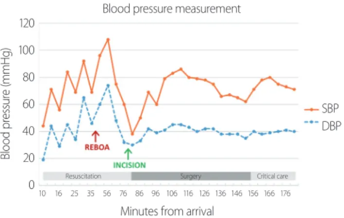
The time from skin puncture to REBOA inflation was 8 minutes. The patient’s hemodynamic status improved after balloon inflation (Fig. 2). Thus, he was wheeled into the operating room 61 minutes after the initial presenta- tion.
Crash laparotomy revealed mesenteric avulsion with small bowel ischemia and a 5-cm partial thickness lacera- tion in the sigmoid colon. Numerous mesenteric arteries were lacerated. Considering these findings, we performed multiple small bowel resections without anastomosis and
Fig. 2. Blood pressure measurements. The arrow indicates the time ofREBOA inflation. REBOA: resuscitative endovascular balloon occlusion of the aorta, SBP: systolic blood pressure, DBP: diastolic blood pressure.
Blood pressure measurement
Blood pressure (mmHg)
120 100 80 60 40 20 0
SBP DBP
Resuscitation Surgery Critical care
Minutes from arrival
10 16 25 35 56 76 86 96 106 116 126 136 146 156 166 176
Fig. 3. Time course during REBOA performance
(time interval from arrival). REBOA: resuscitative endovascular balloon occlusion of the aorta, SBP:
systolic blood pressure.
0:00 Patient arrival
0:21 Transfusion
0:12 Femoral catheter insertion
0:38 REBOA partial inflation (5 mL), Stable hemodynamics (SBP 96 mmHg)
0:30 REBOA insertion
1:01 Transfer to operating romm
1:20 Start of surgery
2:37 End of surgery
3:02 Transfer to the intensive care unit 1:36 REBOA
deflation
Resuscitation Surgery Critical
care
Fig. 4. Postoperative computed tomography scan of the abdomen. (A) A 7-Fr balloon catheter was accurately positioned in the aorta (arrow), and the