Research Report
FISH Karyotype and GISH Meiotic Pairing Analyses of a Stable Intergeneric Hybrid xBrassicoraphanus Line BB#5
Hadassah Roa Belandres 1 , Nomar Espinosa Waminal 1,2,3,4 , Yoon-Jung Hwang 1 , Beom-Seok Park 5 , Soo-Seong Lee 6 , Jin Hoe Huh 2,3,4 , and Hyun Hee Kim 1*
1
Plant Biotechnology Institute, Department of Life Science, Sahmyook University, Seoul 139-742, Korea
2
Department of Plant Science, Seoul National University, Seoul 151-921, Korea
3
Plant Genomics and Breeding Institute, Seoul Natioanl University, Seoul 151-921, Korea
4
Research Institute for Agriculture and Life Sciences, Seoul National University, Seoul 151-921, Korea
5
Agricultural Genome Center, National Academy of Agricultural Science, Rural Development Administration, Suwon 441-707, Korea
6
BioBreeding Institute, Ansung 456-756, Korea
Abstract: xBrassicoraphanus line BB#5, a new synthetic intergeneric hybrid between Brassica rapa L. ssp. pekinensis and Raphanus sativus L. var. rafiphera induced by N-methyl-N-nitroso-urethane mutagenesis in microspore culture, shows high seed fertility and morphological uniformity. Dual-color fluorescence in situ hybridization (FISH) using 5S and 45S rDNA probes and genomic in situ hybridization (GISH) using B. rapa genomic DNA probe were carried out to analyze the chromosome composition and the meiosis pairing pattern compared to its parental lines. The somatic chromosome complement is 2n = 38, which consists of 17 metacentric and two submetacentric chromosomes with lengths of 2.18 to 5.01 µm. FISH karyotype analysis showed five and eight pairs of 5S and 45S rDNA loci. GISH meiosis pairing analysis showed that 19 complete bivalents were most frequent and accounted for 42% of the 100 pollen mother cells examined. Based on chromosome number, size, morphology, rDNA distribution, and meiosis pairing pattern, both parental genomes of B. rapa and R. sativus appear to exist in xBrassicoraphanus line BB#5, demonstrating its genome integrity. Such stable chromosome constitutions and meiotic pairing patterns in somatic and meiotic cells are very rare in natural and synthetic intergeneric hybrids. Chromosomal studies and genetic and phenotypic changes in allopolyploids are discussed. The results presented herein will be usef ul f or f urther genomic study of xBrassicoraphanus lines and their improvement as promising new breeding varieties.
Additional key words: cytogenetic study, intergeneric allotetraploid BB#5
*Corresponding author: [email protected]
※ Received 5 September 2014; Revised 17 September 2014; Accepted 17 September 2014. This study was supported by a grant from the Next-Generation BioGreen21 Program (No. PJ009093), Rural Development Administration, Korea.
Ⓒ 2015 Korean Society for Horticultural Science
Introduction
Brassica and Raphanus species have been cultivated world- wide as important vegetables, fodders, and sources of functional compounds. Owing to their usefulness, many studies have been carried out to investigate breeding of these species (McNaughton, 1979; Namai et al., 1980; Olsson and Ellerstrom, 1980; Prakash et al., 2009).
Synthetic hybrid species between Brassica and Raphanus, xBrassicoraphanus or xRaphanobrassica, have been developed and studied by many researchers using morphological and
cytological methods (Dolstra, 1982; Kato and Tokumasu,
1983; Tokumasu, 1976). In Korea, Lee et al. (1999, 2002)
developed a new intergeneric allotetraploid, xBrassicoraphanus,
by a cross between B. rapa L. ssp. pekinensis and R. sativus
L. and named it ‘Baemoochae’. Initially, this new synthetic
hybrid was so unstable that low seed fertility and poor
uniformity were continued through generations. However,
a microspore mutagenesis using N-nitroso N-methyl urethane
(NMU) created stabilized progenies from this unstable
material (Lee et al., 2011). They also developed another
intergeneric allotetraploid xBrassicoraphanus between Korean
land races of B. rapa L. ssp. pekinensis and R. sativus L.
var. rafiphera and the F1 hybrid was crossed again with a mutagen-induced stabilized line. Using the same microspore mutation technique, another stable line of xBrassicoraphanus showing quite high seed fertility and morphological uni- formity was obtained, which was named BB#5.
Chromosome-based cytogenetic studies using conventional staining method have been applied to Brassica species since 1920 to investigate chromosome composition and genome structure. Chromosome characterization and genome identification have advanced significantly with the develop- ment of molecular cytogenetics through fluorescence in situ hybridization (FISH) and genomic in situ hybridization (GISH) techniques (Capdeville et al., 2008; Fukui, 2005;
Hwang et al., 2010, 2012; Levsky and Singer, 2003; Park et al., 2010). Indeed, these methods have been used to investigate genome structure and inter-genomic relationship of hybrid plants (Jellen et al., 1994; Kenton et al., 1993), allopolyploid species (Cao, 2003; Devi et al., 2005; Yang et al., 19 9 9 ), and recombinant breeding lines (Hwang et al., 2012; Lou et al., 2010; Vasconcelos et al., 2010).
Tandem repeat DNA sequences including 5S and 45S ribosomal DNAs (rDNAs) have primarily been used as cytogenetic markers in FISH (Kato et al., 2004; Lim et al., 2005, 2012). In Brassica, Maluszynska and Heslop-Harrison (1993) first reported the number of 45S rDNA loci in diploids (B. rapa, n = 10, AA genome; B. nigra; n = 8, BB genome; B. oleracea, n = 9, CC genome,) and allo-tetraploids (B. carinata, n = 17, BBCC genome; B. juncea, n = 18, AABB genome; B. napus, n = 19, AACC genome). Genomic dis- tributions of rDNA sites on prometaphase and metaphase chromosomes were described more precisely upon further investigations (Fukui et al., 1998; Hasterok and Muluszynska, 2000a, 2000b; Hasterok et al., 2006; Hwang et al., 2009;
Kim et al., 1998; Snowdon et al., 1997). Some chromosomes of the complements were identified exactly, enabling a detailed karyotype analysis for the genus Brassica.
Lim et al. (2012) analyzed the karyotype of the somatic metaphase of the xBrassicoraphanus line BB#4 by conventional Giemsa-staining, but no cytogenetic studies of the new synthetic intergeneric hybrid line BB#5 have been conducted to date. Here, we carried out FISH karyotype analysis of BB#5 using 5S and 45S rDNA probes, with reference to its parental species B. rapa and R. sativus. In addition, meiotic chromosome pairing patterns were analyzed and the Brassica and Raphanus genomes were discriminated by GISH method.
Materials and Methods Plant Materials
Seeds and flower buds of the xBrassicoraphanus line BB#5 were provided by the Bio Breeding Institute, Ansung, Korea and seeds of its parental species, Brassica. rapa L.
ssp. pekinensis and Raphanus. sativus L. var. rafiphera were provided by the RDA Genebank, Suwon, Korea.
Seeds were sown on moist filter paper in Petri dishes and germinated at 25°C for 48 hours. Approximately 2 cm long root tips were harvested from the germinated seeds, pretreated in 2 mM 8-hydroxyquinoline at 18°C for 5 hours, fixed in aceto-ethanol (1:3 v/v) solution for 2 to 24 hours, and then stored in 70% ethanol until use.
The xBrassicoraphanus flower buds were fixed in the same solution for 24 hours and stored at -20°C in 70% ethanol.
Preparation of Somatic Metaphase Chromosome Spreads A modified version of the method described by Kato et al. (2004) was used to prepare chromosome spreads. Briefly, fixed root tips were thoroughly washed with distilled water, after which the meristem section of the roots was cut out and digested in 2% cellulase (MB Cell, Korea), 1.5%
macerozyme (Maxim Bio, USA) and 1% pectolyase (Sigma, Japan) in 150 mM citrate buffer, pH 4.5 for 9 0 minutes at 37°C. The meristems were then thoroughly washed in ice-cold distilled water, after which the root epidermises were removed and the remaining section containing the dividing cells was pipetted into a tube with chilled aceto- ethanol (1:3 v/v) solution, then suspended by gentle vortexing for 30 seconds. The cells were subsequently collected in the bottom of the tube by centrifugation and re-suspended in aceto- ethanol (9:1 v/v) solution. Finally, the cell suspension was dropped on pre-cleaned glass slides, placed in a humid chamber (37°C) and slowly dried.
Probe Preparation
The procedure described by Lim et al. (2005) was used to obtain the 5S rDNA sequences of genomic DNA from B. rapa L. ssp. pekinensis and the fragment was labelled with dig-11-dUTP (Roche, Germany) by nick translation.
The 45S rDNA sequence, containing a 9-kb fragment of 18S-5.8S-25SrDNA genes from wheat (Gerlach and Bedbrook, 1979) was isolated and labelled with biotin-16-dUTP (Roche, Germany) by nick translation. The 200-500 bp labelled DNA fragments were used as probes.
For GISH analysis, total genomic DNA was extracted and
purified from young leaves of B. rapa L. ssp. pekinensis
and R. sativus L. using the CTAB- extraction method (Allen et al., 2006). The genomic DNA from B. rapa was labelled with dig-11-dUTP (Roche, Germany) by nick translation and used as a probe, while the unlabelled DNA of R. sativus was fragmented at 100°C for 7 minutes and used as a block. The lengths of the probe and the blocking DNA fragments were determined by agarose-gel electrophoresis within the range of 200-500 bp, after which samples were stored at -20°C until use.
Fluorescence in Situ Hybridization (FISH) and Genomic in Situ Hybridization (GISH)
The FISH procedure described by Lim et al. (2005) and Waminal et al. (2012) was used in this study, with some modifications. Briefly, slides were pretreated with RNase A buffer (100 µg・mL
-1RNase A in 2x SSC) at 37°C for 1 hour, after which they were soaked in 0.01 M HCl for 2 minutes. Next, samples were treated with 10% (w/v) pepsin in a 1:100 dilution of 0.01 M HCl for 10 minutes at 37°C and then washed for 5 minutes with 2x SSC. The slides were subsequently post-fixed with 4% paraformaldehyde in 2x SSC, washed with 2x SSC, and dehydrated in ethanol with a series of different concentrations. A hybridization mixture that consisted of 50% formamide, 10% dextransulfate, 2x SSC, 200 ng salmon sperm DNA or 5 ng・µL
-1unlabelled genomic DNA for GISH, and 50 ng・µL
-1of each DNA probe (5S rDNA and 45S rDNA for FISH, or labelled genomic DNA for GISH) was prepared and then adjusted to 40 µL per slide with nuclease free water. The FISH/GISH mixture was denatured at 90°C and cooled on ice for another 10 minutes, then mounted on slides and denatured at 80°C for 2.5 minutes, after which it was placed in an incubator at 37°C overnight (to 16 hours). The next day, the slides were washed in 2x SSC for 30 minutes at RT, 0.1x SSC for 35 minutes at RT and finally in 2x SSC for 15 minutes at 42°C. Dig- labelled and biotin- labelled probes were detected with anti-dig-FITC conjugate (Sigma, USA) and streptavidin-Cy3 conjugate antibodies (Zymed Lab., USA), respectively. The antibodies were reacted on slides at 37°C for 1 hour, then washed in the detection buffer at 37°C.
Next, the slides were dehydrated in ethanol series (70, 90, and 100%, 3 minutes each) and air-dried. A 40 µL aliquot of 1:100 DAPI [f.c. 1 µg・mL
-1(Roche, Germany)]
in Vectashield (Vector Lab., Inc., USA) reagent was mounted to counterstain the chromosomes and covered with a glass coverslip.
Meiotic Chromosome Pairing Analysis
Meiotic chromosomes were prepared according to Zhong
et al. (1996) and Ge and Li (2007) with minor modifications.
Young flower buds from 1.6 to 2.0 mm and anthers from 0.8 to 1.0 mm long were used. Fixed flower buds were washed with distilled water for at least 10 minutes. One anther from a flower bud was squashed in 60% acetic acid, air-dried, stained with 4',6-diamidino-2-phenylindole (DAPI), and analyzed using a fluorescence microscope.
Microsporocytes in prophase I were selected, and the remaining anthers were then treated with an enzyme mixture of 2% cellulase (MB Cell, Korea), 1% cytohelicase (Yakult, Japan), and 1% pectolyase (Sigma, Japan) for 90 minutes at 37°C. Next, the treated anthers were washed in distilled water for 5 minutes, transferred to a glass slide, then squashed after adding 60% acetic acid and air-dried.
Meiotic chromosomes entering late prophase I were scored and meiotic pairing configurations and frequency were analysed using 100 pollen mother cells (PMCs) from different flower buds.
Imaging and Karyotype Analysis
The FISH images of each chromosome were measured three times to obtain the mean values. Homologous chro- mosomes were confirmed based on chromosome length, morphology and, FISH signal patterns. The chromosome arrangements of B. rapa and R. sativus were conducted according to the rDNA distribution patterns, according to the methods described by Lim et al. (2005) and Hwang et al. (2012), respectively. The overall chromosome size of xBrassicoraphanus line BB#5 was estimated by measuring the lengths of short and long arms of chromosomes, excluding satellites and nucleolar organizing regions due to considerable variations in their size in each satellite chromosome and the chromosomes were arranged from longest to shortest. Images were captured using an Olympus BX-51 fluorescence microscope equipped with a charge- coupled device (CCD) camera (CoolSNAP™ cf) and analyzed using the Genus software (version 3.1). Final images were enhanced using Adobe Photoshop CS3. The method described by Levan et al. (1964) was used to classify the chromosome types. Homologous chromosomes were determined based on their lengths, morphological characteristics, and rDNAs signals.
Results
Karyotype Analysis
The mitotic chromosome number of Brassica rapa L.
ssp. pekinensis was 2n = 20, (Fig. 1A), with lengths ranging
from 2.75 to 4.75 µm and a total length of 36.22 µm (Table
A B C
D E F
1 2 3 4 5 6 7 8 9 10 11 12 13 14 15 16 17 18 19
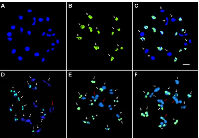
Fig. 1. FISH metaphases and their chromosome arrangement showing 5S and 45S rDNA signals in B. rapa L. ssp. pekinensis, 2n = 20 (A and D), R. sativus L. var. rafiphera, 2n = 18 (B and E), and xBrassicoraphanus line BB#5, 2n = 38 (C and F).
The 5S and 45S rDNA loci are shown as green and red signals, respectively. The white arrows indicate the satellite chromosomes showing the nucleolar organizing region. Scale bar, 5 µm.
Table 1. Summary of FISH karyotype analyses of intergeneric hybrid xBrassicoraphanus line BB#5 and its two parental lines, Brassica rapa L. ssp. pekinensis and Raphanus sativus L. var. rafiphera.
Species Chr. no.
(2n)
Chr. length (µm) rDNA signals
Karyotype formula (2n)
zShortest Longest Total 5S 45S
B. rapa L. 20 2.75 4.76 36.22 3 5 2m + 2sm
*+ 2m + 2m + 2m + 2m + 2sm + 2m + 2m + 2m
R. sativus L. 18 2.04 3.31 24.66 2 3 2m + 2sm
*+ 2m + 2m + 2m + 2m + 2m + 2m + 2m
xBrassicoraphanus
(BB#5) 38 2.18 5.01 65.68 5 8 2m + 2m + 2m + 2m + 2m + 2m + 2m + 2sm +
2m + 2m + 2m + 2m + 2sm
*+ 2m
*+ 2m + 2m + 2m + 2m + 2m
z
m, metacentric; sm, submetacentric. The chromosomes in the karyotypic formula were arranged according to the chromosome number assigned in Fig. 2.
*