Cordycepin의 치밀결합 강화 및 MMPs의 활성 억제를 통한 HCT116 인체대장암세포의 이동성 및 침윤성의 억제
정진우1․최영현2†
1대구경북과학기술원 중앙기기센터
2동의대학교 한의과대학 생화학교실 및 항노화연구소 및 블루바이오소재개발센터
Cordycepin Inhibits Migration and Invasion of HCT116 Human Colorectal Carcinoma Cells by Tightening of Tight Junctions and
Inhibition of Matrix Metalloproteinase Activity
Jin Woo Jeong1 and Yung Hyun Choi2†
1Center for Core Research Facilities, Daegu Gyeongbuk Institute of Science & Technology, Daegu 711-873, Korea
2Dept. of Biochemistry, Dongeui University College of Oriental Medicine, Busan 614-052 and Anti-Aging Research Center & Blue-Bio Industry RIC, Dongeui University, Busan 614-714, Korea
ABSTRACT Cordycepin is the major functional component of Cordyceps species and is widely used in traditional oriental medicine. Cordycepin has been shown to possess many pharmacological properties, such as enhancement of immune function along with anti-inflammatory, antioxidant, anti-aging, and anti-cancer effects. Here, we investigated the inhibitory effects of cordycepin on cell migration and invasion, which are two critical cellular processes that are often deregulated during metastasis, using HCT116 human colorectal carcinoma cells. According to our data, cordycepin at non-cytotoxic concentrations markedly inhibited the motility and invasiveness of HCT116 cells in a time-dependent manner. RT-PCR and Western blotting results indicated that cordycepin reduced the levels of claudin proteins, which are major components of tight junctions (TJs), and induced tightening of TJs. Cordycepin also attenuated the expression and activities of matrix metalloproteinases (MMPs)-2 and -9, whereas levels of tissue inhibitor of metalloproteinases (TIMPs)-1 and -2 were simultaneously elevated. These findings suggest that cordycepin reduces the migration and invasion of HCT116 cells by modulating the activities of TJs and MMPs.
Key words: cordycepin, invasion, tight junctions, claudin, MMP
Received 10 September 2013; Accepted 30 October 2013
†Corresponding author.
E-mail: [email protected], Phone: +82-51-850-7413
서 론
전이는 암이 발병 부위로부터 인체의 다른 곳으로 이동하 여 증식하는 현상으로 암에 의한 사망률을 증가시키는 가장 큰 요인이다. 상피세포에는 세포들 사이에는 gap junction, tight junction(TJ), adherens junction(AJ) 및 desmosome 과 같은 몇 가지 종류의 특화된 세포간의 구조를 가지는데, 그중 TJ는 세포 사이의 치밀한 이음부로서 다양한 분자와 이온의 이동을 차단하는 역할을 한다(1,2). 그러나 많은 상 피조직 유래 암세포들의 경우 TJ 체계가 무질서해지거나 붕괴됨으로써 상피 전기 저항성(transepithelial electrical resistance, TER)으로 대변되는 TJ의 전기적 저항이 감소 되어 있거나 상피곁세포투과성(transepithelial paracellular permeability, TPP)이 증가되어 있다. Claudin family에 속
하는 주요 필수 막단백질들은 TJ의 근간을 이루며, 동형 혹 은 이형의 복합체를 형성함으로써 다양한 물질들의 세포사 이 유동성을 조절하는 장벽 역할을 수행한다(3-5). 최근 연 구들에 의하면 claudin 단백질들은 많은 상피세포 유래 암세 포에서 비정상적으로 높게 발현되며, 이는 암의 발생, 진행 및 전이와 밀접한 연관성이 있는 것으로 알려졌다(5-7). 따 라서 TJ 복합체에서 이들 단백질들은 암의 증식뿐만 아니라 전이를 조절할 수 있는 주요 생물지표(biomarker)로서 역할 을 할 가능성이 높아지고 있다.
Zinc-dependent endopeptidase family에 속하는 기질 금속단백분해효소(matrix metalloproteinases, MMPs)는 염증성 조직의 파과와 혈관신생 및 암세포의 전이에 핵심적 인 역할을 하는 것으로 알려졌다. 침윤성 암세포는 이들 효 소를 사용하여 세포외기질(extracellular matrix, ECM)의 분해와 전이 동안 기저체 막의 붕괴를 촉진함으로써 암세포 의 침윤과 전이를 증대시킨다(8,9). MMPs 중 특히 gelati- nase A와 B로 알려진 MMP-2와 -9가 다양한 암세포에서 활성이 비정상적으로 높게 관찰되며, 암세포의 침윤과 전이
에 중요한 역할을 하는 것으로 알려졌다. 일반적으로 MMP- 2는 섬유아세포와 다양한 상피세포에서 MMP-9는 염증성 세포에서 우선적으로 높은 활성을 보이지만, 두 효소 모두 침윤-전이 잠재력과 깊은 상관성이 있다(10,11). MMPs의 억제인자로 알려진 금속단백분해효소 조직억제제(tissue inhibitors of metalloproteinases, TIMPs)는 MMPs의 발 현을 조절하는 데 중요한 역할을 한다. 그들은 MMPs에 직 접 결합하여 그들의 효소적 활성을 저해함으로써 ECM의 파괴를 억제하는 것으로 알려졌다(12,13). 따라서 MMPs와 TIMPs 사이의 적절한 균형 조절을 위한 MMP 저해제 및 TIMP 활성제는 암세포의 전이를 차단할 수 있는 표적 물질 이 될 수 있다.
Cordycepin(3′-deoxyadenosine)은 전통 동양의학에서 오랫동안 사용되어 온 버섯류인 Cordyceps militaris에서 유래된 nucleoside adenosine 유도체의 일종이다(14,15).
구조적으로 cordycepin은 ribose moiety의 3′ 부분에 산소 가 결여된 특징으로 인하여 RNA 합성과정에 삽입됨으로써 RNA 사슬의 연장을 차단시킨다(16). 암 화학요법제로서 cordycepin의 역할과 작용기전에 대하여 많은 연구가 이루 어지고 있는데, 예를 들어 구강암세포(17), 흑색종암세포 (18) 및 갑상선암세포(19) 등과 같은 고형암세포에서 cor- dycepin은 apoptosis를 촉진시킬 뿐만 아니라, 혈구암세포 에서도 apoptosis를 유발(20,21)하는 것으로 밝혀진 바 있 다. 특히 갑상선암세포의 apoptosis 유발에는 칼슘 의존적 caspase의 활성이 관여하였으며(19), 백혈병세포에서의 apoptosis 유발에는 활성산소종(reactive oxygen spe- cies, ROS) 생성 의존적임이 알려진 바 있다(20). 최근 cor- dycepin의 암세포 전이 억제 가능성에 관한 연구로는 전립 선암세포(22), 방광암세포(23) 및 유방암세포(24) 등에서 이루어진 바 있고 in vivo 실험계에서도 확인된 바 있다(25).
암세포의 전이 억제에 관한 선행 결과들을 종합해보면, MMPs의 발현 및 활성 증가가 기본적인 기전으로 설명되고 있지만, 여전히 더욱 구체적이고 추가적인 연구가 필요한 실정이다. 따라서 본 연구에서는 cordycepin의 암세포 전이 억제 효능에 관한 추가적인 기전 해석의 일환으로 현재 연구 가 상대적으로 미비한 인체 대장암세포(HCT116)를 이용하 여 이들 세포의 이동성과 침윤성에 미치는 효과와 관련 유전 자들의 발현 변화를 조사하였다.
재료 및 방법
세포배양 및 MTT assay
HCT116 세포는 American Type Culture Collection (Rockville, MD, USA)에서 구입하였으며, 10% fetal bo- vine serum(FBS, Gibco BRL, Gaithersburg, MD, USA) 및 1% penicillin–streptomycin이 함유된 RPMI 1640 배지 를 사용하여 37°C, 5% CO2 조건에서 배양하였다. Cordy- cepin은 Sigma-Aldrich Co.(St. Louis, MO, USA)에서 구
입하였으며, 증류수에 1 mg/mL 농도로 녹여 적정 농도로 희석하여 처리하였다. HCT116 세포의 증식에 미치는 cor- dycepin의 영향은 3-(4,5-dimethyl-2 thiazolyl)-2,5-di- phenyl-2H-tetrazolium bromide(MTT, Sigma-Aldrich Co.)가 미토콘드리아 효소에 의하여 MTT-formazan을 형 성하는 원리에 기반을 둔 MTT assay를 이용하여 조사하였 다.
Wound healing migration assay
HCT116 세포를 20 μg/mL의 rat tail collagen이 코팅된 dish(BD Biosciences, Bedford, MA, USA)에 적정 시간 동안 배양 후 파이펫 팁을 이용한 scraping으로 wounded 영역을 만들었다. 정상 배지로 2~3회 수세하고 1% FBS가 함유된 배지로 교체 후 적정 농도의 cordycepin을 처리하였 다. Cordycepin 처리 48시간 후 도립현미경으로 이용하여 40배의 배율로 wounded 영역으로의 세포 이동성의 정도를 비교 관찰하였다.
TER의 측정
STX-2 chopstick electrode가 짝으로 형성된 EVOM Epithelial Tissue Voltohmmeter(World Precision In- struments, Sarasota, FL, USA)를 이용하여 TER 값을 측 정하였다. 이를 위하여 HCT116 세포를 Transwell®
(Corning Costar Corp., Corning, NY, USA)의 8.0 μm pore size insert(upper chamber)로 seeding한 후 100%
confluence를 이룰 때까지 배양하였다. 이후 cordycepin을 48시간 동안 처리하고 Grant-Tschudy 및 Wira(26)의 방 법에 준하여 upper 및 lower chamber에 electrode를 넣어 전기 저항도를 측정하였다.
In vitro invasiveness assay
HCT116 세포의 침윤성에 미치는 cordycepin의 영향을 조사하기 위하여 matrigel invasion assay를 사용하였다.
이를 위하여 정상 조건 및 적정 농도의 cordycepin이 6시간 동안 처리된 HCT116 세포를 cordycepin이 함유된 FBS- free 배지로 처리한 matrigel-coated filter의 apical side 에 분주하였다. 이때 basolateral chamber에는 20% FBS 가 함유된 배지를 분주하였으며, 48시간 후 filter의 하단부 로 이동한 세포를 hematoxylin 및 eosin Y 염색액으로 염색 하고 계수하였다.
RNA의 분리 및 reverse transcription-polymerase chain reaction(PCR)
전사 수준에서 특정 유전자들의 발현 변화를 조사하기 위 하여 RNeasy kit(Qiagen, San Diego, CA, USA)를 이용하 여 RNA를 분리한 후, AMV reverse transcriptase(Amer- sham Corp., Arlington Heights, IL, USA)를 이용하여 complementary DNA를 합성하였다. 각각의 해당 유전자에
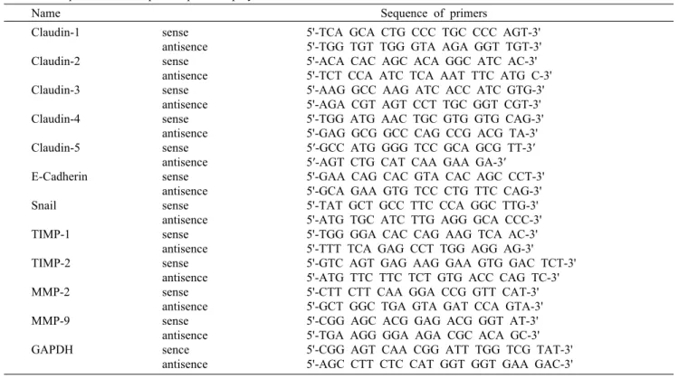
Table 1. Sequences of the primer pairs employed in the RT-PCR reactions
Name Sequence of primers Claudin-1
Claudin-2 Claudin-3 Claudin-4 Claudin-5 E-Cadherin Snail TIMP-1 TIMP-2 MMP-2 MMP-9 GAPDH
sense antisence sense antisence sense antisence sense antisence sense antisence sense antisence sense antisence sense antisence sense antisence sense antisence sense antisence sence antisence
5'-TCA GCA CTG CCC TGC CCC AGT-3' 5'-TGG TGT TGG GTA AGA GGT TGT-3' 5'-ACA CAC AGC ACA GGC ATC AC-3' 5'-TCT CCA ATC TCA AAT TTC ATG C-3' 5'-AAG GCC AAG ATC ACC ATC GTG-3' 5'-AGA CGT AGT CCT TGC GGT CGT-3' 5'-TGG ATG AAC TGC GTG GTG CAG-3' 5'-GAG GCG GCC CAG CCG ACG TA-3' 5′-GCC ATG GGG TCC GCA GCG TT-3′
5′-AGT CTG CAT CAA GAA GA-3′
5'-GAA CAG CAC GTA CAC AGC CCT-3' 5'-GCA GAA GTG TCC CTG TTC CAG-3' 5'-TAT GCT GCC TTC CCA GGC TTG-3' 5'-ATG TGC ATC TTG AGG GCA CCC-3' 5'-TGG GGA CAC CAG AAG TCA AC-3' 5'-TTT TCA GAG CCT TGG AGG AG-3' 5'-GTC AGT GAG AAG GAA GTG GAC TCT-3' 5'-ATG TTC TTC TCT GTG ACC CAG TC-3' 5'-CTT CTT CAA GGA CCG GTT CAT-3' 5'-GCT GGC TGA GTA GAT CCA GTA-3' 5'-CGG AGC ACG GAG ACG GGT AT-3' 5'-TGA AGG GGA AGA CGC ACA GC-3' 5'-CGG AGT CAA CGG ATT TGG TCG TAT-3' 5'-AGC CTT CTC CAT GGT GGT GAA GAC-3'
Table 2. Antibodies used in the present study
Antibody Origin Dilution Company
Claudin-1 Claudin-2 Claudin-3 Claudin-4 Claudin-5 E-Cadherin Snail MMP-2 MMP-9 TIMP-1 TIMP-2 Actin
rabbit polyclonal rabbit polyclonal rabbit polyclonal mouse monoclonal mouse monoclonal mouse monoclonal rabbit polyclonal rabbit polyclonal rabbit polyclonal rabbit polyclonal rabbit polyclonal mouse monoclonal
1 : 1,000 1 : 1,000 1 : 1,000 1 : 1,000 1 : 1,000 1 : 1,000 1 : 500 1 : 1,000 1 : 1,000 1 : 1,000 1 : 1,000 1 : 1,000
Invitrogen Invitrogen Invitrogen Invitrogen Invitrogen
Santa Cruz Biotechnology Santa Cruz Biotechnology Santa Cruz Biotechnology Santa Cruz Biotechnology Santa Cruz Biotechnology Santa Cruz Biotechnology Santa Cruz Biotechnology 적절한 primer(Table 1)를 이용한 PCR은 Mastercycler
(Eppendorf, Hamburg, Germany)를 사용하여 수행하였 다. PCR 반응 산물을 1% agarose를 이용하여 분리한 후, ethidium bromide(EtBr) 염색을 통하여 발현의 정도를 비 교하였다.
단백질의 분리와 Western blot analysis
번역 수준에서 해당 유전자들의 발현 변화 조사를 위한 총 단백질은 25 mM Tris–Cl(pH 7.5), 250 mM NaCl, 5 mM ethylendiaminetetra acetic acid, 1% nonidet P-40, 0.1 mM sodium orthovanadate, 2 μg/mL leupeptin 및 100 μg/mL phenylmethylsulfonyl fluoride가 함유된 lysis buffer를 이용하여 분리하였다. 분리된 단백질들의 농도를 측정한 후, Western blot analysis를 위해 동량의 단백질들
을 sodium dodecyl sulfate(SDS)–polyacrylamide gel electrophoresis를 이용하여 분리하고 nitrocellulose mem- brane(Schleicher & Schuell, Keene, NH, USA)으로 전이 시켰다. 각각의 membrane을 적정 항체 및 enhanced chemiluminescence(ECL, Amersham Corp.) 용액을 이용 하여 단백질들의 발현 변화를 조사하였다. 본 실험에 사용된 1차 항체들(Table 2)은 Santa Cruz Biotechnology Inc.
(Santa Cruz, CA, USA) 및 Invitrogen Co.(Carlsbad, CA, USA)에서 구입하였으며, 2차 항체들은 Amersham Corp.
에서 구입하였다.
Gelatin zymographic analysis를 통한 MMP 활성 측정 적정 농도의 cordycepin을 48시간 처리한 세포 배양액을 수거하여 zymography용 buffer(Invitrogen Co.)와 혼합한
(A) Time (h)
0 12 24 48
0 20 40 60 80 100 120
0 10 25 50
Cordycepin (μg/mL)
Cell viability (%) .
(B)
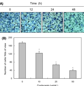
Fig. 1. Effects of cordycepin on cell motility and viability in HCT116 cells. (A) Cells were grown to confluency on 30-mm cell culture dishes; a scratch was then made through the cell layer using a pipette tip. After washing with PBS, serum free media (to prevent cell proliferation) containing either vehicle or cordycepin (50 μg/mL) was added for the indicated times.
Photographs of the wounded area were taken for evaluation of cell movement into the wounded area. (B) Cells were seeded at an initial density of 2.5×10⁵ cells per 60-mm plate, incubated for 24 h, and treated with various concentrations of cordycepin for 48 h. Cell viability was measured using an MTT assay. Each point represents the mean±SD of three independent experiments.
A Student's t-test was used for determination of significance.
후 precast gel(10% polyacrylamide and 0.1% gelatin)을 이용하여 전기영동 후 gel을 2.5% Triton X-100에 수세하 고 buffer(50 mM Tris–HCl, 150 mM NaCl, 5 mM CaCl2, 1 μM ZnCl2, 0.02% NaN3, pH 7.5)에 24시간 침전시켰다.
추가적으로 0.5%(w/v) Coomassie brilliant blue G-250 (Bio-Rad, Hercules, CA, USA)로 염색한 후 methanol /acetic acid/water(3:1:6) 용액을 이용하여 탈색시켰으며 상위 band를 MMP-9로, 하위 band를 MMP-2의 활성 정도 로 비교하였다.
통계 처리
모든 실험결과는 평균±표준편차로 표시하였고 Sigma- Plot(Systat Software Inc., San Jose, CA, USA)을 이용하 였으며, Student t-test를 이용하여 통계적 유의성을 얻었 다.
결 과
Cordycepin에 의한 HCT116 세포의 이동성 억제 Cordycepin에 의한 HCT116 세포의 이동성 억제 여부를 조사하기 위하여 실시한 wound healing migration assay 의 결과는 Fig. 1A에 나타낸 바와 같다. 결과에서 알 수 있듯 이 정상 배지에 배양된 세포에 비하여 cordycepin 처리 농 도 의존적으로 HCT116 세포의 이동성이 현저하게 억제되 었음을 알 수 있었다. 이러한 이동성의 억제가 cordycepin 의 세포독성에 의한 것인지의 여부를 조사하기 MTT assay 를 실시한 결과 본 실험에 시용된 농도 범위에서는 유의적인 세포증식 억제 효과가 관찰되지 않았다(Fig. 1B).
Cordycepin에 의한 HCT116 세포의 침윤성 억제 및 TER의 증가
Cordycepin에 의한 HCT116 세포의 이동성 억제가 침윤 성 억제와 연관성 여부를 조사하기 위하여 matrigel in- vasion assay를 실시한 결과는 Fig. 2에 나타낸 바와 같이 cordycepin 처리 농도 증가에 따라 현저한 침윤 억제 효과 를 보여주었다. 한편 세포와 세포 사이의 밀착 정도를 측정 하는 가장 보편적인 방법 중의 하나인 TER은 최근 세포연접 중 TJ의 견고성을 대별할 수 있기에 세포 해리의 억제 정도 를 측정하는 도구로서 활용되고 있다. 따라서 이러한 cor- dycepin의 암세포 이동성 및 침윤성의 저해 효과가 TJ의 기능 변화와 관련이 있는지를 조사하기 위하여 TJ의 견고성 을 나타내는 TER의 값(26)을 조사한 결과, cordycepin 처리 에 의하여 유의적으로 증가하였음을 알 수 있었다(Fig. 3).
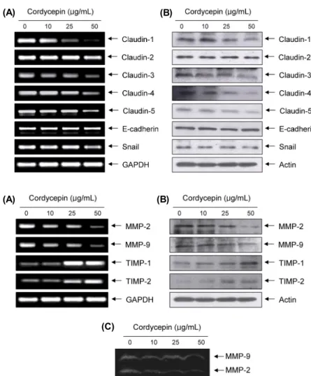
Claudin family의 발현에 미치는 cordycepin의 영향 이상에서 관찰된 cordycepin에 의한 TER 증가와 TJ 구 성 인자들의 발현 변화와 연계성을 조사한 결과, cordyce- pin 처리 농도 의존적으로 조사된 5종류의 claudin family
인자들(claudin-1, -2, -3, -4 및 -5) 중에서 claudin-2를 제외하고는 전사 및 번역수준에서 모두 감소하였다(Fig. 4).
또한 zinc finger transcription factor의 일종으로 epi- thelial-mesenchymal transition(EMT) 조절자에 해당되 는 snail(27,28)의 발현 역시 cordycepin 처리에 의하여 감 소되었으며, TJ뿐만 아니라 AJ의 형성을 조절하는 type Ⅰ transmembrane glycoprotein의 일종인 E-cadherin(29, 30)의 발현에는 큰 변화가 없었다(Fig. 4).
Cordycepin에 의한 MMPs의 발현 및 활성 억제 ECM의 분해에 결정적인 역할을 하는 MMPs의 발현과 활성에 미치는 cordycepin의 영향을 조사한 결과, MMP-2 뿐만 아니라 MMP-9의 발현도 전사 및 번역 수준에서 모두 감소되었으며, zymography에 의한 MMP-2 및 MMP-9의 발현도 cordycepin 처리 농도 의존적으로 모두 감소되었다.
그러나 MMPs의 억제인자에 해당하는 TIMPs의 경우 cor- dycepin의 처리에 의하여 모두 발현이 증가되었다(Fig. 5).
(A)
Time (h)
0 12 24 48
0 40 80 120 160 200
0 10 25 50
Cordycepin (μg/mL)
Number of cells/ filds of view
*
*
*
(B)
Fig. 2. Decrease of cell invasion by cordycepin in HCT116 cells.
Cells pretreated with the indicated concentration of cordycepin for 6 h were plated onto the apical side of matrigel coated filters in serum-free medium containing either vehicle or cordycepin.
Medium containing 20% FBS was placed in the basolateral chamber to act as a chemoattractant. After 48 h, cells on the apical side were wiped off using a Q-tip. Next, cells on the bottom of the filter were stained using hematoxylin and eosin Y (A), and then counted (B). Data are shown as the mean of triplicate samples and represent invasive cell numbers compared with those of control cells. *P<0.05 versus untreated control.
0 40 80 120 160 200
0 10 25 50
Cordycepin (μg/mL) Transendothelial Resistance . (Ω×cm2) .
*
*
Fig. 3. Increase of TER values by cordycepin in HCT116 cells.
Cells were treated with different concentrations of cordycepin for 48 h, and TER values were measured using an EVOM epi- thelial tissue voltohmmeter, as described in the materials and methods section. Results are shown as the mean±SD of three independent experiments (*P<0.05 versus untreated control).
고 찰
암세포의 이동과 침윤은 전이과정에 필수적인 현상으로 항암활성을 지니는 물질들의 전이 억제 여부를 평가하는 중 요한 기준으로 사용되어지고 있다. 최근 세포의 극성 소실과 삼투성의 변화가 상피세포 암화 및 전이 단계의 특징임이 밝혀지면서, 이들 기능 유지에 중요한 역할을 하는 TJ와 암
세포 전이와의 연계성에 관심이 높아지고 있다(1,2). 이와 연계되어 몇몇 선행연구들에서 항암활성을 가지는 물질들 의 암세포 이동성 및 침윤성 억제가 TPP의 상승과 연관되어 있음이 보고된 바 있다(2,31,32). 본 연구의 결과에서도 세 포독성이 없는 범위에서 cordycepin에 의한 HCT116 세포 이동성 및 침윤성 억제(Fig. 1 및 2)가 TER의 증가와 연관성 이 있었는데(Fig. 3), 이는 cordycepin이 TJ의 누출을 억제 하거나 역전시켰음을 의미하는 결과이다. TJ의 누출이 암세 포의 전이과정과 밀접한 연관성을 지닌다는 점에서 TJ의 견고성 유지는 항전이의 척도로 사용된다는 점을 고려할 경 우, cordycepin은 최소한 TJ의 활성을 증가시킴으로써 항 전이 활성을 나타낸다고 할 수 있다.
TJ의 구성 인자인 claudin family에 속하는 단백질들은 상호간에 복합체를 형성하여 TPP를 조절하는데(2,5,12), TJ 구성 단백질들의 비정상화와 수반된 TJ의 붕괴는 암세포 의 침윤과 전이에서 초기 단계에 나타나는 현상이다(6,7).
특히 claudin-3과 -4는 유방암과 난소암을 포함한 다양한 종양조직에서 과발현되어 있으며, 이들 단백질의 발현을 차 단할 경우 반대로 암세포의 침윤력이 억제되었음을 보고한 바 있다(31,33). 최근 간암세포의 침윤력 획득에 claudin-1 이 중요한 역할을 함이 밝혀졌는데, 이는 MMP-2의 발현 증가에 따른 활성 증가와 연관성이 있었으며 claudin-1의 발현을 인위적으로 차단할 경우 간암세포의 침윤력이 완벽 하게 차단되었음이 밝혀졌다(34). 이러한 결과들은 claudin 단백질들이 암세포의 검출과 진단을 위한 biomarker로서의 활용 가능성을 보여주는 것이며, 본 연구의 결과에서 제시한 claudin family들의 발현 저하(Fig. 4)는 cordycepin의 항 전이 효능이 TJ의 기능 강화와 연계성이 있음을 의미하는 것이다. 또한 TJ 형성에는 E-cadherin이 조절인자로서 작 용하며(35), 비록 암세포의 종류에 따라 다양하지만 EMT의 조절인자인 snail은 E-cadherin의 발현을 억제하면서 세포 와 세포 사이의 접착력을 낮추거나 침윤력을 증가시키는 것 으로 알려져 있다(27,36). 따라서 본 연구에서 관찰된 cor- dycepin이 E-cadherin의 발현에는 큰 영향을 미치지 못하 였으나 snail의 발현은 억제한 점(Fig. 4)에 대해서는 추가적 인 연구가 필요한 부분이라고 생각된다.
MMPs는 정상 기관의 발생과 조직 재생에 필수적인 효소 이지만, 암세포의 전이에도 중요한 역할을 담당하고 있다.
특히 MMP-2와 -9는 암세포의 침윤과 암조직 주변의 혈관 신생에 필수적이므로, 이들의 합성과 활성 차단은 종양 전이 및 증식 억제를 위한 필수적인 방법이다(10,11). MMPs의 활성은 TIMPs에 의하여 상대적으로 차단될 수 있으므로 MMPs에 대한 TIMPs의 상대적인 발현 증가는 암세포 침윤 력의 억제를 측정하는 주요 인자로 활용이 가능하다(4,19).
따라서 cordycepin이 이들 인자들의 발현에 어떤 영향을 미치는지를 조사한 결과, MMP-2와 MMP-9의 mRNA 및 단백질의 발현뿐만 아니라 그들의 효소적 활성도 억제되었 음을 알 수 있었다. 그러나 cordycepin은 TIMPs의 발현을
(A) (B) Fig. 4. Effects of cordycepin on expression of claudins, E-cadherin, and snail in HCT116 cells.
(A) Cells were treated with the indicated concen- trations of cordycepin for 48 h. Total RNA was isolated and reverse-transcribed using the in- dicated primers. Resulting cDNAs were then sub- jected to PCR and the reaction products were subjected to electrophoresis in a 1% agarose gel and visualized by EtBr staining. GAPDH was used as an internal control. (B) Cells grown under the same conditions as (A) were sampled, lysed, and 50 μg of proteins were separated by electro- phoresis on SDS-polyacrylamide gels. Western blotting was then performed using the indicated antibodies, and an ECL detection system. Actin was used as an internal control.
(A) (B)
(C)
Fig. 5. Inhibition of MMP expression and their ac- tivities, and induction of TIMPs expression by cor- dycepin in HCT116 cells. (A) Total RNA was iso- lated from cells grown under the same conditions as Fig. 4 and reverse-transcribed for RT-PCR. (B) Western blotting was then performed using the cell lysates, indicated antibodies, and an ECL detection system. Actin was used as an internal control. (C) After incubation with cordycepin for 48 h, the me- dium was collected, and the activities of MMP-2 and -9 were measured by zymography.
처리 농도 의존적으로 매우 증가시켰다(Fig. 5). 따라서 cordycepin의 TIMPs 발현 증가와 동반된 MMPs의 생성 및 활성 억제는 ECM의 분해를 차단함으로써 HCT116 세포 의 이동성과 침윤력을 억제하였을 것으로 생각된다.
결론적으로 cordycepin에 의한 HCT116 인체 대장암세 포의 이동성과 침윤력의 차단은 TJ 활성의 증가와 MMPs 발현의 억제에 의한 것임을 보여주는 것이다. 물론 이러한 결과가 대장암세포 특이적인 현상이라고는 할 수 없으며 동 물 실험을 포함한 추가적인 연구가 필요하지만, 본 연구의 결과는 cordycepin이 항전이 인자로서 항암활성을 나타낸 다는 귀중한 실험적 증거로서 활용될 것으로 생각한다.
요 약
Cordycepin은 C. militaris의 주요 생리활성 물질로서 인체 면역기능 강화, 항염증, 항산화, 항노화 및 항암활성을 포함 한 다양한 약리효능이 있는 것으로 알려져 있다. 본 연구에 서는 HCT116 대장암세포를 이용하여 암전이의 주요 과정 인 암세포의 이동성 및 침윤성에 미치는 cordycepin의 효능 에 관하여 조사하였다. 본 연구의 결과에 의하면 세포독성이 없는 범위에서 cordycepin은 HCT116 세포의 이동성과 침
윤성을 유의적으로 억제하였다. RT-PCR 및 Western blotting 결과에 의하면 cordycepin은 TJs의 주요 구성인자 인 claudin family 인자들의 발현을 억제하였으며, 이는 TJ 의 전기적 저항성의 증대와 연관이 있었다. Cordycepin은 또한 MMP-2 및 -9의 발현과 활성을 저해함과 동시에 TIMP-1 및 -2의 발현은 증가시켰다. 따라서 cordycepin에 의한 HCT116 대장암세포의 전이능 억제는 TJ의 견고성 증 대와 MMPs의 활성 억제와 연관성이 있음을 알 수 있었다.
감사의 글
이 논문은 2010년도 정부(교육과학기술부)의 재원으로 한 국연구재단의 지원을 받아 수행된 연구임(2012046358).
REFERENCES
1. Schneeberger EE, Lynch RD. 2004. The tight junction: a multifunctional complex. Am J Physiol Cell Physiol 286:
C1213-1228.
2. Soler AP, Miller RD, Laughlin KV, Carp NZ, Klurfeld DM, Mullin JM. 1999. Increased tight junctional permeability is associated with the development of colon cancer. Carcino- genesis 20: 1425-1431.
3. Angelow S, Yu AS. 2007. Claudins and paracellular trans- port: an update. Curr Opin Nephrol Hypertens 16: 459-464.
4. Utech M, Brüwer M, Nusrat A. 2006. Tight junctions and cell-cell interactions. Methods Mol Biol 341: 185-195.
5. Morin PJ. 2005. Claudin proteins in human cancer: promis- ing new targets for diagnosis and therapy. Cancer Res 65:
9603-9606.
6. Turksen K, Troy TC. 2011. Junctions gone bad: claudins and loss of the barrier in cancer. Biochim Biophys Acta 1816:
73-79.
7. Singh AB, Sharma A, Dhawan P. 2010. Claudin family of proteins and cancer: an overview. J Oncol 2010: 541957.
8. Vihinen P, Ala-aho R, Kähäri VM. 2005. Matrix metal- loproteinases as therapeutic targets in cancer. Curr Cancer Drug Targets 5: 203-220.
9. John A, Tuszynski G. 2001. The role of matrix metallo- proteinases in tumor angiogenesis and tumor metastasis.
Pathol Oncol Res 7: 14-23.
10. Mook OR, Frederiks WM, Van Noorden CJ. 2004. The role of gelatinases in colorectal cancer progression and meta- stasis. Biochim Biophys Acta 1705: 69-89.
11. Gibbs DF, Warner RL, Weiss SJ, Johnson KJ, Varani J.
1999. Characterization of matrix metalloproteinases pro- duced by rat alveolar macrophages. Am J Respir Cell Mol Biol 20: 1136-1144.
12. Uzui H, Harpf A, Liu M, Doherty TM, Shukla A, Chai NN, Tripathi PV, Jovinge S, Wilkin DJ, Asotra K, Shah PK, Rajavashisth TB. 2002. Increased expression of membrane type 3-matrix metalloproteinase in human atherosclerotic plaque: role of activated macrophages and inflammatory cytokines. Circulation 106: 3024-3030.
13. Lambert E, Dassé E, Haye B, Petitfrère E. 2004. TIMPs as multifacial proteins. Crit Rev Oncol Hematol 49: 187-198.
14. Paterson RR. 2008. Cordyceps: a traditional Chinese medi- cine and another fungal therapeutic biofactory? Phytochem- istry 69: 1469-1495.
15. Cunningham KG, Manson W, Spring FS, Hutchinson SA.
1950. Cordycepin, a metabolic product isolated from cul- tures of Cordyceps militaris (Linn.) Link. Nature 166: 949.
16. Müller WE, Weiler BE, Charubala R, Pfleiderer W, Leser- man L, Sobol RW, Suhadolnik RJ, Schröder HC. 1991.
Cordycepin analogues of 2',5'-oligoadenylate inhibit human immunodeficiency virus infection via inhibition of reverse transcriptase. Biochemistry 30: 2027-2033.
17. Wu WC, Hsiao JR, Lian YY, Lin CY, Huang BM. 2007.
The apoptotic effect of cordycepin on human OEC-M1 oral cancer cell line. Cancer Chemother Pharmacol 60: 103-111.
18. Chen LS, Stellrecht CM, Gandhi V. 2008. RNA-directed agent, cordycepin, induces cell death in multiple myeloma cells. Br J Haematol 140: 682-691.
19. Chen Y, Chen YC, Lin YT, Huang SH, Wang SM. 2010.
Cordycepin induces apoptosis of CGTH W-2 thyroid carci- noma cells through the calcium-calpain-caspase 7-PARP pathway. J Agric Food Chem 58: 11645-11652.
20. Jeong JW, Jin CY, Park C, Hong SH, Kim GY, Jeong YK, Lee JD, Yoo YH, Choi YH. 2011. Induction of apoptosis by cordycepin via reactive oxygen species generation in hu- man leukemia cells. Toxicol In Vitro 25: 817-824.
21. Thomadaki H, Tsiapalis CM, Scorilas A. 2008. The effect of the polyadenylation inhibitor cordycepin on human Molt- 4 and Daudi leukaemia and lymphoma cell lines. Cancer
Chemother Pharmacol 61: 703-711.
22. Jeong JW, Jin CY, Park C, Han MH, Kim GY, Moon SK, Kim CG, Jeong YK, Kim WJ, Lee JD, Choi YH. 2012.
Inhibition of migration and invasion of LNCaP human pros- tate carcinoma cells by cordycepin through inactivation of Akt. Int J Oncol 40: 1697-1704.
23. Lee EJ, Kim WJ, Moon SK. 2010. Cordycepin suppresses TNF-alpha-induced invasion, migration and matrix metal- loproteinase-9 expression in human bladder cancer cells.
Phytother Res 24: 1755-1761.
24. Noh EM, Youn HJ, Jung SH, Han JH, Jeong YJ, Chung EY, Jung JY, Kim BS, Lee SH, Lee YR, Kim JS. 2010. Cordyce- pin inhibits TPA-induced matrix metalloproteinase-9 ex- pression by suppressing the MAPK/AP-1 pathway in MCF- 7 human breast cancer cells. Int J Mol Med 25: 255-260.
25. Nakamura K, Konoha K, Yoshikawa N, Yamaguchi Y, Kagota S, Shinozuka K, Kunitomo M. 2005. Effect of cor- dycepin (3'-deoxyadenosine) on hematogenic lung meta- static model mice. In Vivo 19: 137-141.
26. Grant-Tschudy KS, Wira CR. 2005. Effect of oestradiol on mouse uterine epithelial cell tumour necrosis factor-alpha release is mediated through uterine stromal cells. Immunol- ogy 115: 99-107.
27. Yokoyama K, Kamata N, Hayashi E, Hoteiya T, Ueda N, Fujimoto R, Nagayama M. 2001. Reverse correlation of E-cadherin and snail expression in oral squamous cell carci- noma cells in vitro. Oral Oncol 37: 65-71.
28. Usami Y, Satake S, Nakayama F, Matsumoto M, Ohnuma K, Komori T, Semba S, Ito A, Yokozaki H. 2008. Snail-as- sociated epithelial-mesenchymal transition promotes oeso- phageal squamous cell carcinoma motility and progression.
J Pathol 215: 330-339.
29. McLachlan RW, Yap AS. 2007. Not so simple: the com- plexity of phosphotyrosine signaling at cadherin adhesive contacts. J Mol Med 85: 545-554.
30. Nelson WJ, Nusse R. 2004. Convergence of Wnt, β-catenin, and cadherin pathways. Science 303: 1483-1487.
31. Rangel LB, Agarwal R, D'Souza T, Pizer ES, Alò PL, Lan- caster WD, Gregoire L, Schwartz DR, Cho KR, Morin PJ.
2003. Tight junction proteins claudin-3 and claudin-4 are frequently overexpressed in ovarian cancer but not in ovar- ian cystadenomas. Clin Cancer Res 9: 2567-2575.
32. Gitter AH, Bendfeldt K, Schmitz H, Schulzke JD, Bentzel CJ, Fromm M. 2000. Epithelial barrier defects in HT-29/B6 colonic cell monolayers induced by tumor necrosis factor-α.
Ann N Y Acad Sci 915: 193-203.
33. Agarwal R, D'Souza T, Morin PJ. 2005. Claudin-3 and clau- din-4 expression in ovarian epithelial cells enhances invasion and is associated with increased matrix metalloproteinase-2 activity. Cancer Res 65: 7378-7385.
34. Yoon CH, Kim MJ, Park MJ, Park IC, Hwang SG, An S, Choi YH, Yoon G, Lee SJ. 2010. Claudin-1 acts through c-Abl-protein kinase Cdelta (PKCdelta) signaling and has a causal role in the acquisition of invasive capacity in hu- man liver cells. J Biol Chem 285: 226-233.
35. Van Itallie CM, Anderson JM. 2006. Claudins and epithelial paracellular transport. Annu Rev Physiol 68: 403-429.
36. Grotegut S, von Schweinitz D, Christofori G, Lehembre F.
2006. Hepatocyte growth factor induces cell scattering through MAPK/Egr-1-mediated upregulation of snail. EMBO J 25:
3534-3545.